Skip to main content Accessibility help
×
Hostname: page-component-76fb5796d-wq484 Total loading time: 0 Render date: 2024-04-26T17:39:05.574Z Has data issue: false hasContentIssue false

1 - Past, Present, and Future Electron Microscopy of Liquid Specimens

from Part I - Technique

Published online by Cambridge University Press:  22 December 2016

Frances M. Ross
Affiliation:
IBM T. J. Watson Research Center, New York
Get access

Summary

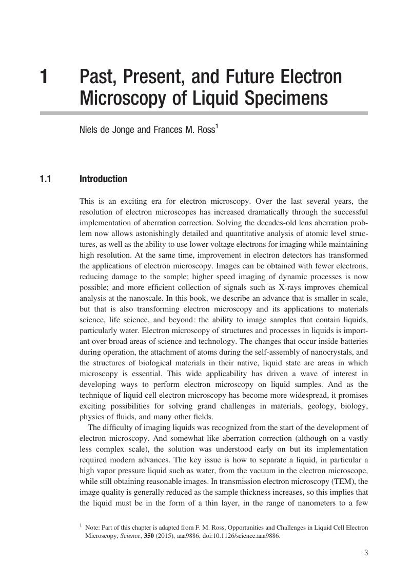
Image of the first page of this content. For PDF version, please use the ‘Save PDF’ preceeding this image.'
Type
Chapter
Information
Publisher: Cambridge University Press
Print publication year: 2016

Access options

Get access to the full version of this content by using one of the access options below. (Log in options will check for institutional or personal access. Content may require purchase if you do not have access.)

References

de Jonge, N. and Ross, F. M., Electron microscopy of specimens in liquid. Nat. Nanotechnol., 6 (2011), 695704.Google Scholar
Liao, H. G. and Zheng, H., Liquid cell transmission electron microscopy, Annu. Rev. Phys. Chem., 67 (2016), 719747.Google Scholar
Butler, E. P. and Hale, K. F., Chapter 6 in Dynamic Experiments in the Electron Microscope (Amsterdam: North-Holland, 1981).Google Scholar
Parsons, D. F., Structure of wet specimens in electron microscopy. Science, 186 (1974), 407414.CrossRefGoogle ScholarPubMed
Ruska, E., Beitrag zur uebermikroskopischen Abbildungen bei hoeheren Drucken. Kolloid Z., 100 (1942), 212219.Google Scholar
Helveg, S., López-Cartes, C., Sehested, J. et al., Atomic-scale imaging of carbon nanofibre growth. Nature, 427 (2004), 426429.CrossRefGoogle ScholarPubMed
Parsons, D. F., Matricardi, V. R., Moretz, R. C. and Turner, J. N., Electron microscopy and diffraction of wet unstained and unfixed biological objects. Adv. Biol. Med. Phys., 15 (1974), 161270.Google Scholar
Huang, J. Y., Zhong, L., Wang, C. M. et al., In situ observation of the electrochemical lithiation of a single SnO2 nanowire electrode. Science, 330 (2010), 15151520.CrossRefGoogle ScholarPubMed
Wang, C. M., Xu, W., Liu, J. et al., In situ transmission electron microscopy and spectroscopy studies of interfaces in Li ion batteries: challenges and opportunities. J. Mater. Res., 25 (2010), 15411547.Google Scholar
Wang, C.-M., Liao, H.-G. and Ross, F. M., Observation of materials processes in liquids by electron microscopy, MRS Bulletin, 40 (2015), 4652.CrossRefGoogle Scholar
Abrams, I. M. and McBain, J. W., A closed cell for electron microscopy. J. Appl. Phys., 15 (1944), 607609.Google Scholar
Heide, H. G., Elektronenmikroskopie von Objekten unter Atmosphaerendruck oder unter Drucken, welche Austricknen verhindern. Naturwissenschaften, 47 (1960), 313317.Google Scholar
Heide, H. G., Electron microscopic observation of specimens under controlled gas pressure. J. Cell Biol., 13 (1962), 147152.CrossRefGoogle ScholarPubMed
Double, D. D., Some studies of the hydration of Portland cement using high voltage (1MV) electron microscopy. Mater. Sci. Eng., 12 (1973), 2934.CrossRefGoogle Scholar
Daulton, T. L., Little, B. J., Lowe, K. and Jones-Meehan, J., In situ environmental cell–transmission electron microscopy study of microbial reduction of chromium(VI) using electron energy loss spectroscopy. Microsc. Microanal., 7 (2001), 470485.CrossRefGoogle ScholarPubMed
Chiou, W.-A. et al., In situ TEM study of DNA/gold nanoparticles in liquid environment. Microsc. Microanal., 5 (Suppl. 2) (1999), MSA.Google Scholar
Fukami, A., Fukushima, K. and Kohyama, N., Observation technique for wet clay minerals using film-sealed environmental cell equipment attached to high-resolution electron microscope. In Bennett, R. et al., eds., Microstructure of Fine-Grained Sediments (New York: Springer, 1991) pp. 321331.Google Scholar
Gai, P. L., Development of wet environment TEM (wet-ETEM) for in situ studies of liquid-catalyst reactions on the nanoscale. Microsc Microanal., 8 (2002), 2128.Google Scholar
Sugi, H. T., Akimoto, K., Sutoh, S. et al., Dynamic electron microscopy of ATP-induced myosin head movement in living muscle filaments. Proc. Natl. Acad. Sci. USA, 94 (1997), 43784392.Google Scholar
Taylor, K. A. and Glaeser, R. M., Electron microscopy of frozen hydrated biological specimens. J. Ultrastruct. Res., 55 (1976), 448456.Google Scholar
Frank, J., Three-Dimensional Electron Microscopy of Macromolecular Assemblies: Visualization of Biological Molecules in Their Native State (Oxford: Oxford University Press, 2006).Google Scholar
Lucic, V., Foerster, F. and Baumeister, W., Structural studies by electron tomography: from cells to molecules. Annu. Rev. Biochem., 74 (2005), 833865.CrossRefGoogle ScholarPubMed
Stahlberg, H. and Walz, T., Molecular electron microscopy: state of the art and current challenges. ACS Chem. Biol., 3 (2008), 268281.Google Scholar
Williamson, M. J., Tromp, R. M., Vereecken, P. M., Hull, R. and Ross, F. M., Dynamic microscopy of nanoscale cluster growth at the solid-liquid interface. Nat. Mater., 2 (2003), 532536.CrossRefGoogle ScholarPubMed
Franks, R., Morefield, S., Wen, J. et al., A study of nanomaterial dispersion in solution by wet-cell transmission electron microscopy. J. Nanosci. Nanotechnol., 8 (2008), 44044407.Google Scholar
Liu, K.-L., Wu, C.-C., Huang, Y.-J. et al., Novel microchip for in situ TEM imaging of living organisms and bio-reactions in aqueous conditions. Lab Chip, 8 (2008), 19151921.Google Scholar
Zheng, H. M., Claridge, S. A., Minor, A. M., Alivisatos, A. P. and Dahmen, U., Nanocrystal diffusion in a liquid thin film observed by in situ transmission electron microscopy. Nano Lett., 9 (2009), 24602465.CrossRefGoogle Scholar
Grogan, J. M. and Bau, H. H., The Nanoaquarium: a platform for in situ transmission electron microscopy in liquid media. J. Microelectromech. Syst., 19 (2010), 885894.Google Scholar
Leenheer, A. J., Sullivan, J. P., Shaw, M. J. and Harris, C. T., A sealed liquid cell for in situ transmission electron microscopy of controlled electrochemical processes. J. Microelectromech. Syst., 24 (2015), 10611068.Google Scholar
Tanase, M., Winterstein, J., Sharma, R. et al., High-resolution imaging and spectroscopy at high pressure: a novel liquid cell for the TEM. Microsc. Micranal., 21 (2015), 16291638.CrossRefGoogle Scholar
Mueller, C., Harb, M., Dwyer, J. R. and Miller, R. J. Dwayne, Nanofluidic cells with controlled pathlength and liquid flow for rapid, high-resolution in situ imaging with electrons. J. Phys. Chem. Lett., 4 (2013), 23392347.CrossRefGoogle Scholar
den Heijer, M., Shao, I., Radisic, A., Reuter, M. C. and Ross, F. M., Patterned electrochemical deposition of copper using an electron beam. APL Materials, 2 (2014), 022101.Google Scholar
Yuk, J. M., Park, J., Ercius, P. et al., High-resolution EM of colloidal nanocrystal growth using graphene liquid cells. Science, 336 (2012), 6164.CrossRefGoogle ScholarPubMed
Ring, E. A. and de Jonge, N., Microfluidic system for transmission electron microscopy. Microsc. Microanal., 16 (2010), 622629.CrossRefGoogle ScholarPubMed
Jungjohann, K. L., Evans, J. E., Aguiar, J., Arslan, I. and Browning, N. D., Atomic-scale imaging and spectroscopy for in situ liquid scanning transmission electron microscopy. Microsc. Microanal., 18 (2012), 621627.CrossRefGoogle ScholarPubMed
Holtz, M. E., Yu, Y., Gunceler, D. et al., Nanoscale imaging of lithium ion distribution during in situ operation of battery electrode and electrolyte. Nano Lett., 14 (2014), 14531459.Google Scholar
Zaluzec, N. J., Burke, M. G., Haigh, S. J. and Kulzick, M. A., X-ray energy-dispersive spectrometry during in situ liquid cell studies using an analytical electron microscope. Microsc. Microanal., 20 (2014), 323329.Google Scholar
Lewis, E. A., Haigh, S. J., Slater, T. J. A. et al., Real-time imaging and local elemental analysis of nanostructures in liquids. Chem. Commun., 50 (2014), 1001910022.CrossRefGoogle ScholarPubMed
Kuwabata, S., Kongkanand, A., Oyamatsu, D. and Torimoto, T., Observation of ionic liquid by scanning electron microscope. Chem. Lett., 35 (2006), 600601.Google Scholar
Arimoto, S., Sugimura, M., Kageyama, H., Torimoto, T. and Kuwabata, S., Development of new techniques for scanning electron microscope observation using ionic liquid. Electrochim. Acta, 53 (2008), 62286234.CrossRefGoogle Scholar
Swift, J. A. and Brown, A. C., An environmental cell for the examination of wet biological specimens at atmospheric pressure by transmission scanning electron microscopy. J. Phys. E, 3 (1970), 924926.Google Scholar
Danilatos, G. D., Review and outline of environmental SEM at present. J. Microsc., 162 (1991), 391402.Google Scholar
Moncrieff, D. A., Barker, P. R. and Robinson, V. N. E., Electron scattering by gas in the scanning electron microscope. J. Phys. D, 12 (1979), 481488.Google Scholar
Stokes, D. J., Principles and Practice of Variable Pressure/Environmental Scanning Electron Microscopy (VP-ESEM) (Chichester: John Wiley & Sons, 2008).CrossRefGoogle Scholar
Bogner, A., Thollet, G., Basset, D., Jouneau, P. H. and Gauthier, C., Wet STEM: a new development in environmental SEM for imaging nano-objects included in a liquid phase. Ultramicroscopy, 104 (2005), 290301.Google Scholar
Bogner, A., Jouneau, P.-H., Thollet, G., Basset, D. and Gauthier, C., A history of scanning electron microscopy developments: towards “wet-STEM” imaging. Micron, 38 (2007), 390401.CrossRefGoogle ScholarPubMed
Peckys, D. B., Baudoin, J. P., Eder, M., Werner, U. and de Jonge, N., Epidermal growth factor receptor subunit locations determined in hydrated cells with environmental scanning electron microscopy. Sci. Rep., 3 (2013), 26212626.Google Scholar
Masenelli-Varlot, K., Malchere, A., Ferreira, J. et al., Wet-STEM tomography: principles, potentialities and limitations. Microsc. Microanal., 20 (2014), 366375.CrossRefGoogle ScholarPubMed
Novotny, F., Wandrol, P., Proska, J. and Slouf, M., In situ wetSTEM observation of gold nanorod self-assembly dynamics in a drying colloidal droplet. Microsc. Microanal., 20 (2014), 385393.Google Scholar
Jansson, A., Nafari, A., Sanz-Velasco, A. et al., Novel method for controlled wetting of materials in the environmental scanning electron microscope. Microsc. Microanal., 19 (2013), 3037.Google Scholar
Jansson, A., Boissier, C., Marucci, M. et al., Novel method for visualizing water transport through phase-separated polymer films. Microsc. Microanal., 20 (2014), 394406.CrossRefGoogle ScholarPubMed
Barkay, Z., Wettability study using transmitted electrons in environmental scanning electron microscope. Appl. Phys. Lett., 96 (2010), 183109.CrossRefGoogle Scholar
Barkay, Z., In situ imaging of nano-droplet condensation and coalescence on thin water films. Microsc. Microanal., 20 (2014), 317322.Google Scholar
Thiberge, S., Nechushtan, A., Sprinzak, D. et al., Scanning electron microscopy of cells and tissues under fully hydrated conditions. Proc. Natl. Acad. Sci. USA, 101 (2004), 33463351.Google Scholar
Hell, S. W., Far-field optical nanoscopy. Science, 316 (2007), 11531158.Google Scholar
Jensen, E., Kobler, C., Jensen, P. S. and Molhave, K.. In-situ SEM microchip setup for electrochemical experiments with water based solutions. Ultramicroscopy, 129 (2013), 6369.CrossRefGoogle ScholarPubMed
Kraus, J., Reichelt, R., Günther, S. et al., Photoelectron spectroscopy of wet and gaseous samples through graphene membranes. Nanoscale, 6 (2014), 1439414403.CrossRefGoogle ScholarPubMed
Yang, W., Zhang, Y., Hilke, M. and Reisner, W., Dynamic imaging of Au-nanoparticles via scanning electron microscopy in a graphene wet cell. Nanotechnology, 26 (2015), 315703.Google Scholar
Yang, L., Yu, X.-Y., Zhu, Z., Thevuthasan, T. and Cowin, J. P., Making a hybrid microfluidic platform compatible for in situ imaging by vacuum-based techniques. J. Vac. Sci. Technol. A, 29 (2011), 061101.Google Scholar
Wojcik, M., Hauser, M., Li, W., Moon, S. and Xu, K., Graphene-enabled electron microscopy and correlated super-resolution microscopy of wet cells. Nat. Commun., 6 (2015), 7384.Google Scholar
Liv, N., Zonnevylle, A. C., Narvaez, A. C. et al., Simultaneous correlative scanning electron and high-NA fluorescence microscopy. PLOS One, 8 (2013), e55707.Google Scholar
Liv, N., Lazić, I., Kruit, P. and Hoogenboom, J. P., Scanning electron microscopy of individual nanoparticle bio-markers in liquid. Ultramicroscopy, 143 (2014), 9399.CrossRefGoogle ScholarPubMed
Nishiyama, H., Suga, M., Ogura, T. et al., Atmospheric scanning electron microscope observes cells and tissues in open medium through silicon nitride film. J. Struct. Biol., 169 (2010), 438449.Google Scholar
Nawa, Y., Inami, W., Chiba, A. et al., Dynamic and high-resolution live cell imaging by direct electron beam excitation. Opt. Express, 20 (2012), 56295635.CrossRefGoogle ScholarPubMed
Vidavsky, N., Addadi, S., Mahamid, J. et al., Initial stages of calcium uptake and mineral deposition in sea urchin embryos. Proc. Natl. Acad. Sci. USA, 111 (2014), 3944.Google Scholar
Creemer, J. F., Helveg, S., Hoveling, G. H. et al., Atomic-scale electron microscopy at ambient pressure. Ultramicroscopy, 108 (2008), 993998.Google Scholar
Jensen, E. and Molhave, K., Monolithic chip system with a microfluidic channel for in situ electron microscopy of liquids. Microsc. Microanal., 20 (2014), 445451.CrossRefGoogle ScholarPubMed
Radisic, A., Vereecken, P. M., Hannon, J. B., Searson, P. C. and Ross, F. M., Quantifying electrochemical nucleation and growth of nanoscale clusters using real-time kinetic data. Nano Lett., 6 (2006), 238242.Google Scholar
Verch, A., Pfaff, M. and De Jonge, N., Exceptionally slow movement of gold nanoparticles at a solid:liquid interface investigated by scanning transmission electron microscopy. Langmuir, 31 (2015), 69566964.Google Scholar
Krueger, M., Berg, S., Stone, D. A. et al., Drop-casted self-assembling graphene oxide membranes for scanning electron microscopy on wet and dense gaseous samples. ACS Nano, 5 (2011), 1004710054.Google Scholar
Schneider, N. M., Norton, M. M., Mendel, B. J. et al., Electron–water interactions and implications for liquid cell electron microscopy. J. Phys. Chem. C, 118 (2014), 2237322382.Google Scholar
Grogan, J. M., Schneider, N. M., Ross, F. M. and Bau, H. H., Bubble and pattern formation in liquid induced by an electron beam. Nano Lett., 14 (2014), 359364.CrossRefGoogle ScholarPubMed
Zheng, H. M., Smith, R. K., Jun, Y. W. et al., Observation of single colloidal platinum nanocrystal growth trajectories. Science, 324 (2009), 13091312.CrossRefGoogle ScholarPubMed
van de Put, M. W., Carcouet, C. C., Bomans, P. H. et al., Writing silica structures in liquid with scanning transmission electron microscopy. Small, 11 (2015), 585590.Google Scholar
Sutter, E., Jungjohann, K., Bliznakov, S. et al., In situ liquid-cell electron microscopy of silver-palladium galvanic replacement reactions on silver nanoparticles. Nat. Commun., 5 (2014), 4946.Google Scholar
Unocic, R. R., Sacci, R. L., Brown, G. M. et al., Quantitative electrochemical measurements using in situ ec-S/TEM devices. Microsc. Microanal., 20 (2014), 452461.Google Scholar
Mehdi, B. L., Gu, M., Parent, L. R. et al., In situ electrochemical transmission electron microscopy for battery research. Microsc. Microanal., 20 (2014), 484492.Google Scholar
Radisic, A., Ross, F. M. and Searson, P. C., In situ study of the growth kinetics of individual islands during electrodeposition of copper. J. Phys. Chem. B, 110 (2006), 78627868.Google Scholar
Radisic, A., Vereecken, P. M., Searson, P. C. and Ross, F. M., The morphology and nucleation kinetics of copper islands during electrodeposition. Surf. Sci., 600 (2006), 18171826.CrossRefGoogle Scholar
Schneider, N. M., Park, J. H. and Grogan, J. M., Visualization of active and passive control of morphology during electrodeposition. Microsc. Microanal., 20 (2014), 15301531.CrossRefGoogle Scholar
White, E. R., Singer, S. B., Augustyn, V. et al., In situ transmission electron microscopy of lead dendrites and lead ions in aqueous solution. ACS Nano, 6 (2012), 63086317.Google Scholar
Sun, M., Liao, H.-G., Niu, K. and Zheng, H., Structural and morphological evolution of lead dendrites during electrochemical migration. Sci. Rep., 3 (2013), 2227.Google Scholar
Zeng, Z., Liang, W.-I., Liao, H.-G. et al., Visualization of electrode-electrolyte interfaces in LiPF6/EC/DEC electrolyte for lithium ion batteries via in-situ TEM. Nano Lett., 14 (2014), 17451750.Google Scholar
Mehdi, B. L., Qian, J., Nasybulin, E. et al., Observation and quantification of nanoscale processes in lithium batteries by operando electrochemical (S)TEM. Nano Lett., 15 (2015), 21682173.Google Scholar
Sacci, R. L., Black, J. M., Balke, N. et al., Nanoscale imaging of fundamental Li battery chemistry: solid-electrolyte interphase formation and preferential growth of lithium metal nanoclusters. Nano Lett., 15 (2015), 20112018.CrossRefGoogle ScholarPubMed
Leenheer, A. J., Jungjohann, K. L., Zavadil, K. R., Sullivan, J. P. and Harris, C. T., Lithium electrodeposition dynamics in aprotic electrolyte observed in situ via transmission electron microscopy, ACS Nano, 9 (2015), 43794389.Google Scholar
Gu, M., Parent, L. R., Mehdi, L. et al., Demonstration of an electrochemical liquid cell for operando transmission electron microscopy observation of the lithiation/delithiation behavior of Si nanowire battery anodes. Nano Lett., 13 (2013), 61066112.Google Scholar
Sacci, R. L., Dudney, N. J., More, K. L. et al., Direct visualization of initial SEI morphology and growth kinetics during lithium deposition by in situ electrochemical transmission electron microscopy. Chemical Commun., 50 (2013), 21042107.Google Scholar
Wu, F. and Yao, N., Advances in sealed liquid cells for in-situ TEM electrochemical investigation of lithium-ion battery. Nano Energy, 11 (2015), 196210.Google Scholar
Abellan Baeza, P., Mehdi, B. L., Parent, L. R. et al., Probing the degradation mechanisms in electrolyte solutions for Li-ion batteries by in-situ transmission electron microscopy. Nano Lett., 14 (2014), 12931299.Google Scholar
Chee, S. W.. Duquette, D., Ross, F. M. and Hull, R., Metastable structures in Al thin films prior to the onset of corrosion pitting as observed using liquid cell transmission electron microscopy. Microsc. Microanal., 20 (2014), 462468.Google Scholar
Chee, S. W., Pratt, S. H., Hattar, K. et al., Studying localized corrosion using liquid cell transmission electron microscopy. Chem. Commun., 51 (2015), 168171.Google Scholar
Zhong, X., Burke, M. G., Schilling, S., Haigh, S. J. and Zaluzec, N. J., Novel hybrid sample preparation method for in situ liquid cell TEM analysis. Microsc. Microanal., 20 (S3) (2014), 15141515.CrossRefGoogle Scholar
Woehl, T. J., Evans, J. E., Arslan, I., Ristenpart, W. D. and Browning, N. D., Direct in situ determination of the mechanisms controlling nanoparticle nucleation and growth. ACS Nano, 6 (2012), 85998610.Google Scholar
Liao, H. G., Cui, L. K., Whitelam, S. and Zheng, H. M., Real-time imaging of Pt3Fe nanorod growth in solution. Science, 336 (2012), 10111014.CrossRefGoogle ScholarPubMed
Jungjohann, K. L., Bliznakov, S., Sutter, P. W., Stach, E. A. and Sutter, E. A., In situ liquid cell electron microscopy of the solution growth of Au–Pd core–shell nanostructures. Nano Lett., 13 (2013), 29642970.Google Scholar
De Clercq, A., Dachraoui, W., Margeat, O. et al., Growth of Pt−Pd nanoparticles studied in situ by HRTEM in a liquid cell. J. Phys. Chem. Lett., 5 (2014), 21262130.Google Scholar
Parent, L. R., Robinson, D. R., Woehl, T. J. et al., Direct in situ observation of nanoparticle synthesis in a liquid crystal surfactant template. ACS Nano, 6 (2012), 35893596.Google Scholar
Zhu, G., Jiang, Y., Lin, F. et al., In situ study of the growth of two-dimensional palladium dendritic nanostructures using liquid-cell electron microscopy. Chem Commun., 50 (2014), 94479450.Google Scholar
Kraus, T. and de Jonge, N., Dendritic gold nanowire growth observed in liquid with transmission electron microscopy. Langmuir, 29 (2013), 84278432.Google Scholar
Jiang, Y., Zhu, G., Lin, F. et al., In situ study of oxidative etching of palladium nanocrystals by liquid cell electron microscopy. Nano Lett., 14 (2014), 37613765.Google Scholar
Wu, J., Gao, W., Yang, H. and Zuo, J.-M., Imaging shape-dependent corrosion behavior of Pt nanoparticles over extended time using a liquid flow cell and TEM. Microsc. Microanal., 20 (S3) (2014), 15081509.Google Scholar
Park, J. H., Schneider, N. M., Grogan, J. M. et al., Control of electron beam-induced Au nanocrystal growth kinetics through solution chemistry. Nano Lett., 15 (2015), 53145320.Google Scholar
Noh, K. W., Liu, Y., Sun, L. and Dillon, S. J., Challenges associated with in-situ TEM in environmental systems: the case of silver in aqueous solutions. Ultramicroscopy, 116 (2012), 3438.Google Scholar
Liao, H.-G. and Zheng, H., Liquid cell transmission electron microscopy study of platinum iron nanocrystal growth and shape evolution. J. Am. Chem. Soc., 135 (2013), 50385043.Google Scholar
Liao, H. G., Zherebetskyy, D., Xin, H. et al., Facet development during platinum nanocube growth. Science, 345 (2014), 916919.Google Scholar
Liu, Y., Tai, K. and Dillon, S. J., Growth kinetics and morphological evolution of ZnO precipitated from solution. Chem. Mater., 25 (2013), 29272933.Google Scholar
Kimura, Y., Niinomi, H., Tsukamoto, K. and García-Ruiz, J. M., In situ live observation of nucleation and dissolution of sodium chlorate nanoparticles by transmission electron microscopy. J. Am. Chem. Soc., 136 (2014), 17621765.Google Scholar
Xin, H. L. and Zheng, H., In Situ observation of oscillatory growth of bismuth nanoparticles. Nano Lett., 12 (2012), 14701474.Google Scholar
Niu, K.-Y., Park, J., Zheng, H. and Alivisatos, A.P., Revealing bismuth oxide hollow nanoparticle formation by the Kirkendall effect. Nano Lett., 13 (2013), 57155719.CrossRefGoogle ScholarPubMed
Evans, J. E., Jungjohann, K. L., Browning, N. D. and Arslan, I., Controlled growth of nanoparticles from solution with in situ liquid transmission electron microscopy. Nano Lett., 11 (2011), 28092813.Google Scholar
Donev, E. U. and Hastings, J. T., Electron-beam-induced deposition of platinum from a liquid precursor. Nano Lett., 9 (2009), 27152718.Google Scholar
Zheng, H. M., Mirsaidov, U. M., Wang, L. W. and Matsudaira, P., Electron beam manipulation of nanoparticles. Nano Lett., 12 (2012), 56445648.Google Scholar
Li, D. S., Nielsen, M. H., Lee, J. R. I. et al., Direction-specific interactions control crystal growth by oriented attachment. Science, 336 (2012), 10141018.Google Scholar
Woehl, T. J., Park, C., Evans, J. E. et al., Direct observation of aggregative nanoparticle growth: kinetic modeling of the size distribution and growth rate. Nano Lett., 14 (2013), 373378.Google Scholar
Grogan, J. M., Rotkina, L. and Bau, H. H., In situ liquid-cell electron microscopy of colloid aggregation and growth dynamics. Phys. Rev. E, 83 (2011), 061405.Google Scholar
Park, J., Zheng, H., Lee, W. C. et al., Direct observation of nanoparticle superlattice formation by using liquid cell transmission electron microscopy. ACS Nano, 6 (2012), 20782085.CrossRefGoogle ScholarPubMed
Liu, Y., Lin, X.-M., Sun, Y. and Rajh, T., In situ visualization of self-assembly of charged gold nanoparticles. J. Am. Chem. Soc., 135 (2013), 37643767.CrossRefGoogle ScholarPubMed
White, E. R., Mecklenburg, M., Singer, S. B., Aloni, S. and Regan, B. C., Imaging nanobubbles in water with scanning transmission electron microscopy. Appl. Phys. Express, 4 (2011), 055201.Google Scholar
Tai, K., Liu, Y. and Dillon, S. J., In situ cryogenic transmission electron microscopy for characterizing the evolution of solidifying water ice in colloidal systems. Microsc. Microanal., 20 (2014), 330337.Google Scholar
Bhattacharya, D., Bosman, M., Mokkapati, V. R. S. S., Leong, F. Y. and Mirsaidov, U., Nucleation dynamics of water nanodroplets. Microsc. Microanal., 20 (2014), 407415.Google Scholar
Ruan, C.-Y., Lobastov, V. A., Vigliotti, F., Chen, S. and Zewail, A. H., Ultrafast electron crystallography of interfacial water. Science, 304 (2004), 8084.Google Scholar
Mirsaidov, U., Ohl, C.-D. and Matsudaira, P., A direct observation of nanometer-size void dynamics in an ultra-thin water film. Soft Matter, 8 (2012), 71087111.Google Scholar
Mirsaidov, U. M., Zheng, H., Bhattacharya, D., Casana, Y. and Matsudaira, P., Direct observation of stick-slip movements of water nanodroplets induced by an electron beam. Proc. Natl. Acad. Sci. USA, 109 (2012), 71877190.CrossRefGoogle ScholarPubMed
Norton, M., Park, J. H., Kodambaka, S., Ross, F. M. and Bau, H., Dynamics of sub-micron bubbles growing in a wedge in the low capillary number regime. Bull. Ameri. Phys. Soc., 59 (2014); and Bau, H., Grogan, J. M., Norton, M. and Ross, F. M., On the surface tension of nanobubbles. APS Division of Fluid Dynamics Meeting (2013).Google Scholar
Mattia, D. and Gogotsi, Y., Review: static and dynamic behavior of liquids inside carbon nanotubes. Microfluidics and Nanofluidics, 5 (2008), 289305.Google Scholar
Mirsaidov, U., Mokkapati, V. R. S. S., Bhattacharya, D. et al., Scrolling graphene into nanofluidic channels. Lab Chip, 13 (2013), 28742878.Google Scholar
Ring, E. A. and de Jonge, N., Video-frequency scanning transmission electron microscopy of moving gold nanoparticles in liquid. Micron, 43 (2012), 10781084.Google Scholar
White, E. R., Mecklenburg, M., Shevitski, B., Singer, S. B. and Regan, B. C., Charged nanoparticle dynamics in water induced by scanning transmission electron microscopy. Langmuir, 28 (2012), 36953698.Google Scholar
Lu, J. Y., Aabdin, Z., Loh, N. D., Bhattacharya, D. and Mirsaidov, U., Nanoparticle dynamics in a nanodroplet. Nano Lett., 14 (2014), 21112115.Google Scholar
Chen, Q., Smith, J. M., Park, J. et al., 3D motion of DNA-Au nanoconjugates in graphene liquid cell electron microscopy. Nano Lett., 13 (2013), 45564561.Google Scholar
Cazade, P.-A., Hartkamp, R. and Coasne, B., Structure and dynamics of an electrolyte confined in charged nanopores. J. Phys. Chem. C, 118 (2014), 50615072.Google Scholar
Kim, H. I., Kushmerick, J. G., Houston, J. E. and Bunker, B. C., Viscous “interphase” water adjacent to oligo(ethylene glycol)-terminated monolayers. Langmuir, 19 (2003), 92719275.Google Scholar
Kashyap, S., Woehl, T. J., Liu, X., Mallapragada, S. K. and Prozorov, T., Nucleation of iron oxide nanoparticles mediated by Mms6 protein in situ, ACS Nano, 8 (2014), 90979106.Google Scholar
Nielsen, M. H., Lee, J. R. I., Hu, Q. N., Han, T. Y. J., and De Yoreo, J. J., Structural evolution, formation pathways and energetic controls during template-directed nucleation of CaCO3. Faraday Discuss., 159 (2012), 105121.Google Scholar
Nielsen, M. H., Aloni, S. and De Yoreo, J. J., In situ TEM imaging of CaCO3 nucleation reveals coexistence of direct and indirect pathways. Science, 345 (2014), 11581162.Google Scholar
Smeets, P. J. M., Cho, K. R., Kempen, R. G. E., Sommerdijk, N. A. J. M. and De Yoreo, J. J., In situ TEM shows ion binding is key to directing CaCO3 nucleation in a biomimetic matrix. Nat. Mater., 14 (2015), 394399.Google Scholar
Woehl, T. J., Kashyap, S., Firlar, E. et al., Correlative electron and fluorescence microscopy of magnetotactic bacteria in liquid: toward in vivo imaging. Sci. Rep., 4 (2014), 6854.Google Scholar
de Jonge, N., Peckys, D. B., Kremers, G. J. and Piston, D. W., Electron microscopy of whole cells in liquid with nanometer resolution. Proc. Natl. Acad. Sci. USA, 106 (2009), 21592164.Google Scholar
Peckys, D. B., Korf, U. and de Jonge, N., Local variations of HER2 dimerization in breast cancer cells discovered by correlative fluorescence and liquid electron microscopy. Sci. Adv., 1 (2015), e1500165.Google Scholar
Peckys, D. B. and de Jonge, N., Visualization of gold nanoparticle uptake in living cells with liquid scanning transmission electron microscopy. Nano Lett., 11 (2011), 17331738.Google Scholar
Dukes, M. J., Peckys, D. and de Jonge, N., Correlative fluorescence- and scanning transmission electron microscopy of quantum dot labeled proteins on whole cells in liquid. ACS Nano, 4 (2010), 41104116.Google Scholar
Pohlmann, E. S., Patel, K., Guo, S. et al., Real-time visualization of nanoparticles interacting with glioblastoma stem cells. Nano Lett., 15 (2015), 23292335.Google Scholar
Peckys, D. B., Baudoin, J.-P., Eder, M., Werner, U. and de Jonge, N., Epidermal growth factor receptor subunit locations determined in hydrated cells with environmental scanning electron microscopy. Sci. Rep., 3 (2013), 2626.Google Scholar
Peckys, D. B., Mazur, P., Gould, K. L. and de Jonge, N., Fully hydrated yeast cells imaged with electron microscopy. Biophys. J., 100 (2011), 25222529.Google Scholar
Peckys, D. B. and de Jonge, N., Liquid scanning transmission electron microscopy: imaging protein complexes in their native environment in whole eukaryotic cells. Microsc. Microanal., 20 (2014), 189198.Google Scholar
Glaeser, R., Comment on electron microscopy of biological specimens in liquid water. Biophys. J., 103 (2012), 163164.Google Scholar
Kirk, S. E., Skepper, J. N. and Donald, A. M., Application of environmental scanning electron microscopy to determine biological surface structure. J. Microsc., 233 (2009), 205224.Google Scholar
Sugi, H., Minoda, H., Inayoshi, Y. et al., Direct demonstration of the cross-bridge recovery stroke in muscle thick filaments in aqueous solution by using the hydration chamber. Proc. Natl. Acad. Sci. USA, 105 (2008), 1739617401.Google Scholar
Sugi, H., Chaen, S., Akimoto, T. et al., Electron microscopic recording of myosin head power stroke in hydrated myosin filaments. Sci. Rep., 5 (2015), 15700.Google Scholar
Mohanty, N., Fahrenholtz, M., Nagaraja, A., Boyle, D. and Berry, V., Impermeable graphenic encasement of bacteria. Nano Lett., 11 (2011), 12701275.Google Scholar
Park, J., Park, H., Ercius, P. et al., Direct observation of wet biological samples by graphene liquid cell transmission electron microscopy. Nano Lett., 15 (2015), 47374744.Google Scholar
Dukes, M. J., Thomas, R., Damiano, J. et al., Improved microchip design and application for in situ transmission electron microscopy of macromolecules. Microsc. Microanal., 20 (2014), 338345.Google Scholar
Degen, K., Dukes, M., Tanner, J. R. and Kelly, D. F., The development of affinity capture devices: a nanoscale purification platform for biological in situ transmission electron microscopy. RSC Adv., 2 (2012), 24082412.Google Scholar
Gilmore, B. L., Showalter, S. P., Dukes, M. J. et al., Visualizing viral assemblies in a nanoscale biosphere. Lab Chip, 13 (2013), 216219.Google Scholar
Wang, C., Qiao, Q., Shokuhfar, T. and Klie, R. F., High-resolution electron microscopy and spectroscopy of ferritin in biocompatible graphene liquid cells and graphene sandwiches. Adv. Mater., 26 (2014), 34103414.Google Scholar
Hoppe, S. M., Sasaki, D. Y., Kinghorn, A. N. and Hattar, K., In-situ transmission electron microscopy of liposomes in an aqueous environment. Langmuir, 29 (2013), 99589961.Google Scholar
Proetto, M. T., Rush, A. M., Chien, M.-P. et al., Dynamics of soft nanomaterials captured by transmission electron microscopy in liquid water. J. Am. Chem. Soc., 136 (2014), 11621165.Google Scholar
Plamper, F. A., Gelissen, A. P., Timper, J. et al., Spontaneous assembly of miktoarm stars into vesicular interpolyelectrolyte complexes. Macromol. Rapid Commun., 34 (2013), 855860.Google Scholar
Mirsaidov, U. M., Zheng, H., Casana, Y. and Matsudaira, P., Imaging protein structure in water at 2.7 nm resolution by transmission electron microscopy. Biophys. J., 102 (2012), L15–17.Google Scholar
Varano, A. C., Rahimi, A., Dukes, M. J. et al., Visualizing virus particle mobility in liquid at the nanoscale, Chem. Commun., 51 (2015), 1617616179.CrossRefGoogle Scholar
Maraloiu, V. A., Hamoudeh, M., Fessi, H. and Blanchin, M. G., Study of magnetic nanovectors by Wet-STEM, a new ESEM mode in transmission. J. Coll. Interf. Sci., 352 (2010), 386392.Google Scholar
Adachi, K., Freney, E. J., Buseck, P. R., Shapes of internally mixed hygroscopic aerosol particles after deliquescence, and their effect on light scattering. Geophys. Res. Lett., 38 (2011), L13804.CrossRefGoogle Scholar
de Gennes, P. G., Wetting: statics and dynamics. Rev. Mod. Phys., 57 (1985), 827863.Google Scholar
Liu, J., Wei, B., Sloppy, J. D. et al., Direct imaging of electrochemical deposition of poly(3,4-ethylene dioxythiophene) (PEDOT) by transmission electron microscopy. Macro Lett., 4 (2015), 897900.Google Scholar
Sadki, S., Schottland, P., Brodie, N. and Sabouraud, G., The mechanisms of pyrrole electropolymerization. Chem. Soc. Rev., 29 (2000), 283293.Google Scholar
Ross, F. M., Controlling nanowire structures through real time growth studies. Rep. Prog. Phys., 73 (2010), 114501114522.Google Scholar
Zhang, L., Miller, B. K. and Crozier, P. A., Atomic level observation of surface amorphization in anatase nanocrystals during light irradiation in water vapor. Nano Lett., 13 (2013), 679684.Google Scholar
Zhu, G.-Z., Prabhudev, S., Yang, J. et al., In situ liquid cell TEM study of morphological evolution and degradation of Pt–Fe nanocatalysts during potential cycling. J. Phys. Chem. C, 118 (2014), 2211122119.Google Scholar
Browning, N. D., Bonds, M. A., Campbell, G. H. et al., Recent developments in dynamic transmission electron microscopy. Curr. Opin. Solid State Mater. Sci., 16 (2012), 2330.Google Scholar
Mourik, M. W., van Engelen, W. J., Vredenbregt, E. J. D. and Luiten, O. J., Ultrafast electron diffraction using an ultracold source. Struct. Dynam., 1 (2014), 034302.Google Scholar
de Jonge, N., System and methods for live cell transmission electron microscopy. US Patent Application 13,299,241 (2011).Google Scholar
Danev, R. and Nagayama, K., Transmission electron microscopy with Zernike phase plate. Ultramicroscopy, 88 (2001), 243252.Google Scholar
Simon, P., Lichte, H., Formanek, P. et al., Electron holography of biological samples. Micron, 39 (2008), 229256.Google Scholar

Save book to Kindle

To save this book to your Kindle, first ensure coreplatform@cambridge.org is added to your Approved Personal Document E-mail List under your Personal Document Settings on the Manage Your Content and Devices page of your Amazon account. Then enter the ‘name’ part of your Kindle email address below. Find out more about saving to your Kindle.

Note you can select to save to either the @free.kindle.com or @kindle.com variations. ‘@free.kindle.com’ emails are free but can only be saved to your device when it is connected to wi-fi. ‘@kindle.com’ emails can be delivered even when you are not connected to wi-fi, but note that service fees apply.

Find out more about the Kindle Personal Document Service.

Available formats
×

Save book to Dropbox

To save content items to your account, please confirm that you agree to abide by our usage policies. If this is the first time you use this feature, you will be asked to authorise Cambridge Core to connect with your account. Find out more about saving content to Dropbox.

Available formats
×

Save book to Google Drive

To save content items to your account, please confirm that you agree to abide by our usage policies. If this is the first time you use this feature, you will be asked to authorise Cambridge Core to connect with your account. Find out more about saving content to Google Drive.

Available formats
×