Skip to main content Accessibility help
×
Hostname: page-component-76fb5796d-skm99 Total loading time: 0 Render date: 2024-04-25T12:55:13.587Z Has data issue: false hasContentIssue false

Examination 3

Published online by Cambridge University Press:  05 March 2012

Usman Shaikh
Affiliation:
Royal Liverpool University Hospital
John Curtis
Affiliation:
University Hospital Aintree
Rebecca Hanlon
Affiliation:
University Hospital Aintree
David White
Affiliation:
University Hospital Aintree
Andrew Dunn
Affiliation:
Royal Liverpool University Hospital
Andrew Healey
Affiliation:
University Hospital Aintree
Jane Belfield
Affiliation:
Royal Liverpool University Hospital
Elizabeth Kneale
Affiliation:
University Hospital Aintree
Peter Dangerfield
Affiliation:
University of Liverpool
Hilary Fewins
Affiliation:
Liverpool Heart and Chest Hospital
Get access

Summary

AP radiograph of the pelvis

  1. Lesser trochanter of the right femur. The iliopsoas tendon attaches here. This is a powerful flexor of the hip.

  2. Greater trochanter of the right femur. Gluteus medius and gluteus minimis attach here. These tendons act to perform hip abduction and lateral rotation. They can produce avulsion fractures of the greater trochanter in trauma.

  3. Left L5 transverse process. The ilio-lumbar ligament attaches here. Traction of this ligament in pelvic trauma can cause an avulsion fracture of the transverse process. It also acts as an anatomical landmark on MRI for identifying the L5 vertebral body.

  4. Pubic symphysis. It is a secondary cartilaginous joint.

  5. Left inferior pubic ramus. Adductor magnus and adductor brevis attach here acting to adduct the hip.

Axial T2-weighted lumbar spine through L5

  1. Left L5 nerve. At the level of the L5/S1 disc, the L5 nerve has already left the neural exit foramen. It may become compromised by a far lateral L5 disc herniation in this position.

  2. Nucleus pulposus of L5/S1 disc. This soft central component of the disc is surrounded by the tough outer annulus fibrosus. Annular defects result in herniation of the nucleus pulposus referred to as protrusions or extrusions, based upon their morphology. On T2-weighted images the nucleus pulposus is of high signal and the annulus fibrosus is of low signal intensity.

  3. Left lamina of L5 vertebra. Each lamina fuses in the midline to form the spinous process. The lamina is partly or completely resected (laminectomy) during lumbar disc surgery to facilitate access to the disc.

  4. Right psoas major muscle. This is a powerful hip flexor. In the clinical setting of lumbar discitis it is common to see infection tracking from the disc space into the psoas muscle to form a psoas abscess.

  5. Right S1 nerve.

Type
Chapter
Information
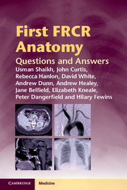
First FRCR Anatomy
Questions and Answers
, pp. 78 - 84
Publisher: Cambridge University Press
Print publication year: 2012

Access options

Get access to the full version of this content by using one of the access options below. (Log in options will check for institutional or personal access. Content may require purchase if you do not have access.)

Save book to Kindle

To save this book to your Kindle, first ensure coreplatform@cambridge.org is added to your Approved Personal Document E-mail List under your Personal Document Settings on the Manage Your Content and Devices page of your Amazon account. Then enter the ‘name’ part of your Kindle email address below. Find out more about saving to your Kindle.

Note you can select to save to either the @free.kindle.com or @kindle.com variations. ‘@free.kindle.com’ emails are free but can only be saved to your device when it is connected to wi-fi. ‘@kindle.com’ emails can be delivered even when you are not connected to wi-fi, but note that service fees apply.

Find out more about the Kindle Personal Document Service.

Available formats
×

Save book to Dropbox

To save content items to your account, please confirm that you agree to abide by our usage policies. If this is the first time you use this feature, you will be asked to authorise Cambridge Core to connect with your account. Find out more about saving content to Dropbox.

Available formats
×

Save book to Google Drive

To save content items to your account, please confirm that you agree to abide by our usage policies. If this is the first time you use this feature, you will be asked to authorise Cambridge Core to connect with your account. Find out more about saving content to Google Drive.

Available formats
×