Skip to main content Accessibility help
×
  • Cited by 3
Publisher:
Cambridge University Press
Online publication date:
December 2009
Print publication year:
2008
Online ISBN:
9780511551734

Book description

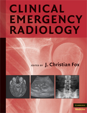
Imaging represents the most dynamic sub-discipline of emergency medicine, as recent technological advances have revealed. The use of ultrasound, MRI, and CT scans has revolutionized the way that acute injuries and conditions are managed in the emergency room. More sophisticated imaging modalities are commonplace now, enabling acute conditions such as cardiac arrest, aortic aneurysm, and fetal trauma to be diagnosed within seconds. This book is a new clinical resource in the field of emergency radiology and covers both the technical applications and interpretation of all imaging studies utilized in the emergency room, including x-rays, MRI, CT, and contrast angiography. The full spectrum of conditions diagnosed within each modality is covered in detail, and examples of normal radiologic anatomy, patterns, and anomalies are also included. With over 2,000 images to comprehensively cover every aspect of radiology in the emergency room, it is a standard reference for emergency physicians.

Refine List

Actions for selected content:

Select all | Deselect all
  • View selected items
  • Export citations
  • Download PDF (zip)
  • Save to Kindle
  • Save to Dropbox
  • Save to Google Drive

Save Search

You can save your searches here and later view and run them again in "My saved searches".

Please provide a title, maximum of 40 characters.
×

Contents


Page 2 of 2


  • 26 - Soft Tissue Ultrasound
    pp 358-366
  • View abstract

    Summary

    The use of ultrasound in the evaluation of soft tissue structures has many potential applications. When evaluating a soft tissue infection with ultrasound, consideration of several key principles will increase the potential for successful image acquisition. Soft tissue foreign bodies represent a troubling entity for emergency physicians. They often pose remarkable clinical challenges, in identification and removal, and represent a significant component of malpractice claims against emergency physicians. Most soft tissue ultrasound is best performed with a high-frequency (7 to 13 MHz) linear probe. The detection of foreign bodies may prove to be of difficulty for the novice sonologist. The differentiation between peritonsillar cellulitis and abscess can be difficult based solely on clinical findings. As with many soft tissue ultrasounds, appropriate patient preparation will improve both procedural tolerance and image acquisition. The primary limitation in the sonographic evaluation of suspected peritonsillar abscess is patient tolerance.
  • 27 - Ultrasound in Resuscitation
    pp 367-396
  • View abstract

    Summary

    Ultrasound in resuscitation is a necessary tool for evaluating the emergent and unstable patient presenting to the ED. When compared to plain radiography in the diagnosis of free fluid in the thoracic, cardiac, or abdominal cavities, ultrasound is more accurate and time efficient. Severe hypoxia, hypotension, dyspnea, chest pain, ECG abnormalities, tachyarrhythmias, and high-risk conditions such as malignancy and renal failure are a few of the indications to image cardiothoracic structures and their functioning. Cardiac sonography is considered the gold standard of cardiac diagnostic and functional testing. Transesophageal echocardiography (TEE) holds the advantage over transthoracic echocardiography (TTE) of being able to look at the major pulmonary arteries. Unlike other diagnostic imaging modalities, ultrasound is limited by the operator's ability to perform bedside ultrasound. Ultrasound does not allow the physician to distinguish fluid type such as blood and urine, or to evaluate the retroperitoneum.
  • 28 - CT in the ED: Special Considerations
    pp 399-403
  • View abstract

    Summary

    This chapter reviews the renal effects of radiological contrast media and pretreatment options in the patient with underlying renal insufficiency. Many patients will report an allergic reaction to contrast media. Diagnostic imaging, in general, exposes a patient to such a minimal dose of radiation that ED physicians often order multiple imaging tests on the same patient without regard to the radiation dose the patient receives. However, when a patient is pregnant or very young, the radiation exposure is more concerning, and the necessity of the imaging test must be balanced by the risk of the dose of radiation. In patients with renal insufficiency, diabetes, adverse reactions to contrast, and pregnancy, CT scans are not completely without risk. A careful consideration of these risks balanced with the clinical benefits of the diagnostic study should guide clinical decision making when considering CT scans in these patients.
  • 29 - CT of the Spine
    pp 404-419
  • View abstract

    Summary

    Computed tomography (CT) of the spine is becoming one of the most common studies in the modern ED. CT is the fastest, cost-effective method of imaging the spine and provides the most accurate information for diagnosis, with the exception of some soft tissue or spinal cord lesions. CT is exactly as its name implies, a mathematical approximation of the multiple densities seen as the X-ray beam rotates around the patient. The pediatric population presents a wide array of pitfalls for the clinician when evaluating the spine. Dislocations and ligamentous injury are more common in the pediatric population; they can be occult on CT and require MRI. They also present with multiple levels of injury, and complete imaging of the spine is indicated if injury is found at any level of the spine. Correct clinical judgment is required prior to sending a patient to the scanner.
  • 30 - CT Imaging of the Head
    pp 420-437
  • View abstract

    Summary

    Head computed tomography (CT) is one of the most common imaging studies ordered from the ED. Head CT is used in the evaluation of the patient who presents with headache, altered mental status, suspected stroke, or other acute neurological abnormalities. Current third-generation CT scanners are very sensitive for the detection of acute hemorrhage and bony injury, the two principal pathologies sought in evaluation of patients with head injury. In the patient with head injury, head CT will readily identify skull fractures, although fractures through the thinnest areas of the base of the skull may be missed in the axial cuts. In patients with altered mental status and other neurological abnormalities, head CT is extremely valuable in detecting pathology that requires emergent intervention. The vast majority of head CTs performed in the ED are done without contrast. Artifact is an important consideration in the interpretation of head CTs.
  • 31 - CT Imaging of the Face
    pp 438-456
  • View abstract

    Summary

    This chapter provides a brief overview of the methods for obtaining computed tomography (CT) images of the face and the facial buttress system of analysis. It presents four main categories of midfacial fractures: orbital blowout, zygoma, maxilla, and mandible. Images in the axial plane are useful in evaluating the zygomatic arch, posterior walls of the maxillary and frontal sinuses, and degree of posterior displacement in Le Fort fractures and the zygomaticomaxillary complex fractures. The zygoma is the second most common fractured midfacial bone after the nasal bones and represents 13% of all craniofacial fractures. Fractures to the maxilla occur unilaterally or bilaterally. Isolated unilateral fractures are uncommon. The use of CT to identify isolated mandibular fractures has been increasing. Non-displaced symphyseal fractures are better viewed using CT because the problem of overlapping spine lucency on plain film is not an issue when using CT.
  • 32 - CT of the Chest
    pp 457-472
  • View abstract

    Summary

    Chest computed tomography (CT) is extremely useful in the assessment of injuries to the aorta, chest wall, lung parenchyma, airway, pleura, and diaphragm. It is very useful in estimating the extent of contusion, which is important in predicting the degree of posttraumatic respiratory insufficiency. The sensitivity of CT in detecting pulmonary contusions is very high. Pneumothorax succeeds rib fractures as the second most common injury seen in chest trauma. CT is able to detect pneumothoraces missed by initial chest radiograph in 5% to 15% of trauma patients. Empyemas are exudates associated with pulmonary infections. On CT, they usually have a regularly shaped lumen and a smooth inner surface. Traumatic rupture of the aorta (TRA) is rarely visualized on CT because patients expire from exsanguination before reaching medical facilities. Posterior dislocations are more easily diagnosed on CT. Scapular fractures are frequently overlooked on the interpretation of chest radiographs.
  • 33 - CT of the Abdomen and Pelvis
    pp 473-481
  • View abstract

    Summary

    Computed tomography (CT) scanning has widely become the diagnostic test of choice for patients presenting with abdominal or pelvic pain and for the stable trauma patient to evaluate for intra abdominal injury. Due to the limitations of plain radiographs, CT scanning is also being increasingly used in cases of bowel obstruction to help delineate the location, severity, and underlying cause of the obstruction. First-generation, single-slice CT scanners have almost been completely replaced with multidetector CT (MDCT) scanners. The faster acquisition of images has reduced motion artifact because the entire abdomen can be scanned on a single breathhold. Conditions in the right upper quadrant that can be easily diagnosed by CT are pancreatitis, ascending cholangitis, perforated hollow viscus, and hepatic tumor or abscess. A major disadvantage of MDCT is the massive amount of data that are generated during image acquisition. This makes efficient transfer of image data to hard copies challenging.
  • 34 - CT Angiography of the Chest
    pp 482-488
  • View abstract

    Summary

    The three most common life-threatening conditions facing patients with chest pain in the ED are myocardial ischemia, pulmonary embolism, and aortic dissection. Coronary heart disease is the leading cause of death in the United States, responsible for about 817,000 deaths each year. Multidetector Computed Tomography (MDCT) coronary angiography has emerged as a highly sensitive tool to diagnose both obstructive and non-obstructive coronary artery disease. The sensitivity and specificity of CT for the detection of pulmonary embolism exceeds 90%, and a negative spiral CT is associated with a good prognosis when anticoagulation is withheld. The mortality of untreated aortic dissection involving the ascending aorta (Stanford type A dissection) can approach 1% to 2% per hour after symptom onset. During CT pulmonary angiography, the major limitation to imaging is proper timing of the contrast bolus and scan acquisition to maximize visualization of the central and peripheral pulmonary arteries.
  • 35 - CT Angiography of the Abdominal Vasculature
    pp 489-496
  • View abstract

    Summary

    The progress made by transitioning from axial imaging of soft tissue (with single-detector Computed Tomography (CT) scanners) to volumetric imaging (by multidetector CTs (MDCTs)) has revolutionized vascular studies. CT angiography (CTA) of the abdomen can truly alter management and dispositions. Leaking or dissecting abdominal aortas, along with similar lesions in the iliac vessels, are among the most anxiety-provoking diagnoses for the emergency medicine physician. The search for the cause of intractable chronic hypertension often includes a study to determine whether renal artery stenosis is present. CTA is well-suited to this task. CTA has been shown to be useful in both arterial and venous occlusion in the mesenteric circulation, and to demonstrate whether bowel injury from vascular compromise is present. Data acquisition of thrombus in the inferior vena cava (IVC) or its contributaries from the pelvis and proximal lower extremities can be useful in guiding therapy regarding thrombolysis versus stent placement.
  • 36 - CT Angiography of the Head and Neck
    pp 497-505
  • View abstract

    Summary

    Noninvasive imaging techniques are playing an ever-increasing role in the diagnosis and management of patients with lesions of the vascular structures of the head and neck. Stroke is the most important clinical indications for which patients undergo emergent imaging. Computed tomography angiography (CTA) is accurate, more rapid, less expensive, and requires less staffing than surgical exploration or conventional angiography. Initial noncontrast CT followed immediately by CTA and CTP provides a rapid yet thorough assessment of potential intracranial hemorrhage, stroke mimics, large evolved infarct, arterial clot and stenosis, infarct size and location, and penumbra. Many patients who present with signs of trauma to the head, neck, or great vessels receive noncontrast CT imaging as part of the initial diagnostic workup. Technical factors such as slice thickness, length of coverage, kilovolt and milliampere settings, and bolus delay time can influence the accuracy and speed with which a CTA is obtained.
  • 37 - CT Angiography of the Extremities
    pp 506-514
  • View abstract

    Summary

    Computed tomography angiography (CTA) and magnetic resonance angiography (MRA) are used instead of conventional angiography for many cases requiring evaluation of the blood vessels. This chapter discusses the indications, diagnostic capabilities, and limitations of CTA of the upper and lower extremities, followed by images of important pathological findings. CTA, like conventional angiography, should be performed after traumatic injuries in patients whose injured extremity is pulseless, has a neurological deficit, has an expanding hematoma, or has a bruit or thrill. It can be used to detect most vascular lesions, including thrombus, aneurysm, arteriovenous fistulas, and injury to the vessel wall. CTA is useful in detecting traumatic injuries, with specificities from 87% to 98%. Suitable images require multidetector scanners and appropriate reconstruction software. Since CTA is not performed in real time but produces static images, it may be difficult to delineate vascular occlusion and other vascular injuries from vasospasm.
  • 38 - The Physics of MRI
    pp 517-520
  • View abstract

    Summary

    All magnetic resonance imaging (MRI) instruments contain a homogeneous magnetic field that is required to establish longitudinal magnetization of the protons within it. Many instruments achieve this by using permanent magnets that directly create magnetic fields, which are oriented along an axis extending between the two poles of a magnet. The secret of MRI lies in nuclear magnetic resonance (NMR) technology first pioneered by Felix Bloch and Edward Purcell in 1946. The intensity of MRI signals traditionally correlates to three characteristics of the tissue being imaged: proton density, T1 relaxation time, and T2 relaxation time. Properties of the tissue's molecular environment directly influence the T1 and T2 relaxation values. The issue of using MRI in pregnancy often arises as well, particularly when the safety of CT is called into question due to the risk of exposing a growing fetus to ionizing radiation.
  • 39 - MRI of the Brain
    pp 521-537
  • View abstract

    Summary

    Magnetic resonance imaging (MRI) represents a breakthrough in medical diagnostics and research, and is becoming especially valuable in the evaluation of neurological and musculoskeletal pathology. It remains indispensable in the evaluation of brain tumors, strokes, and chronic demyelinating disorders, including multiple sclerosis. The fundamental concept of MRI is based on resonance; specifically, nuclei of certain atoms, most commonly hydrogen atoms, resonate when placed in powerful magnetic fields. A growing area in the emergent setting that uses MRI involves aortic dissection. MRI is superior to computed tomography (CT) in detecting injuries to soft tissues such as the brain and spinal cord. The lack of widespread availability of MRI and the high cost compared to CT scanning make its use limited in the ED. Additionally, the risk of allergic reaction to the contrast agent (gadolinium) does exist in MRI.
  • 40 - MRI of the Spine
    pp 538-559
  • View abstract

    Summary

    Magnetic resonance imaging (MRI) has become accepted as the most sensitive imaging modality for the study of spine pathology. MRI has several major advantages over other imaging modalities that often make it the most sensitive diagnostic tool for common spine pathology. MRI does not use high-energy ionizing radiation like its counterparts, computed tomography (CT) and X-rays, which are associated with long-term cancer risks. Plain films or CT may be able to visualize bone fractures or dislocations in trauma patients, but are unable to detect associated traumatic disc herniations and occult ligamentous injuries that can only be picked up by MRI. MRI is also the best modality to detect early infection and to evaluate the full extent of potentially life-threatening lesions affecting the spine, such as osteomyelitis, discitis, epidural abscess, and myelitis. MR safety continues to evolve with the proliferation of more sophisticated implant and monitoring devices.
  • 41 - MRI of the Heart and Chest
    pp 560-567
  • View abstract

    Summary

    A growing body of research and technological advancement suggests that magnetic resonance imaging (MRI) of the chest and heart will play a prominent role in future screening and monitoring of appropriate patients. MRI has the advantage of obtaining high-resolution 3D images non-invasively without the use of contrast. MRI of the chest and heart with its high spatial resolution and tissue blood contrast may identify incidental structures such as cardiac masses, congenital malformations, or pulmonary tumors. Cost and on-call availability are the major limitations of using MRI of the chest and heart regularly. Plain films and ultrasound have benefited from drastic reductions in cost and easy accessibility in EDs. The second significant limitation is the time required to image patients using MRI. Medical equipment for intubation, ECG leads, and patient lines have been engineered to be MRI compatible.
  • 42 - MRI of the Abdomen
    pp 568-585
  • View abstract

    Summary

    Magnetic resonance imaging (MRI) is known for its superior ability to diagnose pathology of the abdomen due to its intrinsically high contrast. MRI is also increasingly used in the diagnosis of acute pancreatitis. There is now strong evidence that non-enhanced MRI is as effective as contrast-enhanced computed tomography in assessing the severity of acute pancreatitis. In addition, in patients presenting with malignant hypertension, MR angiography has emerged as an exquisitely sensitive modality for the diagnosis of renal artery stenosis. Despite the wide range of possible applications of MRI, there are several limitations that prevent its use in a wide range of patients. MRI scans take a significant amount of time, and the risk of an unstable patient decompensating during the scan far outweighs the possible benefits of the imaging. Patients selected for MRI must be able to lie still for the duration of the scan to prevent movement artifacts.
  • 43 - MRI of the Extremities
    pp 586-632
  • View abstract

    Summary

    Magnetic resonance imaging (MRI) is an excellent imaging modality for visualizing soft tissue and bony pathology. It is exquisitely sensitive to bone marrow edema and can be used to evaluate for occult bony injuries that cannot be seen on radiography, or sometimes even on computed tomography (CT). Trauma is one of the most common reasons for a patient to present to the ED. Fractures and dislocations are common, and are almost always associated with injuries to intraarticular structures and surrounding soft tissues. MRI is more sensitive for early soft tissue or bone marrow edema. Trauma to ligaments, tendons, muscles, neurovascular bundles and intraarticular structures is best assessed with MRI. MRI scanners with higher field strength, new coil technology, fast pulse sequences, and increased use of contrast agents have expanded the use of MRI in musculoskeletal applications. MRI is non-invasive, involves no radiation, and is therefore ideal for younger patients.

Page 2 of 2


Metrics

Full text views

Total number of HTML views: 0
Total number of PDF views: 0 *
Loading metrics...

Book summary page views

Total views: 0 *
Loading metrics...

* Views captured on Cambridge Core between #date#. This data will be updated every 24 hours.

Usage data cannot currently be displayed.