
Book contents
- Placental–Fetal Growth Restriction
- Placental–Fetal Growth Restriction
- Copyright page
- Contents
- Contributors
- Foreword
- Preface
- Glossary and Commonly Used Abbreviations
- Section 1 Basic Principles
- Chapter 1 What Is Optimal Fetal Growth?
- Chapter 2 Definition of Fetal Growth Restriction and Uteroplacental Insufficiency
- Chapter 3 Differential Diagnosis of Fetal Growth Restriction
- Chapter 4 Fetal Growth Restriction and Hypertensive Diseases of Pregnancy
- Chapter 5 Assisted Reproduction Techniques (ART) and Fetal Growth
- Chapter 6 Fetal Growth Restriction Study Design and Outcomes
- Chapter 7 Analysis of National and International Guidelines on Placental–Fetal Growth Restriction
- Section 2 Maternal Cardiovascular Characteristics and the Placenta
- Section 3 Screening for Placental–Fetal Growth Restriction
- Section 4 Prophylaxis and Treatment
- Section 5 Characteristics of Fetal Growth Restriction
- Section 6 Management of Fetal Growth Restriction
- Section 7 Postnatal Aspects of Fetal Growth Restriction
- Index
- References
Chapter 3 - Differential Diagnosis of Fetal Growth Restriction
from Section 1 - Basic Principles
Published online by Cambridge University Press: 23 July 2018

Book contents
- Placental–Fetal Growth Restriction
- Placental–Fetal Growth Restriction
- Copyright page
- Contents
- Contributors
- Foreword
- Preface
- Glossary and Commonly Used Abbreviations
- Section 1 Basic Principles
- Chapter 1 What Is Optimal Fetal Growth?
- Chapter 2 Definition of Fetal Growth Restriction and Uteroplacental Insufficiency
- Chapter 3 Differential Diagnosis of Fetal Growth Restriction
- Chapter 4 Fetal Growth Restriction and Hypertensive Diseases of Pregnancy
- Chapter 5 Assisted Reproduction Techniques (ART) and Fetal Growth
- Chapter 6 Fetal Growth Restriction Study Design and Outcomes
- Chapter 7 Analysis of National and International Guidelines on Placental–Fetal Growth Restriction
- Section 2 Maternal Cardiovascular Characteristics and the Placenta
- Section 3 Screening for Placental–Fetal Growth Restriction
- Section 4 Prophylaxis and Treatment
- Section 5 Characteristics of Fetal Growth Restriction
- Section 6 Management of Fetal Growth Restriction
- Section 7 Postnatal Aspects of Fetal Growth Restriction
- Index
- References
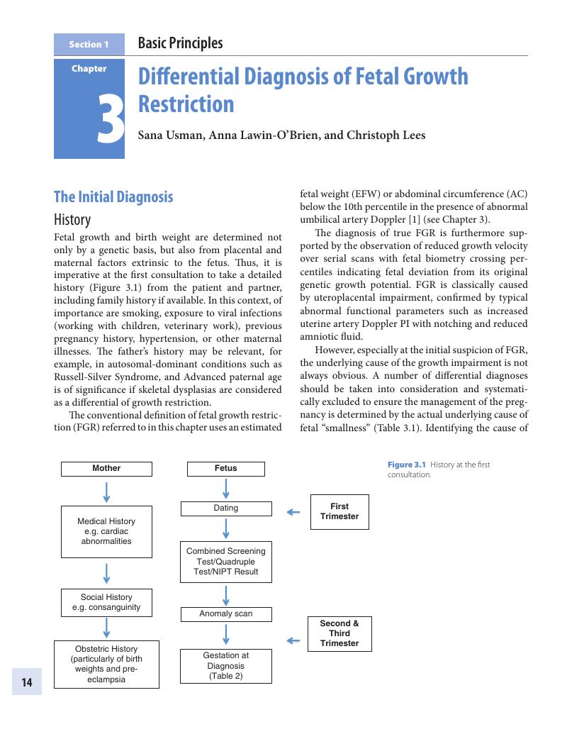
Summary
A summary is not available for this content so a preview has been provided. Please use the Get access link above for information on how to access this content.
- Type
- Chapter
- Information
- Placental-Fetal Growth Restriction , pp. 14 - 30Publisher: Cambridge University PressPrint publication year: 2018
References
Lees, C, Marlow, N, Arabin, B, Bilardo, CM, Brezinka, C, Derks, JB, Duvekot, J, Frusca, T, Diemert, A, Ferrazzi, E, Ganzevoort, W, Hecher, K, Martinelli, P, Ostermayer, E, Papageorghiou, AT, Schlembach, D, Schneider, KTM, Thilaganathan, B, Todros, T, Van Wassenaer Leemhuis, A, Valcamonico, A, Visser, GHA, Wolf, H. Perinatal morbidity and mortality in early‐onset fetal growth restriction: Cohort outcomes of the trial of randomized umbilical and fetal flow in Europe (TRUFFLE). Ultrasound Obstet Gynecol 2013;42(4):400–8. doi:10.1002/uog.13190.CrossRefGoogle ScholarPubMed
Mongelli, M, Gardosi, J. Fetal growth velocity. Lancet 1999;353(9170):2156. doi:10.1016/S0140-6736(05)75590–2.Google Scholar
Lin, CC, Santolaya-Forgas, J. Current concepts of fetal growth restriction: Part I. Causes, classification, and pathophysiology.Obstet Gynecol 1998;92(6):1044–55.Google ScholarPubMed
Papageorghiou, AT, Ohuma, EO, Altman, DG, Todros, T. International Fetal and Newborn Growth Consortium for the 21st Century (INTERGROWTH-21st). International standards for fetal growth based on serial ultrasound measurements: the Fetal Growth Longitudinal Study of the INTERGROWTH-21st Project. Lancet 2014 Sep 6;384(9946):869–79. doi: 10.1016/S0140-6736(14)61490–2.CrossRefGoogle Scholar
Gardosi, J, Figueras, F, Clausson, B, Francis, A. The customised growth potential: An international research tool to study the epidemiology of fetal growth. Paediatr Perinat Epidemiol 2011;25(1):2–10. doi:10.1111/j.1365-3016.2010.01166.x.Google Scholar
Unterscheider, J, Daly, S, Geary, MP, Kennelly, MM, McAuliffe, FM, O’Donoghue, K, Hunter, A, Morrison, JJ, Burke, G, Dicker, P, Tully, EC, Malone, FD. Optimizing the definition of intrauterine growth restriction: The multicenter prospective PORTO study. Am J Obstet Gynecol 2013;208(4):290.e1-.e6. doi:10.1016/j.ajog.2013.02.007.Google Scholar
Yaron, Y, Heifetz, S, Ochshorn, Y, Lehavi, O, Orr-Urtreger, A. Decreased first trimester PAPP-A is a predictor of adverse pregnancy outcome. Prenat Diagn 2002;22(9):778–82. doi:10.1002/pd.407.CrossRefGoogle ScholarPubMed
Goetzl, L, Krantz, D, Group, NBS. Low first-trimester PAPP-a identifies pregnancies requiring IUGR screening. Am J Obstet Gynecol December 2003;189(6): Supplement, Page S215CrossRefGoogle Scholar
Krantz, D, Goetz, L, Simpson, JL. Association of extreme first-trimester free human chorionic gonadotropin-beta, pregnancy-associated plasma protein A, and nuchal translucency with intrauterine growth restriction and other adverse pregnancy outcomes. Am J Obstet Gynecol 2004 Oct;191(4):1452–8.Google Scholar
Albaiges, G. One-stage screening for pregnancy complications by color Doppler assessment of the uterine arteries at 23 weeks’ gestation. Obstet Gynecol 2000;96(4):559–64.Google Scholar
Campbell, S, Black, RS, Lees, CC, Armstrong, V, Peacock, JL. Doppler ultrasound of the maternal uterine arteries: Disappearance of abnormal waveforms and relation to birthweight and pregnancy outcome. http://dxdoiorg/101080/j1600-04122000079008631x. 2009;79(8):631–4. doi:10.1080/j.1600-0412.2000.079008631.xGoogle Scholar
Kingdom, JCP, Burrell, SJ, Kaufmann, P. Pathology and clinical implications of abnormal umbilical artery Doppler waveforms. Ultrasound Obstet Gynecol 1997;9(4):271–86. doi:10.1046/j.1469-0705.1997.09040271.x.CrossRefGoogle ScholarPubMed
Ott, WJ. Diagnosis of intrauterine growth restriction: Comparison of ultrasound parameters. Am J Perinatol 2002;19(3):133–7. doi:10.1055/s-2002–25313.Google Scholar
Rowlands, DJ, Vyas, SK. Longitudinal study of fetal middle cerebral artery flow velocity waveforms preceding fetal death. BJOG 1995;102(11):888–90. doi:10.1111/j.1471-0528.1995.tb10876.x.Google Scholar
Baschat, AA, Hecher, K. Fetal growth restriction due to placental disease. Semin Perinatol 2004;28(1):67–80.Google Scholar
Lees, CC, Marlow, N, Van Wassenaer-Leemhuis, A, Arabin, B, Bilardo, CM, Brezinka, C, Calvert, S, Derks, JB, Diemert, A, Duvekot, JJ, Ferrazzi, E, Frusca, T, Ganzevoort, W, Hecher, K, Martinelli, P, Ostermayer, E, Papageorghiou, AT, Schlembach, D, Schneider, KTM, Thilaganathan, B, Todros, T, Valcamonico, A, Visser, GHA, Wolf, H. 2 year neurodevelopmental and intermediate perinatal outcomes in infants with very preterm fetal growth restriction (TRUFFLE): A randomised trial. Lancet March 2015. doi:10.1016/S0140-6736(14)62049-3.CrossRefGoogle ScholarPubMed
Ferrazzi, E, Bozzo, M, Rigano, S, Bellotti, M, Morabito, A, Pardi, G, Battaglia, FC, Galan, HL. Temporal sequence of abnormal Doppler changes in the peripheral and central circulatory systems of the severely growth‐restricted fetus. Ultrasound Obstet Gynecol 2002;19(2):140–6. doi:10.1046/j.0960-7692.2002.00627.x.Google Scholar
Hecher, K, Campbell, S, Doyle, P, Harrington, K, Nicolaides, K. Assessment of fetal compromise by Doppler ultrasound investigation of the fetal circulation. Arterial, intracardiac, and venous blood flow velocity studies. Circulation 1995;91(1):129–38. doi:10.1161/01.CIR.91.1.129.Google Scholar
Vergani, P, Roncaglia, N, Locatelli, A, Andreotti, C, Crippa, I, Pezzullo, JC, Ghidini, A. Antenatal predictors of neonatal outcome in fetal growth restriction with absent end-diastolic flow in the umbilical artery. Am J Obstet Gynecol 2005;193(3 Pt 2):1213–18. doi:10.1016/j.ajog.2005.07.032.Google Scholar
Khoury, MJ, Erickson, JD, Cordero, JF, McCarthy, BJ. Congenital malformations and intrauterine growth retardation: A population study. Pediatrics 1988;82(1):83–90.Google Scholar
Rosenthal, GL, Wilson, PD, Permutt, T, Boughman, JA, Ferencz, C. Birth weight and cardiovascular malformations: A population-based study. The Baltimore-Washington Infant Study. Am J Epidemiol 1991;133(12):1273–81.CrossRefGoogle Scholar
Rosenthal, GL. Patterns of prenatal growth among infants with cardiovascular malformations: possible fetal hemodynamic effects. Am J Epidemiol 1996;143(5):505–13.Google Scholar
Spiers, PS. Does growth retardation predispose the fetus to congenital malformation? Lancet 1982;1(8267):312–14.Google ScholarPubMed
Capper, A. The fate and development of the immature and of the premature child: A clinical study. Review of the Literature and Study of Cerebral Hemorrhage in the New-Born Infant. Am J Dis Child 1928;35(2):262–88. doi:10.1001/archpedi.1928.01920200094012.Google Scholar
Hussain, U, Daemen, A, Missfelder-Lobos, H, De, Moor, B, Timmerman, D, Bourne, T, Lees, C. Umbilical artery pulsatility index and fetal abdominal circumference in isolated gastroschisis. Ultrasound Obstet Gynecol 2011;38(5):538–42. doi:10.1002/uog.8947.CrossRefGoogle ScholarPubMed
Carroll, SG, Kuo, PY, Kyle, PM, Soothill, PW. Fetal protein loss in gastroschisis as an explanation of associated morbidity. Am J Obstet Gynecol 2001;184(6):1297–301. doi:10.1067/mob.2001.114031.CrossRefGoogle ScholarPubMed
Norman, SM, Odibo, AO, Longman, RE, Roehl, KA, Macones, GA, Cahill, AG. Neural tube defects and associated low birth weight. Am J Perinatol 2012;29(6):473–6. doi:10.1055/s-0032-1304830.Google Scholar
Scott, KE, Usher, R. Fetal malnutrition: Its incidence, causes, and effects. Am J Obstet Gynecol 1966;94(7):951–63.Google Scholar
Snijders, RJ, Sherrod, C, Gosden, CM, Nicolaides, KH. Fetal growth retardation: Associated malformations and chromosomal abnormalities. Am J Obstet Gynecol 1993;168(2):547–55.Google Scholar
Wilkins-Haug, L, Roberts, DJ, Morton, CC. Confined placental mosaicism and intrauterine growth retardation: A case-control analysis of placentas at delivery. Am J Obstet Gynecol 1995;172(1):44–50. doi:10.1016/0002-9378(95)90082–9.Google Scholar
Boghassian, NS et al. Anthropometric charts for infants with trisomies 21, 18, or 13 born between 22 weeks gestation and term: The VON charts. Am J Med Genet A 2012 Feb;158A(2):322–32. doi: 10.1002/ajmg.a.34423. Epub 2012 Jan 13.CrossRefGoogle Scholar
Morris, JK, Cole, TJ, Springett, AL, Dennis, J. Down syndrome birth weight in England and Wales: Implications for clinical practice. Am J Med Genet A 2015;167A(12):3070–5. doi:10.1002/ajmg.a.37366.Google ScholarPubMed
Yeo, L, Guzman, ER, Day-Salvatore, D, Walters, C, Chavez, D, Vintzileos, AM. Prenatal detection of fetal trisomy 18 through abnormal sonographic features. J Ultrasound Med 2003;22(6):581–90–quiz591–2.CrossRefGoogle ScholarPubMed
Snijders, RJ, Sebire, NJ, Nayar, R, Souka, A, Nicolaides, KH. Increased nuchal translucency in trisomy 13 fetuses at 10–14 weeks of gestation. Am J Med Genet 1999;86(3):205–7.Google Scholar
Kroes, I, Janssens, S, Defoort, P. Ultrasound features in trisomy 13 (Patau syndrome) and trisomy 18 (Edwards syndrome) in a consecutive series of 47 cases. Facts Views Vis Obgyn 2014;6(4):245–9.Google Scholar
Abu-Amero, S, Wakeling, EL, Preece, M, Whittaker, J, Stanier, P, Moore, GE. Epigenetic signatures of Silver-Russell syndrome. J Med Genet 2010;47(3):150–4. doi:10.1136/jmg.2009.071316.Google Scholar
Prickett, AR, Ishida, M, Böhm, S, Frost, JM, Puszyk, W, Abu-Amero, S, Stanier, P, Schulz, R, Moore, GE, Oakey, RJ. Genome-wide methylation analysis in Silver-Russell syndrome patients. Hum Genet 2015;134(3):317–32. doi:10.1007/s00439-014-1526-1.Google Scholar
Price, SM, Stanhope, R, Garrett, C, Preece, MA, Trembath, RC. The spectrum of Silver-Russell syndrome: A clinical and molecular genetic study and new diagnostic criteria. J Med Genet 1999;36(11):837–42.Google Scholar
Wakeling, EL, Amero, SA, Alders, M, Bliek, J, Forsythe, E, Kumar, S, Lim, DH, MacDonald, F, Mackay, DJ, Maher, ER, Moore, GE, Poole, RL, Price, SM, Tangeraas, T, Turner, CLS, Van, Haelst, MM, Willoughby, C, Temple, IK, Cobben, JM. Epigenotype–phenotype correlations in Silver-Russell syndrome. J Med Genet 2010;47(11):jmg.2010.079111-jmg.2010.079768. doi:10.1136/jmg.2010.079111.Google Scholar
Paladini, D, Volpe, P. Ultrasound of Congenital Fetal Anomalies: Differential Diagnosis and Prognostic Indicators. 2014.Google Scholar
Chen, M, Hwu, W-L, Kuo, S-J, Chen, C-P, Yin, P-L, Chang, S-P, Lee, D-J, Chen, T-H, Wang, B-T, Lin, CC. Subtelomeric rearrangements and 22q11.2 deletion syndrome in anomalous growth-restricted fetuses with normal or balanced G-banded karyotype. Ultrasound Obstet Gynecol 2006;28(7):939–43. doi:10.1002/uog.3884.Google Scholar
Volpe, P, Marasini, M, Caruso, G, Marzullo, A, Buonadonna, AL, Arciprete, P, Di Paolo, S, Volpe, G, Gentile, M. 22q11 deletions in fetuses with malformations of the outflow tracts or interruption of the aortic arch: Impact of additional ultrasound signs. Prenat Diagn 2003;23(9):752–7. doi:10.1002/pd.682.Google Scholar
Tavormina, PL, Shiang, R, Thompson, LM, Zhu, YZ, Wilkin, DJ, Lachman, RS, Wilcox, WR, Rimoin, DL, Cohn, DH, Wasmuth, JJ. Thanatophoric dysplasia (types I and II) caused by distinct mutations in fibroblast growth factor receptor 3. Nat Genet 1995;9(3):321–8. doi:10.1038/ng0395-321.Google Scholar
Vanhoenacker, FM, Van der Aa, N, Blaumeiser, B. The French telephone receiver sign in thanatophoric dysplasia. JBR-BTR 2009;92(1):63.Google Scholar
Langer, LO, Yang, SS, Hall, JG, Sommer, A, Kottamasu, SR, Golabi, M, Krassikoff, N. Thanatophoric dysplasia and cloverleaf skull. Am J Med Genet Suppl 1987;3:167–79.Google Scholar
Picone, O, Simon, I, Benachi, A, Brunelle, F, Sonigo, P. Comparison between ultrasound and magnetic resonance imaging in assessment of fetal cytomegalovirus infection. Prenat Diagn 2008;28(8):753–8. doi:10.1002/pd.2037.Google Scholar
Lazzarotto, T, Guerra, B, Lanari, M, Gabrielli, L, Landini, MP. New advances in the diagnosis of congenital cytomegalovirus infection. J Clin Virol 2008;41(3):192–7. doi:10.1016/j.jcv.2007.10.015.Google Scholar
Feldman, B, Yinon, Y, Tepperberg, Oikawa, M, Yoeli, R, Schiff, E, Lipitz, S. Pregestational, periconceptional, and gestational primary maternal cytomegalovirus infection: Prenatal diagnosis in 508 pregnancies. Am J Obstet Gynecol 2011;205(4):342.e1–342.e6. doi:10.1016/j.ajog.2011.05.030.CrossRefGoogle ScholarPubMed
Ruellan Eugene, G, Barjot, P, Campet, M, Vabret, A, Herlicoviez, M, Muller, G, Levy, G, Guillois, B, Freymuth, F, Freymuth, F. Evaluation of virological procedures to detect fetal human cytomegalovirus infection: Avidity of IgG antibodies, virus detection in amniotic fluid and maternal serum. J Med Virol 1996;50(1):9–15. doi:10.1002/(SICI)1096–9071(199609)50:1<9::AID-JMV3>3.0.CO;2–5.Google Scholar
Guerra, B, Lazzarotto, T, Quarta, S, Lanari, M, Bovicelli, L, Nicolosi, A, Landini, MP. Prenatal diagnosis of symptomatic congenital cytomegalovirus infection. Am J Obstet Gynecol 2000;183(2):476–82. doi:10.1067/mob.2000.106347.Google Scholar
Gouarin, S, Gault, E, Vabret, A, Cointe, D, Rozenberg, F, Grangeot-Keros, L, Barjot, P, Garbarg-Chenon, A, Lebon, P, Freymuth, F. Real-time PCR quantification of human cytomegalovirus DNA in amniotic fluid samples from mothers with primary infection. J Clin Microbiol 2002;40(5):1767–72. doi:10.1128/JCM.40.5.1767-1772.2002.Google Scholar
Picone, O, Costa, J-M, Leruez-Ville, M, Ernault, P. Cytomegalovirus (CMV) glycoprotein B genotype and CMV DNA load in the amniotic fluid of infected fetuses. Prenat Diagn 2004.Google Scholar
Nedelec, O, Bellagra, N, Devisme, L, Hober, D, Wattré, P, Dewilde, A. [Congenital human cytomegalovirus infection: Value of human cytomegalovirus DNA quantification in amniotic fluid]. Ann Biol Clin (Paris) 2002;60(2):201–7.Google Scholar
Revello, MG, Lazzarotto, T, Guerra, B, Spinillo, A, Ferrazzi, E, Kustermann, A, Guaschino, S, Vergani, P, Todros, T, Frusca, T, Arossa, A, Furione, M, Rognoni, V, Rizzo, N, Gabrielli, L, Klersy, C, Gerna, G, CHIP Study Group. A randomized trial of hyperimmune globulin to prevent congenital cytomegalovirus. N Engl J Med 2014;370(14):1316–26. doi:10.1056/NEJMoa1310214.Google Scholar
Jacquemard, F, Yamamoto, M, Costa, J-M, Romand, S, Jaqz-Aigrain, E, Dejean, A, Daffos, F, Ville, Y. Maternal administration of valaciclovir in symptomatic intrauterine cytomegalovirus infection. BJOG 2007;114(9):1113–21. doi:10.1111/j.1471-0528.2007.01308.x.Google Scholar
Kimberlin, DW, Jester, PM, Sánchez, PJ. Valganciclovir for symptomatic congenital cytomegalovirus disease. N Engl J Med 2015;372(10):933–43. doi:10.1056/NEJMoa1404599.Google Scholar
Lipitz, S, Yinon, Y, Malinger, G, Yagel, S, Levit, L, Hoffman, C, Rantzer, R, Weisz, B. Risk of cytomegalovirus-associated sequelae in relation to time of infection and findings on prenatal imaging. Ultrasound Obstet Gynecol 2013;41(5):508–14. doi:10.1002/uog.12377.Google Scholar
Farkas, N, Hoffmann, C, Ben-Sira, L, Lev, D, Schweiger, A, Kidron, D, Lerman-Sagie, T, Malinger, G. Does normal fetal brain ultrasound predict normal neurodevelopmental outcome in congenital cytomegalovirus infection? Prenat Diagn 2011;31(4):360–6. doi:10.1002/pd.2694.Google Scholar
Malinger, G, Lev, D, Lerman-Sagie, T. Imaging of fetal cytomegalovirus infection. Fetal Diagn Ther 2011;29(2):117–26. doi:10.1159/000321346.Google Scholar
Yinon, Y, Farine, D, Yudin, MH. Screening, diagnosis, and management of cytomegalovirus infection in pregnancy. Obstet Gynecol Surv 2010;65(11):736–43. doi:10.1097/OGX.0b013e31821102b4.Google Scholar
Dunn, D, Wallon, M, Peyron, F, Petersen, E, Peckham, C, Gilbert, R. Mother-to-child transmission of toxoplasmosis: risk estimates for clinical counselling. Lancet 1999;353(9167):1829–33. doi:10.1016/S0140-6736(98)08220-8.Google Scholar
Romand, et al. Usefulness of quantitative polymerase chain reaction in amniotic fluid as early prognostic marker of fetal infection with Toxoplasma gondii. Am J Obstet Gynecol March 2004;190(3):797–802.Google Scholar
Malinger, G, Werner, H, Rodriguez, Leonel, JC, Rebolledo, M, Duque, M, Mizyrycki, S, Lerman, Sagie, T, Herrera, M. Prenatal brain imaging in congenital toxoplasmosis. Prenat Diagn 2011;31(9):881–6. doi:10.1002/pd.2795.Google Scholar
Berrébi, A, Assouline, C, Bessières, M-H, Lathière, M, Cassaing, S, Minville, V, Ayoubi, J-M. Long-term outcome of children with congenital toxoplasmosis. Am J Obstet Gynecol 2010;203(6):552.e1-e6. doi:10.1016/j.ajog.2010.06.002.Google Scholar
Tookey, PA. Review of antenatal rubella susceptibility screening and the standard criteria for screening. Institute of Child Health May 2012:1–11.Google Scholar
Hardelid, P, Cortina-Borja, M, Williams, D, Tookey, PA, Peckham, CS, Cubitt, WD, Dezateux, C. Rubella seroprevalence in pregnant women in North Thames: Estimates based on newborn screening samples. J Med Screen 2009;16(1):1–6. doi:10.1258/jms.2009.008080.Google Scholar
Robertson, SE, Featherstone, DA, Gacic-Dobo, M, Hersh, BS. Rubella and congenital rubella syndrome: Global update. Rev Panam Salud Publica 2003;14(5):306–15.Google Scholar