


Non-digestible carbohydrates such as dietary fibres and oligosaccharides have a variety of beneficial effects on our health. Epilactose (4-O-β-galactopyranosyl-d-mannose) is a rare non-digestible disaccharide, which can be produced in considerable amounts by heating and alkali treatment of cows’ milk( Reference Cataldi, Angelotti and Bufo 1 ). Recently, we have developed a method for the mass-production of epilactose using cellobiose 2-epimerase (EC 5.1.3.11) from the ruminal strain Ruminococcus albus NE1( Reference Ito, Taguchi and Hamada 2 ). Although our previous studies have found that epilactose enhances the absorption of intestinal Ca and Fe in rodents( Reference Suzuki, Nishimukai and Shinoki 3 ), information on its biological functions remains quite limited. As previous studies have demonstrated that some non-digestible carbohydrates ameliorate metabolic disorders such as obesity, epilactose also may have a potential to prevent these disorders.
Obesity and related diseases, such as diabetes and CVD, have emerged as major health problems in many countries, particularly in the West( Reference Kahn, Hull and Utzschneider 4 ). It is known that obesity and diabetes are the results of a complex interaction between genetic and environmental factors. In particular, the intake of a fat-enriched diet and intake of excessive calories are primary factors associated with the increased occurrence and further progression of metabolic disorders. At the same time, the consumption of cereals, a major source of non-digestible carbohydrates, is falling in Western countries, and a number of important relationships between the intestinal fermentation of these carbohydrates and the regulation of metabolic disorders have been suggested( Reference Johns, Lindroos and Jebb 5 – Reference Roberfroid, Gibson and Hoyles 7 ). It appears that the release of bacterial metabolites – that is, SCFA including acetic, propionic and butyric acids – plays a number of important roles in metabolic regulation. Previous studies have shown that luminal SCFA change the synthesis of gastrointestinal peptides such as peptide YY and glucagon-like peptide-1, which control food intake and glucose tolerance( Reference Cani and Delzenne 8 , Reference Delzenne, Daubioul and Neyrinck 9 ). In addition, the SCFA transported to the circulatory system reportedly act as regulators of energy metabolism and adiposity( Reference Hara, Kashihara and Ichimura 10 , Reference Kimura, Ozawa and Inoue 11 ). However, the precise roles of SCFA in the regulation of metabolic disorders and energy metabolisms remain to be clarified.
Energy metabolism is organised by different systems or mechanisms in mammals. The uncoupling protein (UCP) is located in the mitochondrial inner membrane, where it uncouples the oxidation of fuels, mainly fatty acids, from ATP production, so that the energy associated with fuel oxidation is simply released as heat. UCP-1 is highly expressed in brown adipose tissue, which specialises in the production of heat through non-shivering thermogenesis( Reference Ricquier and Bouillaud 12 ). Therefore, UCP-1 activation could serve as a novel therapeutic approach for the treatment of obesity. In addition, recent studies have demonstrated that the ectopic expression of UCP-1 in the skeletal muscle of mice increases whole-body energy expenditure, resulting in the restoration of insulinaemia, cholestelolaemia and adiposity( Reference Li, Nolte and Ju 13 , Reference Couplan, Gelly and Goubern 14 ). Although the basal density of UCP-1 in skeletal muscle is lower than that in brown adipose tissue, evidence suggests that increased UCP-1 in skeletal muscle could make a significant contribution to whole-body energy expenditure due to the large muscle mass.
The purpose of present study was to investigate the preventive effect of epilactose on obesity and metabolic dysregulation in mice fed high-fat (HF) diets in relation to intestinal fermentation. Further, the direct effects of SCFA on UCP-1 expression in differentiated myogenic-like cells were also examined.
Methods
Animals (Expt 1 and 2)
All animal studies were pre-approved by the Hiroshima University Animal Committee, and the mice were maintained in accordance with the Hiroshima University guidelines for the care and use of laboratory animals (no. C11-24).
Male C57/BL6 mice (5 weeks old; Charles River) were housed in cages in a room with controlled temperature (22 (sem 2)°C), relative humidity (40–60 %) and lighting (light 08.00–20.00 hours) throughout the study. The body weight and food intake of the mice were measured every day. The mice were allowed to acclimatise to the laboratory environment with free access to the control diet (AIN-93G formula; Table 1)( Reference Reeves, Nielsen and Fahey 15 ) and distilled water for 1 week.
Table 1 Composition of diets (Expt 1 and 2)

* Epilactose was added to diets at 10 % through substitution of an equal amount of maize starch.
† Maize starch (α-maize starch; Chuou Shokuryo Co. Ltd).
‡ Casein (ALACID; New Zealand Daily Board).
§ Crystallised cellulose (Ceolus PH102; Asahi Chemical Industry).
|| Mineral and vitamin mixtures were prepared according to the AIN-93G formulation.
In Expt 1, mice were randomly divided into the following four groups; the standard diet (Std)-control (Cont), Std-epilactose (Epi), HF-Cont and HF-Epi groups (n 7–8 per group). The Std-Epi and Std-Cont groups were fed a diet containing 7 % soyabean oil with and without 10 % epilactose by weight (Table 1). The HF-Epi and HF-Cont groups were fed diets containing 7 % soyabean oil and 33 % lard with and without 10 % epilactose. Epilactose was added to the experimental diets by substitution for an equal amount of starch. The mice had free access to each experimental diet for 8 weeks. On day 54 of the experimental period, mice were fasted for 8 h, and blood samples were collected from the tail vein to measure plasma glucose, insulin and TAG levels. At the end of the experiment, the mice were sacrificed by exsanguination under diethyl ether anaesthesia. The livers and fat pads (mesenteric, epididymal and retroperitoneal) were removed and weighed, and the epididymal fat pads were fixed with 4 % paraformaldehyde and embedded in paraffin for haematoxylin–eosin staining. Adipocyte size was determined by averaging the diameters of at least fifty adipocytes from the fat pad of each mice. The caecal contents were also collected for organic acid analyses as described below.
In Expt 2, mice were randomly divided into the following three groups: the Std-Cont, HF-Cont and HF-Epi groups (n 7–8 per group). Experimental diets and periods were the same as those in Expt 1. To evaluate the effects of epilactose on lipid absorption and excretion, faeces samples were collected continuously for 3 d from day 51 after the start of feeding the test diets. At the end of the experiment, the liver, fat pads, gastrocnemius muscle and suprascapular brown adipose tissue were collected and weighed for quantitative RT-PCR analysis as described below.
Measurements of plasma glucose, insulin and TAG
The plasma glucose and TAG concentrations were assayed by enzymatic methods using commercially available kits (Glucose CII and TAG E Test Wako; Wako Pure Chemical Industries). The plasma insulin concentration was assayed using an ELISA kit (Ultra Sensitive Mouse Insulin ELISA Kit; Morinaga Institute of Biological Science).
Organic acid analysis
The caecal contents were diluted with four volumes of distilled water and homogenised using a polytron-type homogeniser. A 5 μl of 25 mm crotonic acid as an internal standard was added to 70 μl of caecal contents and the mixture was centrifuged at 15 700 g for 10 min at 4°C. The resultant supernatant was de-proteinised with 1 % sulphosalicylic acid and diluted with two volumes of distilled water. The samples were filtrated and applied to a UPLC/MS system.
Organic acids (acetic, propionic, n-butyric, iso-butyric, n-valeric, iso-valeric, lactic, succinic and crotonic acids) were identified and quantified using a UPLC/MS system equipped with an electric spray ionisation (ESI) interface (Acquity UPLC; Waters Co. Ltd). The temperature of the capillary heater and vaporisation heater was maintained at 120 and 400°C, respectively. The flow rate of the sheath gas (N) was 150 l/h. LC/ESI-MS was carried out in scan mode from (m/z) +50 to 2000 and in selected ion recording (SIR) mode from (m/z) +59·0 for acetic acid, (m/z) +73·0 for propionic acid, (m/z) +87·0 for n- and iso-butyric acid, (m/z) 101·1 for n- and iso-valeric acid, (m/z) 89·0 for lactic acid, (m/z) 117·0 for succinic acid and (m/z) 85·1 for crotonic acid. The UPLC system was fitted with a 1·8 μm C18 column (ACQUITY UPLC HSS T3, 2·1×100 mm; Waters Co. Ltd) set at 40°C. Solvents A (water–formic acid, 100:0·1) and B (methanol–formic acid, 100:0·1) were run at a flow rate of 0·2 ml/min using a gradient from 0 up to 5 % solvent B for 2 min, followed by 0 % solvent B for 2 min, from 5 up to 60 % solvent B for 8 min, from 60 up to 80 % solvent B for 1 min and from 80 up to 10 % solvent B for 2 min and then reduced linearly back to 0 % solvent B over the next 1 min and subsequently maintained at the initial condition for 5 min. The injection volume was 5 μl. Concentrations of individual organic acids were calculated from the peak area in the chromatogram detected with SIR relative to the internal standard (crotonic acid).
Measurement of faecal TAG
Faeces samples collected in Expt 2 were freeze-dried and milled. Total lipids were extracted from powdered faeces (about 100 mg) using the Bligh & Dyer method( Reference Bligh and Dyer 16 ). TAG levels in the extracts were estimated by an enzymatic method as described above.
Quantitative RT-PCR analysis
Total RNA from the mouse tissues and C2C12 cells were isolated using TRI reagent (Sigma) and reverse-transcribed using a ReverTra Ace qPCR RT kit (TOYOBO) according to the manufacturer’s instructions. Real-time qPCR was performed using a Step One Real-Time PCR system (Life Technologies) and a KAPA SYBR FAST qPCR kit (KAPA BIOSYSTEMS). The primer sequences used for the PCR are shown in online Supplementary Table S1. Data were analysed by the ΔΔC t method and presented as fold changes in gene expression after normalisation to the internal control (GAPDH gene expression level).
Immunoblot analysis
Mouse tissues (50 mg) were homogenised in 1 ml lysis buffer containing 1 % (w/v) SDS, 1 % (v/v) TritonX-100 and 1 % (w/v) sodium deoxycholate in 30 mmol/l-Tris with protease and phosphatase inhibitors (pH 7·4) using a polytron-type homogeniser. Protein concentrations were measured using the BCA method (Pierce Biotechnology). Tissue extracts were mixed with a half volume of Laemmli sample buffer (3× concentrated) containing 6 % (w/v) SDS, 30 % (v/v) glycerol, 15 % (v/v) 2-β-mercaptoethanol and 0·02 % (w/v) bromphenol blue in 188 mmol/l-Tris, (pH 6·8) and heated to 100°C for 5 min( Reference Laemmli 17 ). Proteins (30 μg) were separated by SDS-PAGE and transferred to PVDF membranes. Membranes were blotted for UCP-1 and β-actin using specific antibodies in combination with HRP-conjugated anti-mouse or anti-rabbit IgG antibodies. The blots were developed using the ECL chemiluminescence method (Perkin Elmer). Quantification was performed by densitometric analysis of specific bands on the immunoblots using Image J software.
Cell culture (Expt 3)
To examine the direct effects of organic acids on UCP-1 expression in the skeletal muscle, a mouse myoblastic-like C2C12 cell line was used. C2C12 cells (CRL-1772; American Type Culture Collection) were propagated and maintained using DMEM supplemented with 10 % FBS as described previously( Reference Okazaki, Ohshima and Yoshizawa 18 ). The cells were seeded into twenty-four-well plates (1·9 cm2; Thermo Scientific Inc.) at a density of 0·44×105 cells/cm2. After the cells reached confluence, the medium was replaced with myogenic medium consisting of DMEM supplemented with 2 % horse serum to induce myogenic differentiation. All the experiments were conducted on day 8 after the start of differentiation. Cultures were used between passage 7 and 11, and the medium was refreshed every 2 d. The experiment was repeated three times, and a representative analysis is shown in the result section.
Three individual organic acids – acetic, propionic and n-butyric acids – were administered into the medium (0·3–3·0 mm-acetic acid, 0·1–1·0 mm-propionic acid and 0·01–0·1 mm-n-butyric acid). These concentrations were chosen based on the physiological levels present in the blood of rodents( Reference Jakobsdottir, Jadert and Holm 19 ). The total RNA and protein contents of cells incubated with organic acids for 24 h were determined to examine the UCP-1 gene and protein expressions as described above.
Statistical analysis
All the values are expressed as means with their standard errors. The significance of differences between groups was assessed using the Tukey–Kramer post hoc test. A difference with a P value<0·05 was considered significant. Statistical analyses were performed using JMP software (version 12; SAS Institute Inc.).
Results
Feeding epilactose prevents high-fat-diet-induced metabolic disorders (Expt 1)
Feeding HF diets induced increases in body weight and the weight of the three fat pads (epididymal, retroperitoneal and mesenteric adipose tissues, Tables 2 and 3). The body weight gain and total fat pad weights were increased approximately 1·6-fold by HF diets (P<0·01). Supplemental feeding with epilactose effectively prevented these increases, and the body weight gain and individual fat pad weights in the HF-Epi group were lower than those in the HF-Cont group (P<0·01) without any difference in the food intake between the two groups. There were no differences in liver weight among the four groups. Feeding epilactose did not have any effect on these parameters in mice fed the standard diets.
Table 2 Body weight, food intake and caecum weight of mice (Expt 1) (Mean values with their standard errors; n 7–8)

a,b,cMean values with unlike superscript letters were significantly different (P<0·05).
Table 3 Liver and white adipose tissue weights (Expt 1) (Mean values with their standard errors; n 7–8)

a,bMean values with unlike superscript letters were significantly different (P<0·05).
Plasma glucose, insulin and TAG concentrations in mice were measured to examine metabolic dysregulation (Table 4). Although no differences were detected in the plasma glucose concentration among the groups, the HF diets increased plasma insulin concentrations to 1·3-fold (P<0·01), and this increase was attenuated by feeding epilactose (P<0·01). Homoeostasis model assessment for insulin resistance, an indicator of insulin resistance, in the HF-Cont group was nearly double than that in mice fed the standard diets (P<0·01), but the HF-Epi group showed a marked increase in value similar to those of the Std-Cont and Std-Epi groups. Plasma TAG concentrations in the Std-Epi and HF-Cont groups tended to be lower than that in the Std-Cont group, and TAG concentrations in the HF-Epi group was 24 % lower than that in the Std-Cont group (P<0·01).
Table 4 Plasma glucose, insulin, TAG and Homoeostasis model assessment for insulin resistance (HOMA-IR) in fasted mice (Expt 1) (Mean values with their standard errors; n 7–8)
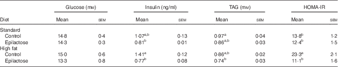
a,bMean values with unlike superscript letters were significantly different (P<0·05).
Haematoxylin–eosin staining of the epididymal fat pads showed that feeding HF diets led to hypertrophy of the adipocytes, indicating excessive fat accumulation in the cells (Fig. 1). The morphology of adipocytes in the HF-Epi group was observed to be similar to those in the two standard diet groups. The average diameter of adipocytes in the HF-Epi group was approximately 30 % lower than that in the HF-Cont group (P=0·03).

Fig. 1 Haematoxylin–eosin staining images and adipocyte diameters of the epididymal adipose tissue of mice in Expt 1. (A) Representative haematoxylin–eosin staining images of epididymal adipose tissue of mice are shown. A black bar indicates 100 μm. (B) Quantification of adipocyte diameters is shown. Values are means (n 7–8), with their standard errors represented by vertical bars. a,bMean values with unlike letters were significantly different (P<0·05). ![]() , Control;
, Control; ![]() , Epilactose.
, Epilactose.
Feeding epilactose leads to the accumulation of organic acid pools in the caecum (Expt 1)
Intestinal bacterial metabolites in the form of organic acids were determined in the caecum, as epilactose is known to exhibit a prebiotic property. The levels of all organic acids measured, except for lactic and succinic acids, were higher in the Std-Epi group compared with the Std-Cont group (Fig. 2). Acetic, propionic, n-valeric and iso-valeric acid levels in the HF-Epi group were 3- to 6-fold higher compared with the HF-Cont group (P<0·01). Incremental trends were observed for succinic, n-butyric and iso-butyric acids by the supplemental feeding of epilactose in mice fed the HF diets.

Fig. 2 Organic acid pools in the caecum of mice in Expt 1. Caecum samples were collected at the end of experiment. (A) Pools of acetate, propionate, lactate, succinate and n-butyrate are shown. (B) Pools of iso-butyrate, n-valerate and iso-valerate are shown. Std, standard diet; HF, high-fat diet; Cont, control; Epi, epilactose. Values are means (n 7–8), with their standard errors represented by vertical bars. a,b,cMean values with unlike letters were significantly different (P<0·05). ![]() , Std-Cont;
, Std-Cont; ![]() , Std-Epi;
, Std-Epi; ![]() , HF-Cont;
, HF-Cont; ![]() , HF-Epi.
, HF-Epi.
Feeding epilactose enhanced uncoupling protein-1 expression in skeletal muscle and brown adipose tissue (Expt 2)
To investigate the mechanisms underlying the epilactose-mediated prevention of HF diet-induced metabolic disorders, another animal study using three experimental groups (Std-Cont, HF-Cont and HF-Epi groups) was conducted, and the expression levels of genes involved in carbohydrate and lipid metabolisms together with energy expenditure in the liver, white adipose tissue, skeletal muscles and brown adipose tissue, were examined. Feeding the HF diet increased the body weight and fat pad weights in mice (P<0·01), whereas supplemental feeding with epilactose prevented these increases in a similar manner to the results observed in Expt 1 (P<0·01, Table 5). No differences were observed in gastrocnemius muscle or suprascapular brown adipose tissue weights among the groups.
Table 5 Body weight gain, food intake and white adipose tissue, brown adipose tissue and gastrocnemius muscle weights (Expt 2) (Mean values with their standard errors; n 7–8)

a,bMean values with unlike superscript letters were significantly different (P<0·05).
The expression levels of two genes involved in fatty acid synthesis – fatty acid synthase (FAS) and acetyl-CoA carboxylase-α – were approximately 60 and 40 % lower in the livers of the HF-Cont and HF-Epi groups than in those of the Std-Cont group (P<0·01, Fig. 3). Gluconeogenesis-related genes – phosphoenolpyruvate carboxykinase 1 and glucose-6-phosphatase – were increased 2- and 3-fold, respectively, by feeding the HF diets (P<0·01). Expressions of these genes in the liver were not influenced by epilactose. In the pathogenesis of metabolic syndrome and obesity, macrophages are chemoattracted by the monocyte chemotactic protein (MCP)-1 to adipocytes, leading to inflammation. Although no differences in lipoprotein lipase, hormone-sensitive lipase and PPARγ gene expression levels were found among the groups, the inflammation markers, MCP-1 (P<0·01), TNF-α (P<0·01) and F4/80 expression levels (P=0·04), were increased 1·5- to 2·5-fold by feeding the HF diets (Fig. 4). Supplemental feeding with epilactose prevented the increases in MCP-1 (P<0·01) and TNF-α expression (P<0·01) and tended to attenuate the increase in F4/80. In addition, UCP-1, Cidea and PPARγ coactivator 1α (PGC-1α) expressions in white adipose tissues were examined, because recent studies have shown that another UCP-1-positive cell, referred to as a beige adipocyte, resides in white adipose tissues and contributes to the energy expenditure. However, no difference was observed in these two gene expressions in white adipose tissues (data not shown). Among the genes examined in the gastrocnemius muscle, the expression of UCP-1, which is involved in the dissipation of energy as heat, thereby enhancing energy expenditure, was influenced by diet (Fig. 5), with UCP-1 expression in the HF-Epi group being more than double compared with the other groups at the mRNA and protein levels (P=0·04). The brown adipose tissue specialises in generating heat through non-shivering thermogenesis using UCP proteins. UCP-1 mRNA and protein expressions in the two HF diet groups were higher than that in the Std-Cont group (P<0·01 and P=0·03), and these UCP-1 expressions in the HF-Epi group were even higher than that in the HF-Cont group (Fig. 6). The UCP-1 expressions in the HF-Epi groups were approximately 25 % higher than those in the HF-Cont groups at the mRNA and protein levels (P=0·02 and 0·03). The expression of PGC-1α, a regulator of UCP-1 transcription, in the HF-Epi group was 50 % higher compared with the expression levels in the other groups (P=0·04).

Fig. 3 Expression levels of fatty acid synthase (FAS) acetyl-CoA carboxylase-α (ACC-α), phosphoenolpyruvate carboxykinase 1 (PEPCK) and glucose-6-phosphatase (G6Pase) in the liver of mice in Expt 2. Livers were collected at the end of experiment. AU, arbitrary units; Std, standard diet; HF, high-fat diet; Cont, control; Epi, epilactose. Values are means (n 7–8), with their standard errors represented by vertical bars. a,bMean values with unlike letters were significantly different (P<0·05). ![]() , Std+Cont;
, Std+Cont; ![]() , HF+Cont;
, HF+Cont; ![]() , HF+Epi.
, HF+Epi.

Fig. 4 Expression levels of hormone-sensitive lipase (HSL), lipoprotein lipase (LPL), PPARγ, monocyte chemotactic protein-1 (MCP-1), TNF-α and F4/80 in the epididymal adipose tissue of mice in Expt 2. Epididymal adipose tissues were collected at the end of experiment. AU, arbitrary units; Std, standard diet; HF, high-fat diet; Cont, control; Epi, epilactose; UCP, uncoupling protein. Values are means (n 7–8), with their standard errors represented by vertical bars. a,bMean values with unlike letters were significantly different (P<0·05). ![]() , Std+Cont;
, Std+Cont; ![]() , HF+Cont;
, HF+Cont; ![]() , HF+Epi.
, HF+Epi.

Fig. 5 Expression levels of uncoupling protein (UCP)-1, UCP-2, UCP-3, PPARγ coactivator 1α (PGC-1α), lipoprotein lipase (LPL) and carnitine palmitoyltransferase-1α (CPT-1α) in the gastrocnemius muscle of mice in Expt 2. Gastrocnemius muscles were collected at the end of experiment. (A) mRNA expression levels of UCP-1, UCP-2, UCP-3, PGC-1α, LPL and CPT-1α in the muscle are shown. (B) The UCP-1 protein expression level is shown. AU, arbitrary units; Std, standard diet; HF, high-fat diet; Cont, control; Epi, epilactose. Values are means (n 7–8), with their standard errors represented by vertical bars. a,bMean values with unlike letters were significantly different, P<0·05. ![]() , Std+Cont;
, Std+Cont; ![]() , HF+Cont;
, HF+Cont; ![]() , HF+Epi.
, HF+Epi.

Fig. 6 Expression levels of uncoupling protein (UCP)-1 and PPARγ coactivator 1α (PGC-1α) in the brown adipose tissue of mice in Expt 2. Brown adipose tissues were collected at the end of experiment. (A) mRNA expression levels of UCP-1 and PGC-1α in the brown adipose tissue are shown. (B) The UCP-1 protein expression level is shown. AU, arbitrary units; Std, standard diet; HF, high-fat diet; Cont, control; Epi, epilactose. Values are means (n 7–8), with their standard errors represented by vertical bars. a,b,cMean values with unlike letters were significantly different (P<0·05). ![]() , Std+Cont;
, Std+Cont; ![]() , HF+Cont;
, HF+Cont; ![]() , HF+Epi.
, HF+Epi.
Faecal excretions of TAG were higher in the two HF groups than in the Std-Cont group (P<0·01), but feeding epilactose did not influence excretion (Std-Cont, 0·58 (sem 0·06); HF-Cont, 2·16(sem 0·15); HF-Epi, 2·35(sem 0·19) mg/3 d). No difference was observed in dry weights of faeces collected for 3 d among groups.
Propionic acid induces uncoupling protein-1 expression in differentiated C2C12 cells
C2C12 cells were used to examine the direct effects of three major organic acids – acetic, propionic and n-butyric acids – on UCP-1 expression. The cells incubated with 0·1 and 0·3 mm-propionic acid showed dose-dependent increases in UCP-1 mRNA expression (P=0·03 and P<0·01, Fig. 7). Similarly, the UCP-1 protein expressions were increased by 0·1 and 0·3 mm-propionic acids (P<0·01). No significant effect were observed after incubation with acetic and n-butyric acids, although an incremental trend was observed in the cells incubated with 0·1 mm-n-butyric acid in comparison with the control treatment.

Fig. 7 Uncoupling protein (UCP)-1 expression in C2C12 cells in Expt 3. Cells were collected after incubation for 24 h with acetate, propionate or n-butyrate. mRNA (A) and protein (B) expressions of UCP-1 in cells are shown. Data are representative of three independent experiments. Values are means (n 6), with their standard errors represented by vertical bars. a,bMean values with unlike letters were significantly different (P<0·05). AU, arbitrary units.
Discussion
Although a previous study demonstrated that supplemental feeding with epilactose did not have an impact on the plasma lipid concentration in rats fed high-sucrose diets( Reference Nishimukai, Watanabe and Taguchi 20 ), some prebiotic oligosaccharides reportedly exhibit an ameliorative effect on diet-induced obesity and metabolic dysregulation( Reference Delzenne, Daubioul and Neyrinck 9 , Reference Cani, Possemiers and Van de Wiele 21 , Reference Xie, Zhu and Zhang 22 ). The present study showed that epilactose effectively prevents metabolic disorders probably through the up-regulation of UCP-1 expression in skeletal muscles and brown adipose tissue in mice fed HF diets. Notably, our results reveal that propionic acid, which is produced in large amounts through the intestinal fermentation of epilactose, induces UCP-1 expression in differentiated myogenic cells.
Obesity and related diseases are the result of a complex interaction of genetic and environmental factors. Among environmental factors, fat-enriched diet and excessive energy intake are closely associated with the occurrence of these diseases, and therefore the development of novel preventive approaches has been eagerly anticipated. Supplemental feeding with epilactose for 8 weeks resulted in a significant reduction in body weight gain and visceral fat pad weight without affecting food intake or faecal TAG excretion in mice fed the HF diets. The histological analysis of epididymal adipose tissues shows that feeding epilactose suppresses the adipocyte hypertrophy induced by the HF diets. White adipose tissue stores redundant energy in the form of TAG droplets during nutritional excess, and obesity is characterised by an increase in the size of adipocytes differentiated from fibroblastic pre-adipocytes in adipose tissue. These findings suggest that supplemental epilactose increases whole-body energy expenditure, resulting in lower body and fat pad weights.
Several studies have demonstrated that UCP proteins have pathophysiological roles in energy metabolism in human as well as in rodents( Reference Cypess, Lehman and Williams 23 , Reference Virtanen, Lidell and Orava 24 ). Our results suggest that supplemental epilactose enhances energy expenditure through increased UCP-1 expression in skeletal muscles and brown adipose tissue. We focused on muscular UCP-1, in particular, as skeletal muscles make up approximately 40 % of the total adult body weight in contrast to the relatively small contribution made by brown adipose tissue( Reference Harper, Green and Brand 25 ). It is likely that whole-body energy expenditure could be significantly increased by the activation of muscular UCP-1. It has been reported that the overexpression of UCP-1 in the skeletal muscle of mice increases energy expenditure, resulting in decreased plasma insulin, glucose, and cholesterol levels as well as decreased adiposity( Reference Li, Nolte and Ju 13 , Reference Couplan, Gelly and Goubern 14 ). In addition, UCP-1 expression in the muscles protects mice from HF diet-induced obesity( Reference Katterle, Keipert and Hof 26 ). These findings re-inforce our hypothesis that the increases in muscular UCP-1 make a significant contribution to the epilactose-mediated prevention of obesity and metabolic dysregulation.
The finding that propionic acid, at a concentration corresponding to physiological levels in the blood of humans and rodents, stimulates UCP-1 expression in C2C12 cells suggests that this SCFA mediates the epilactose-induced increase in UCP-1 in mouse muscle. It has been reported that at least two G protein-coupled receptors (GPR) for SCFA, GPR41 and GPR43 are expressed in muscle cells and C2C12 cells( Reference Nilsson, Kotarsky and Owman 27 , Reference Suzuki, Elias and Seth 28 ). Some studies have demonstrated that GPR41 and GPR43 have an important role in the regulation of energy expenditure and carbohydrate and lipid metabolisms( Reference Hara, Kashihara and Ichimura 10 , Reference Kimura, Ozawa and Inoue 11 ). UCP-1 expression is increased in C2C12 cells incubated with propionic acid at 0·1 and 0·3 mm, and a small, although NS, increase is observed in C2C12 cells incubated with 0.1 mm-n-butyric acid. Functional assays indicate that GPR43 is activated by acetic, propionic and n-butyric acids to a similar degree, whereas the rank order of potencies for GPR41 is propionic>n-butyric≫acetic acids( Reference Suzuki, Elias and Seth 28 ). According to these studies, the propionic acid-mediated increase in UCP-1 expression may occur via GPR41 activation. However, the underlying mechanisms for the epilactose-mediated increases in UCP-1 expression appear to differ between skeletal muscle and brown adipose tissue, as feeding epilactose increases PGC-1α, a transcription regulator of UCP-1, in brown adipose tissue only. Further studies are required to clarify the precise mechanisms for the epilactose-mediated increase in UCP-1 expression.
A growing body of evidence supports the notion that obesity and metabolic disorders are characterised by a chronic state of low-grade inflammation with pro-inflammatory macrophage infiltration into white adipose tissue( Reference Bai and Sun 29 , Reference Exley, Hand and O’Shea 30 ). This macrophage infiltration is triggered by the release of pro-inflammatory adipokines, including MCP-1, from lipid-laden adipocytes. Once macrophages infiltrate into adipose tissue, they interact with adipocytes through TNF-α release to increase pro-inflammatory adipokine secretion and decrease anti-inflammatory adipokine secretion, leading to the progression of metabolic dysregulation such as insulin resistance and hyperlipidaemia. The inhibition of crosstalk between adipocytes and macrophages and the decrease in macrophage infiltration by feeding epilactose are confirmed by the decreased expression of MCP-1, TNF-α and F4/80 in adipose tissue. These results suggest that supplemental feeding with epilactose prevents macrophage infiltration into adipose tissues due to the suppression of lipid accumulation.
In conclusion, supplemental feeding with epilactose – a non-digestible disaccharide – effectively prevents HF diet-induced obesity and metabolic dysregulation probably through increased UCP-1 expression in the skeletal muscles and brown adipose tissue. Propionic acid, an intestinal bacterial metabolite, may be involved in the epilactose-mediated increase in UCP-1 expression in muscles. Although further studies for the direct evaluation of the increased thermogenesis and energy expenditure resulting from feeding epilactose are required, this non-digestible disaccharide appears to have the potential as a novel functional food for the prevention of metabolic disorders.
Acknowledgements
The present study was supported in part by JSPS KAKENHI (T. S., grant no. 25660106). JSPS had no role in the design, analysis or writing of this article.
The authors’ contributions are as follows: Y. M. and T. S. designed and conducted the research and performed the statistical analysis of the data; T. O., W. S., H. M., H. Ma. and S. T. helped in conducting the research; and T. S. wrote the paper and had the primary responsibility for the final content. All the authors read and approved the final version of the manuscript.
The authors declare no conflicts of interest.
Supplementary material
For supplementary material/s referred to in this article, please visit http://dx.doi.org/doi:10.1017/S0007114515003505


