


Prevalence of hypovitaminosis D has been reported to be high and is a public health issue in various regions around the Middle East, including the United Arab Emirates, Oman, Saudi Arabia, Jordan, Iran and Qatar. This major public health problem could affect all individuals across all life stages. The high prevalence of vitamin D deficiency in the region is suggested to be largely the result of limited sun exposure due to religious and cultural practices, prolonged breast-feeding without vitamin D supplementation( Reference Al-Daghri, Al-Attas and Alokail 1 – Reference Mahdy, Al-Emadi and Khanjar 7 ) and also the lack of food fortification policies( Reference Bandeira and Gris 8 ). The levels of serum 25-hydroxycholecalciferol (25(OH)D3) have also been reported to be <25 nmol/l in one-third of individuals in this region( Reference Arabi, El Rassi and El-Hajj Fuleihan 9 ).
There are a number of risk factors that are reported to be associated with vitamin D status in different populations. Generally, it has been reported that younger individuals have higher levels of vitamin D than older ones and males have slightly higher levels than females( Reference Arabi, El Rassi and El-Hajj Fuleihan 9 , Reference Lips, Hosking and Lippuner 10 – Reference Sharif and Rizk 12 ). There are also reports to suggest that the prevalence of vitamin D deficiency is higher during the winter than during the summer months and that winter season is one of the risk factors associated with vitamin D deficiency( Reference Lund and Sorensen 13 , Reference Scharla, Scheidt-Nave and Leidig 14 ). In addition, biochemical markers such as plasma parathyroid hormone (PTH) and serum Ca have also been reported to be associated with vitamin D status( Reference Harkness and Cromer 15 , Reference Lips 16 ). Serum level of 25(OH)D3 plus 25-hydroxyergocalciferol (25(OH)D2), which is known as total 25-hydroxyvitamin D (25(OH)D), has been reported to be the most precise marker of vitamin D status as it reflects cutaneous synthesis as well as total intakes of vitamin D from foods and supplements( Reference Meddeb, Sahli and Chahed 17 ). Based on its review of data of vitamin D needs, a committee of the Institute of Medicine concluded that persons are at risk of vitamin D deficiency with serum total 25(OH)D concentrations of <30 nmol/l and inadequacy at levels ranging from 30 to 50 nmol/l. The committee stated that a serum total 25(OH)D level of 50 nmol/l (optimal) could cover the needs of 97·5 % of a population( 18 ).
In the present study, the associations of vitamin D deficiency and insufficiency with biometric and biochemical markers related to vitamin D metabolism in healthy adults were investigated for the first time in a Bahraini population.
Materials and methods
The present study was conducted between October 2010 and October 2011 among volunteers attending the blood bank centre in Bahrain Defense Force (BDF) Hospital, the second largest hospital in Bahrain. The volunteers were queried to assess their eligibility and willingness to participate in the study. All completed a questionnaire on age, gender, style of clothing, history of any chronic diseases (such as diabetes mellitus, hypertension, obesity, osteoporosis, osteomalacia, liver disease, renal disease, hypo- or hyperparathyroidism), vitamin and mineral deficiency, steroid therapy or any drugs that could interfere with vitamin D metabolism, or vitamin therapy including vitamin D and Ca intake by face-to-face interview. Out of 700 persons who attended the blood bank and were asked to participate in the study, sixty-six were non-Bahrainis. Of the remaining 634, 550 agreed with a response rate of 87 %; however, fifty persons were excluded because of obesity (BMI >25 kg/m2), history of liver, renal, gestational or endocrine disorder, and also taking medications that influence bone metabolism and current vitamin D and Ca intake. All study participants gave written informed consent after being fully informed of the study objectives and procedures and their right to withdraw from the study. The research protocol was approved by the Research and Ethics Committees of the College of Medicine and Medical Sciences, Arabian Gulf University, and the Research and Ethics Committee of BDF Hospital.
Conservative style of clothing was defined as women wearing a traditional long black garment called ‘abayah’ that covers the body from the shoulders down to the feet, with scarf-like cover that covers the hair but not the face.
A blood sample was collected from each participant and serum and plasma were prepared. Serum Ca and P were analysed immediately using a Cobas 6000 analyser system (Roche Ltd, Basel, Switzerland). Remaining serum and plasma samples were then stored at −70°C until further analysis of vitamin D and PTH. Plasma PTH was determined using commercially available ELISA kits (Creative Diagnostics, Shirley, NY, USA). The intra-assay and inter-assay CV for determination of PTH in plasma were <4·5 % and 7·1 % for low control and 3·2 % and 5·2 % for high control, respectively. Serum 25(OH)D3 and 25(OH)D2 concentrations were determined by ultra-performance liquid chromatography mass spectrometry (UPLC-MS/MS) using a commercially available kit (Chromsystem, Munich, Germany). The intra-assay and inter-assay CV for determination of 25(OH)D3 in serum for low control were <2·7 % and 3·9 %, and for high control were 4·2 % and 4·0 %, respectively. The intra-assay and inter-assay CV for determination of 25(OH)D2 in serum for low control were <3·9 % and 5·7 % and for high control were <4·3 % and 4·7 %, respectively.
Statistical analysis
Vitamin D status was assessed according to the 2011 recommendation of the Institute of Medicine that classifies vitamin D deficiency at serum 25(OH)D concentration of <30 nmol/l, insufficiency at levels ranging from 30 to 50 nmol/l and optimal at levels of ≥50 nmol/l( 18 ).
The normality of total 25(OH)D distribution was assessed using the Kolmogorov–Smirnov test. As the distribution was negatively skewed, logarithmic transformation was applied to reduce kurtosis before geometric means were calculated. Thus the logarithmic transformations were used in further statistical analysis.
Student's t test was used to compare the total 25(OH)D concentrations according to all other biometric and biochemical parameters between males and females. Pearson's correlation coefficient was used to examine the correlation between total 25(OH)D and other variables. ANOVA was used to compare the values of biometric and biochemical parameters between participants with optimal, insufficiency and deficiency of vitamin D.
Mantel–Haenszel relative risk analysis was used to determine the association of age, gender, season, hyperparathyroidism, low Ca and style of clothing with vitamin D deficiency and insufficiency in the entire cohort, males and females. Hyperparathyroidism was defined as plasma PTH levels of >65·0 pg/ml and hypocalcaemia was defined as serum Ca of <2·1 nmol/l. Receiver operating characteristic (ROC) curve analysis was used to determine the best biochemical marker to predict vitamin D deficiency. Participants with vitamin D deficiency were considered affected, and those with an optimal amount of vitamin D were considered as controls and not affected. Greater deviation toward the left upper corner of the curve indicates better detection of vitamin D deficiency.
All statistical inferences were made based on a two-sided significance level of P < 0·05 and all statistical analyses were performed using the statistical software package IBM SPSS version 19·0.
Results
Baseline biochemical and biometric characteristics of the entire cohort, males and females are illustrated in Table 1. Among all the variables, plasma PTH and serum P were significantly higher in females than males (P < 0·05), whereas serum total 25(OH)D and serum Ca were significantly lower in females than in males (P < 0·05).
Table 1 Biochemical and biometric characteristics of the entire cohort, males and females; Bahrain, October 2010–October 2011
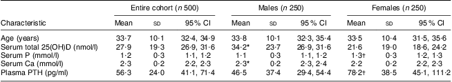
25(OH)D, 25-hydroxyvitamin D; PTH, parathyroid hormone.
Results are expressed as mean and standard deviation, except for total 25(OH)D which is expressed as geometric mean and standard deviation.
*Mean value was significantly higher in males than females (P < 0·05).
†Mean value was significantly higher in females than males (P < 0·05).
Results from the Pearson correlation analysis in the entire cohort showed that, among all variables, serum total 25(OH)D was significantly and positively correlated with age (r = 0·131, P = 0·003), serum Ca (r = 0·174, P = 0·001) and male sex (r = 0·388, P = 0·000) and negatively correlated with plasma PTH (r = −0·19, P = 0·03). In females, serum total 25(OH)D was significantly and positively correlated with age (r = 0·289, P = 0·001) and significantly and negatively correlated with plasma PTH (r = −0·21, P = 0·01); whereas in males among all variables serum total 25(OH)D was significantly and negatively correlated with plasma PTH (r = −0·134, P = 0·03).
Table 2 presents the serum levels of total 25(OH)D in the entire cohort, males and females according to age group, season and clothing style.
Table 2 Serum levels of total 25(OH)D (nmol/l) in the entire cohort, males and females, according to age group, season and clothing style; Bahrain, October 2010–October 2011
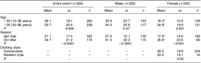
25(OH)D, 25-hydroxyvitamin D.
All results are presented as geometric mean and standard deviation.
*Mean value was significantly lower than that of males (P ≤ 0·0001).
Biochemical and biometric parameters according to vitamin D status in the entire cohort, females and males are illustrated in Table 3. In the entire cohort, ANOVA showed that serum Ca (P = 0·02) was significantly decreased whereas plasma PTH (P = 0·03) was significantly increased between individuals with optimal, insufficiency and deficiency of vitamin D. In females, age (P = 0·04) and serum Ca (P = 0·04) were significantly decreased and plasma PTH (P = 0·004) was significantly increased between individuals with vitamin D insufficiency and deficiency compared with those with optimal total 25(OH)D.
Table 3 Biochemical and biometric characteristics of the entire cohort, males and females, according to vitamin D status; Bahrain, October 2010–October 2011
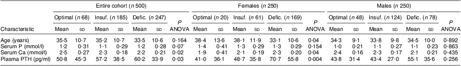
PTH, parathyroid hormone; 25(OH)D, 25-hydroxyvitamin D.
Vitamin D status: optimal, serum 25(OH)D ≥ 50 nmol/; insufficiency (Insuf.), serum 25(OH)D = 30–50 nmol/l; deficiency (Defic.), serum 25(OH)D < 30 nmol/l.
In Table 4, Mantel-Haenszel relative risk analysis showed that, in the entire cohort, the relative risk of vitamin D deficiency was significantly increased with younger age group (P = 0·03), low serum Ca (P < 0·001), warm and hot months of the year (P < 0·0001) and female sex (P = 0·002), while relative risk of hyperparathyroidism was significantly increased with vitamin D deficiency (P = 0·01). In females the relative risk of vitamin D deficiency was significantly increased with younger age group (P = 0·04), low serum Ca (P = 0·001), warm and hot months of the year (P = 0·001) and conservative clothing style (P = 0·04), and relative risk of hyperparathyroidism was significantly increased with vitamin D deficiency (P = 0·03). In males, the relative risk of vitamin D deficiency was increased by 1·6 fold with warm and hot months of the year (P < 0·0001). In the entire cohort (P = 0·03), females (P = 0·03) and males (P = 0·006), the relative risk of vitamin D insufficiency was also significantly increased in warm and hot months of the year.
Table 4 Mantel–Haenszel relative risk analysis for the association of age, high plasma PTH and low serum calcium with vitamin D deficiency and insufficiency in the entire cohort, females and males; Bahrain, October 2010–October 2011
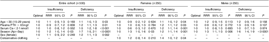
PTH, parathyroid hormone; RRR, relative risk ratio; 25(OH)D, 25-hydroxyvitamin D.
Vitamin D status: optimal, serum 25(OH)D ≥ 50 nmol/; insufficiency, serum 25(OH)D = 30–50 nmol/l; deficiency, serum 25(OH)D < 30 nmol/l.
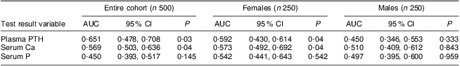
As illustrated in Table 5, results from the ROC curve analyses used to examine the ability of biochemical parameters to detect vitamin D deficiency suggested that the best predictive markers for vitamin D deficiency were plasma PTH and serum Ca in the entire cohort and females.
Table 5 ROC curve analysis for the best predictors of vitamin D deficiency
ROC, receiver operating characteristic; AUC, area under the curve; PTH, parathyroid hormone.
Discussion
Vitamin D status has been investigated in different populations and its deficiency is increasingly recognized worldwide. The present study is the first one to assess vitamin D status in healthy men and women in relation to biochemical parameters including serum Ca and plasma PTH, and also other factors such as age, gender, women's style of clothing and season, in Bahrain.
In adult Bahrainis a clear association of gender with vitamin D status was observed, as total 25(OH)D was significantly lower in females than males and the results from Mantel–Haenszel relative risk analysis showed that female sex was one of the major risk factors associated with vitamin D deficiency. Results from the present study are consistent with published studies suggesting that female sex is one of the most common risk factors associated with vitamin D deficiency and insufficiency in developing countries( Reference Mahdy, Al-Emadi and Khanjar 7 , Reference Bandeira and Gris 8 , Reference Meddeb, Sahli and Chahed 17 , Reference Mishal 19 – Reference MacLaughlin and Holick 21 ). This could be due to the fact that women tend to spend more time indoors than men, in addition to their style of clothing and sun protection and sun avoidance attitude, which could all attribute to the vitamin D deficiency in Bahraini women.
There are reports to suggest that age has an effect on vitamin D status due to the fact that skin production of vitamin D precursor declines with age( Reference Holick 22 ). However, in this Bahraini population, total 25(OH)D was significantly higher in older than in younger individuals. In addition, Pearson correlation coefficients and Mantel–Haenszel relative risk analysis suggested that age was positively and significantly associated with serum total 25(OH)D and the risk of vitamin D deficiency was associated with younger age (<30 years) in the entire cohort and in female participants. These findings are inconsistent with a large number of studies in Europe and the USA suggesting that vitamin D deficiency is associated with older populations rather than younger ones( Reference Haddad, Chyu and Hahn 23 , Reference Lips, Hackeng and Jongen 24 ). Nevertheless, consistent with the present study, some studies in the Middle East have reported an association of vitamin D deficiency with younger age groups( Reference Gannage-Yared, Chemali and Yaacoub 20 , Reference Kashi, Saeedian and Akha 25 ).
Globally, the prevalence of vitamin D deficiency has been found to be higher during the winter than during the summer months, and winter season has been reported to be one of the risk factors associated with vitamin D deficiency( Reference Lund and Sorensen 13 , Reference Scharla, Scheidt-Nave and Leidig 14 ). However, paradoxically, in the present study serum total 25(OH)D concentrations in the entire group of participants of all ages averaged 41·5 nmol/l in October–March and 21·1 nmol/l in April–September. Mantel–Haenszel relative risk analysis revealed that the risks of vitamin D insufficiency and deficiency were significantly associated with warm and hot months of the year, particularly in women, consistent with a number of studies in the Arabian Peninsula( Reference Al-Daghri, Al-Attas and Alokail 1 , Reference Al Anouti, Thomas and Abdel-Wareth 2 , Reference Bandeira and Gris 8 , Reference Mithal, Wahl and Bonjour 26 ). In winter there are lots of outdoor activities which can be seen from October to the end of March, whereas during summer these outdoor activities are very limited owing to the high humidity and extreme heat in Bahrain.
In the present study serum total 25(OH)D levels were significantly lower in women with conservative Middle Eastern clothing style than those with non-conservative style and Mantel–Haenszel relative risk analysis showed that conservative clothing style was another risk factor associated with vitamin D deficiency in women. Vitamin D status is reported to be associated with lifestyle, particularly with type of clothing, and consistent with the present results there are reports to suggest that vitamin D status is higher in women with European-style clothing than in women with conservative clothing style( Reference Mishal 19 , Reference Gannage-Yared, Chemali and Yaacoub 20 , Reference Alagol, Shihadeh and Boztepe 27 ). In addition to a marked gender difference in vitamin D levels, being significantly decreased in females compared with males, plasma PTH was also significantly higher in women than in men. The negative correlation of PTH with total 25(OH)D and the association of vitamin D deficiency with high PTH observed in the present study are consistent with a large number of reported studies( Reference Alagol, Shihadeh and Boztepe 27 – Reference Ooms, Lips and Roos 29 ). When participants were stratified according to their vitamin D levels, Ca and PTH were decreased significantly in participants with vitamin D deficiency compared with those with insufficiency and optimal levels of vitamin D in the entire cohort and females, but not males. In addition, Mantel–Haenszel relative risk analysis showed a significant association of vitamin D deficiency with hyperparathyroidism. Vitamin D supplementation is reported to decrease serum PTH concentration and vitamin D insufficiency and deficiency-induced hyperparathyroidism have been reported to stimulate high bone turnover leading to bone loss( Reference Chapuy, Chapuy and Meunier 30 – Reference Strife and Hug 33 ).
Furthermore, results from ROC curve analyses to examine the ability of biochemical parameters to detect vitamin D deficiency showed that the best predictive markers for vitamin D deficiency in the entire cohort and also females were plasma PTH and serum Ca. A low Ca intake has been reported to aggravate vitamin D deficiency whereas a high Ca intake may reduce the vitamin D requirement( Reference Strife and Hug 33 ). In addition, low Ca intake has also been reported to increase PTH secretion and effectively it may influence vitamin D metabolism. Animal studies suggest that a low Ca intake causes increases in serum PTH and 1,25-dihydroxyvitamin D (1,25(OH)2D) with increased metabolic clearance of 25(OH)D in rats. Therefore low Ca would lead to an increase in serum 1,25(OH)2D from hydroxylation of 25(OH)D, thereby decreasing serum 25(OH)D and inducing or aggravating vitamin D deficiency( Reference Chapuy, Chapuy and Meunier 30 , Reference Clements, Johnson and Fraser 34 ).
Although the prevalence of vitamin D insufficiency and deficiency was very high in our study participants, one of the limitations of the study is that vitamin D status was investigated only in volunteers attending a blood bank. Therefore the study may not fully reflect the vitamin D status in Bahrain, as healthy volunteers attending a blood bank could have higher vitamin D levels than the general population.
Conclusion
The present study is the first one in Bahrainis highlighting the associations of vitamin D status and risk factors associated with vitamin D deficiency and insufficiency, as well as the best indices to investigate vitamin D status. Despite the abundance of sunshine in Bahrain that allows vitamin D synthesis all year round, the study revealed low levels of vitamin D and high prevalence of hypovitaminosis D in adult Bahrainis, mainly in women. This can be a major public health problem that can affect all life stages and all individuals. Urgent public health measures, including education to increase awareness of vitamin D supplements and the role of lifestyle, and also vitamin D fortification of foods, are strongly recommended in Bahrain.
Acknowledgements
Sources of funding: This research work received no specific grant from any funding agency in the public, commercial or not-for-profit sectors. Conflicts of interest: None to declare. Authors’ contributions: J.G. designed the study and wrote the article; N.A.-S. and D.A.D. carried out the collection and analysis of samples; G.A.-K. and A.D. contributed to the design of the study and recruited the volunteers from the blood bank.


