


No CrossRef data available.
Article contents
Morphological Aspects and Microscopic Analyses of Fibrous Tunic and Uveal Components in Bovine Eye
Part of:
Micrographia Collection
Published online by Cambridge University Press: 26 May 2022
Abstract
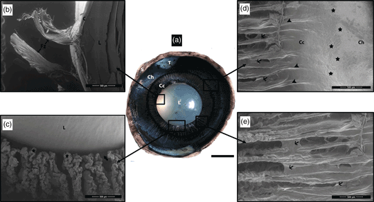
This study aimed to reveal the anatomical features of the bovine eye by scanning electron and light microscopic methods. For this purpose, a total of 40 eyes were evaluated. Gross and microscopic characteristics of the cornea, sclera, ciliary body, choroid, iris, and lens were determined. Bowman's and Descemet's membranes of the cornea were quite dense and prominent. Collagen lamellae of the cornea were wavy in the periphery and more parallel to the basal and metachromatic fibroblasts were noted. Three to four ciliary plicae merged to form ciliary processes. The presence of prominent intermediate bands connecting the ciliary plicae was determined. The zonular fibrils merged and attached to the lens in the form of thick zonular bands. A dense corpora nigra was present at the rectangular pupillary border of the iris. Tapetum fibrosum, consisting of polygonal tapetal cells, was in blue-yellow-green color and covered most of the choroid. A complex drainage system consisting of trabecular meshwork, angular aqueous plexus, ciliary sinus, and scleral venous vessels localized in a fairly wide iridocorneal angle was identified. Identifying structural features of the bovine eye is very important and useful for pathological evaluations, understanding species-specific physiological mechanisms and for operative interventions of ruminant species.
- Type
- Micrographia
- Information
- Copyright
- Copyright © The Author(s), 2022. Published by Cambridge University Press on behalf of the Microscopy Society of America
References
Abuagla, IA, Ali, HA & Ibrahim, ZH (2016). An anatomical study on the eye of the one-humped camel (Camelus dromedarius). Int J Vet Sci 5(3), 137–141.Google Scholar
Adhikary, J (2016). A gross and histomorphological studies of eye in Murrah buffalo. PhD Thesis, West Bengal University of Animal and Fishery Sciences, Department of Veterinary Anatomy, Histology and Embryology, Belgachia, Kolkata.Google Scholar
Akbalık, ME, Erdoğan, S, Sağsöz, H & Saruhan, BG (2015). Morphological study on iridocorneal angle and ciliary body of the Anatolian Shepherd dogs (Canis familiaris). J Vet Med, Kafkas University 21(5), 677–684.Google Scholar
Alina, D, Muste, A, Beteg, F & Briciu, R (2008). Morphological aspect of tapetum lucidum at some domestic animals. Bull UASVM Vet Med 65(2), 166–170.Google Scholar
Alvites, RD, Branquinho, MV, Sousa, AC, Lopes, B, Sousa, P, Mendonça, C, Atayde, LM & Maurício, AC (2021). Small ruminants and its use in regenerative medicine: Recent works and future perspectives. Biology 10(3), 249.CrossRefGoogle ScholarPubMed
Aly, KH (2003). Glycohistochemical, immunohistochemical and electron microscopic examination of the bovine eyeball. PhD Thesis, Ludwig Maximalian University, Munich, Germany.Google Scholar
Aly, KH, Abd-Elhafez, E, Ali, M & Abd-Elmaksoud, A (2009). Histomorphometric analysis of the irides of dogs, camels, buffaloes, and donkeys. Res Vet Sci 86, 1–6.CrossRefGoogle ScholarPubMed
Bacha, JW & Bacha, ML (2012). Color Atlas of Veterinary Histology. Oxford: John Wiley & Sons, Ltd.Google Scholar
Barhaiya, RK, Bhayani, DM & Vyas, YL (2014). Histomorphological study of ciliary body, iris and iridocorneal angle in adult Marwari goat (Capra hircus). J Biomed Life Sci 5(3), 412–418.Google Scholar
Barhaiya, RK, Malsawmkima Vyas, YL & Bhayani, DM (2015). Gross anatomical, histomorphological and biometrical study of the cornea in adult Marwari goat (Capra hircus). Indian J Vet Anat 27(1), 24–26.Google Scholar
Bernstein, MH & Pease, DC (1958). Electron microscopy of the tapetum lucidum of the cat. J Biophys Biochem Cytol 5(1), 35–39.CrossRefGoogle Scholar
Bouhenni, AR, Dunmire, J, Sewell, A & Edward, DP (2012). Animal models of glaucoma. J Biomed Biotechnol 2012, 1–11.CrossRefGoogle ScholarPubMed
Braekevelt, CR (1981). Fine structure of the tapetum lucidum in the domestic ferret. Anat Embryol 163, 201–214.CrossRefGoogle Scholar
Braekevelt, CR (1983). Retinal photoreceptor fine structure in the domestic sheep. Acta Anat (Basel) 116(3), 265–275.CrossRefGoogle ScholarPubMed
Braekevelt, CR (1986 a). Fine structure of the bovine tapetum fibrosum. Anat Histol Embryol 15, 251–222.CrossRefGoogle ScholarPubMed
Braekevelt, CR (1986 b). Fine structure of the tapetum cellulosum of the grey seal (Halichoerus grypus). Acta Anat (Basel) 127, 81–87.CrossRefGoogle Scholar
Braekevelt, CR (1990). Fine structure of the retinal photoreceptors of domestic cat. Anat Histol Embryol 19(1), 67–76.CrossRefGoogle ScholarPubMed
Braekevelt, CR (1993). Fine structure of the tapetum lucidum of the paca (Cuniculus paca). Acta Anat (Basel) 146, 244–250.CrossRefGoogle Scholar
Broom, DM & Corke, MJ (2002). Effects of disease on farm animal welfare. Acta Vet Brno 71, 133–136.CrossRefGoogle Scholar
Coroneo, MT (1990). The bovine eye as a model for the novice cataract surgeon. Osli Retina 21(11), 772–777.CrossRefGoogle ScholarPubMed
De Geest, JP, Lauwers, H, Simoens, P & DeSchaepdrijver, L (1990). The morphology of the equine iridocorneal angle: A light and scanning electron microscopic study. Equine Vet J Suppl 22(10), 30–35.CrossRefGoogle Scholar
Dellmann, HD & Collier, LL (1987). Eye and related structures. In Textbook of Veterinary Histology, Dellmann, HD & Brown, EM (Eds.), pp. 416–433. Philadelphia: Lea & Febiger.Google Scholar
Diesem, C (1975). Sense organs and common integuments. In The Anatomy of the Domestic Animals, Getty, R (Ed.), pp. 202–226. Philadelphia, London: WB Saunders Company.Google Scholar
Dyce, KM, Sack, WO & Wensing, CJG (1987). The sense organs. In Textbook of Veterinary Anatomy, pp. 328–352. Tokyo: WB Saunders Company.Google Scholar
El-Ghazali, HM & Mahdy, EA (2018). Absence or presence of tapetum lucidum: Macro and microscopic investigations in donkey (Equus asinus), cat (Felis domestica) and one-humped camel (Camelus dromedarius). Slov Veterinary Res 55(20), 263–272.Google Scholar
Evangelho, K, Mastronardi, CA & De-La-Torre, A (2019). Experimental models of glaucoma: A powerful translational tool for the future development of new therapies for glaucoma in humans—A review of the literature. Medicina 55(6), 280.CrossRefGoogle ScholarPubMed
Frandson, RD, Wilke, WL & Fails, AD (2009). Anatomy and Physiology of Farm Animals. USA: John Wiley & Sons, Ltd.Google Scholar
Gelatt, KN (2014). Opbhthalmic structures. In Essentials of Veterinary Ophthalmology, Gelatt, KN (Ed.), pp. 12–39. Hoboken, NJ: John Wiley & Sons, Ltd.Google Scholar
Iwata, T & Tomarev, S (2008. Animal models for eye diseases and therapeutics. In Sourcebook of Models for Biomedical Research, Michael Conn, P. (Ed.), pp. 279–287. New Jersey: Humana Press.CrossRefGoogle Scholar
Januschowski, K, Mueller, S, Spitzer, MS, Schramm, C, Doycheva, D, Bartz-Schmidt, KU & Szurman, P (2012). Evaluating retinal toxicity of a new heavy intraocular dye, using a model of perfused and isolated retinal cultures of bovine and human origin. Graef Arch Clin Exp 250(7), 1013–1022.CrossRefGoogle Scholar
Junqueira, LC (1998). Alange Medical Book: Basic Histology. Norwalk, CA: Appelton&Lange.Google Scholar
Kassab, A (2012). Ultrasonographic and macroscopic anatomy of the enucleated eyes of the buffalo (Bos bubalis) and the one-humped camel (Camelus dromedarius) of different ages. Anat Histol Embryol 41, 7–11.CrossRefGoogle ScholarPubMed
Kassab, A, Aoyama, M & Sugita, S (2001). The morphology of the iridocorneal angle in the eye of buffaloes (Bos bubalis): A light and scanning electron microscopic study. Okajimas Folia Anat Jpn 78(4), 145–152.CrossRefGoogle ScholarPubMed
Kassab, A & El-Zoghby, I (2010). Anatomical and histological studies of the aqueous outflow system in the eye of goat (Capra hircus). J Vet Anat 3(2), 13–22.CrossRefGoogle Scholar
König, HE & Liebich, HC (2014). Veterinary Anatomy of Domestic Mammals. Stuttgart: Schattauer.Google Scholar
Kotb, AM (2006). Comparative morphological studies on the vascular tunic of the eyeball in some domestic mammals. M.V.S. Thesis, Assiut University of Department of Anatomy and Histology, Egypt.Google Scholar
Leeson, T & Leeson, R (1970). Histology. Philadelphia, London, Toronto: W.B. Saunders Company.Google Scholar
Leite, AGB, Oliveira, D & Baraldi-Artoni, SM (2013). Morphology of ocular system of domestic animals. Ars Veterinaria 29(1), 42–51.CrossRefGoogle Scholar
Lesiuk, TP & Braekevelt, CR (1983). Fine structure of the canine tapetum lucidum. J Vet Anat 136(1), 157–164.Google ScholarPubMed
Malsawmkima, BR, Vyas, YL & Bhayani, DM (2015). Gross morphological studies on the eyeball of adult Surti buffalo (Bubalus bubalis). Indian J Vet Anat 27(1), 21–23.Google Scholar
Malsawmkima, BRK, Vyas, YL & Bhayani, DM (2014). Histomorphological study on vascular tunics of the adult Surti buffalo (Bubalus bubalis). Int J Interdiscip Multidiscip Stud 2, 24–28.Google Scholar
McMinn, RM (1994). Last's Anatomy Regional and Applied. Edinburgh, New York: Churchill Livingstone.Google Scholar
Merindano, MD, Costa, J, Canals, M, Potau, JM & Ruano, D (2002). A comparative study of Bowman's layer in some mammals: Relationships with other constituent corneal structures. Eur J Anat 6(3), 133–139.Google Scholar
Mohammadi, SF, Mazouri, A, Jabbarvand, M, Rahman-A, N & Mohammadi, A (2011). Sheep practice eye for ophthalmic surgery training in skills laboratory. J Cataract Refract Surg 37(6), 987–991.CrossRefGoogle ScholarPubMed
Nautscher, N, Bauer, A, Steffl, M & Amselgruber, WM (2016). Comparative morphological evaluation of domestic animal cornea. Vet Ophthalmol 19(4), 297–304.CrossRefGoogle ScholarPubMed
Ollivier, FJ, Samuelson, DA, Brooks, DE, Lewis, PA, Kallberg, ME & Komaromy, AM (2004). Comparative morphology of the tapetum lucidum (among selected species). Vet Ophthalmol 7(1), 11–22.CrossRefGoogle Scholar
Patt, DI & Patt, GR (1969). Comparative Vertebrate Histology. New York, Evanston, London: Harper & Row Publishers.Google Scholar
Potter, TJ, Hallowell, GD & Bowen, IM (2008). Ultrasonographic anatomy of the bovine eye. Vet Radiol Ultrasoun 49(2), 172–175.CrossRefGoogle ScholarPubMed
Prince, JH, Diesem, C, Eglitis, I & Ruskell, GL (1960). Anatomy and Histology of the Eye and Orbit in Domestic Animals. Springfield, IL: Charles C. Thomas Publisher.Google Scholar
Rahi, AHS, Sheikh, H & Morgan, G (1980). Histology of the camel eye. Acta Anat 106, 345–350.CrossRefGoogle ScholarPubMed
Reece, WO & Rowe, EW (2017). Functional Anatomy and Physiology of Domestic Animals. Iowa: John Wiley & Sons, Ltd.Google Scholar
Rehfeld, A, Nylander, M & Karnov, K (2017). Compendium of Histology: A Theoretical and Practical Guide. Cham, Switzerland: Springer International Publishing.CrossRefGoogle Scholar
Samuelson, DA (1999). Ophthalmic anatomy. In Veterinary Ophthalmology, Gelatt, KN (Ed.), pp. 41–122. Philadelphia: Lippincott, Williams & Wilknis.Google Scholar
Schnichels, S, Kiebler, T, Hurst, J, Maliha, AM, Löscher, M, Dick, HB, Bartz-Schmidt, KU & Joachim, SC (2019). Retinal organ cultures as alternative research models. Altern Lab Anim 47(1), 19–29.CrossRefGoogle ScholarPubMed
Shinozaki, A, Hosaka, Y, Imagawa, T & Uehara, M (2009). Relationship between distribution of tapetum fibrosum and retinal pigment epithelium in the sheep eye. J Vet Med Sci 72(2), 211–215.CrossRefGoogle ScholarPubMed
Shinozaki, A, Takagi, S, Hosaka, YZ & Uehara, M (2013). The fibrous tapetum of the horse eye. J Anat 223(5), 509–518.CrossRefGoogle ScholarPubMed
Slatter, D (2008). Fundamentals of Veterinary Ophthalmology. Philadelphia: WB Saunders Company Harcourt.Google Scholar
Thompson, LB (2020). An eye simulation for training in the use of ophthalmologic equipment and corneal foreign body removal techniques. Adv Med Educ Pract 11, 155–161.CrossRefGoogle ScholarPubMed
Tortora, GJ & Derrickson, BH (2018). The special senses. In Principles of Anatomy and Physiology, pp. 576–622. New York: John Wiley & Sons.Google Scholar
Trautmann, A & Fiebiger, J (1957). Fundamentals of the Histology of Domestic Animals. New York: Comstock Pub Associates.Google Scholar
Verma, A, Pathak, A, Farooqui, MM, Prakash, A & Kumar, P (2016). Gross and morphometrical observations of eyeball in calf (Bubalus bubalus). Rum Sci 5(2), 169–172.Google Scholar
Vinayak, A, Greene, CE, Moore, PA & Powell-Johnson, G (2004). Clinical resolution of Brucella canis-induced ocular inflammation in a dog. J Am Vet Me Assoc 224(11), 1804–1807.CrossRefGoogle Scholar
Walls, GL (1942). The Vertebrate Eye and its Adaptive Radiation. Bloomfield Hills, MI: The Cranbook Press.Google Scholar
Williams, DL (2004). Lens morphometry determined by B-mode ultrasonography of the normal and cataractous canine lens. Vet Ophthalmol 7, 91–95.CrossRefGoogle ScholarPubMed
Yamaue, Y, Hosaka, YZ & Uehara, M (2015). Spatial relationships among the cellular tapetum, visual streak and rod density in dogs. J Vet Med Sci 77(2), 175–179.CrossRefGoogle ScholarPubMed
Zayed, AEZ, Aly, KH, Ibrahim, IAA & Kotb, AM (2012). Comparative morphology of the iris of donkey (Equus asinus) and buffalo (Bos bubalis). J Vet Anat 5(1), 75–90.CrossRefGoogle Scholar