


No CrossRef data available.
Article contents
Automated ExEm-spFRET Microscope
Published online by Cambridge University Press: 21 February 2022
Abstract
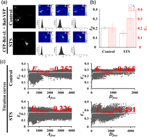
Excitation–emission-spectral unmixing-based fluorescence resonance energy transfer (ExEm-spFRET) microscopy exhibits excellent robustness in living cells. We here develop an automatic ExEm-spFRET microscope with 3.04 s of time resolution for a quantitative FRET imaging. The user-friendly interface software has been designed to operate in two modes: administrator and user. Automatic background recognition, subtraction, and cell segmentation were integrated into the software, which enables FRET calibration or measurement in a one-click operation manner. In administrator mode, both correction factors and spectral fingerprints are only calibrated periodically for a stable system. In user mode, quantitative ExEm-spFRET imaging is directly implemented for FRET samples. We implemented quantitative ExEm-spFRET imaging for living cells expressing different tandem constructs (C80Y, C40Y, C10Y, and C4Y, respectively) and obtained consistent results for at least 3 months, demonstrating the stability of our microscope. Next, we investigated Bcl-xL-Bad interaction by using ExEm-spFRET imaging and FRET two-hybrid assay and found that the Bcl-xL-Bad complexes exist mainly in Bad-Bcl-xL trimers in healthy cells and Bad-Bcl-xL2 trimers in apoptotic cells. We also performed time-lapse FRET imaging on our system for living cells expressing Yellow Cameleon 3.6 (YC3.6) to monitor ionomycin-induced rapid extracellular Ca2+ influx with a time interval of 5 s for total 250 s.
Keywords
- Type
- Software and Instrumentation
- Information
- Copyright
- Copyright © The Author(s), 2022. Published by Cambridge University Press on behalf of the Microscopy Society of America
References
Behera, S, Wang, N, Zhang, C, Schmitz-Thom, I, Strohkamp, S, Schültke, S, Hashimoto, K, Xiong, L & Kudla, J (2015). Analyses of Ca2+ dynamics using a ubiquitin-10 promoter-driven yellow cameleon 3.6 indicator reveal reliable transgene expression and differences in cytoplasmic Ca2+ responses in Arabidopsis and rice (Oryza sativa) roots. New Phytol 206, 751–760. doi:10.1111/nph.13250CrossRefGoogle ScholarPubMed
Belosludtsev, KN, Dubinin, MV, Belosludtseva, NV & Mironova, GD (2019). Mitochondrial Ca2+ transport: Mechanisms, molecular structures, and role in cells. Biochemistry 84, 593–607. doi:10.1134/S0006297919060026Google ScholarPubMed
Butz, ES, Ben-Johny, M, Shen, M, Yang, PS, Sang, L, Biel, M, Yue, DT & Wahl-Schott, C (2016). Quantifying macromolecular interactions in living cells using FRET two-hybrid assays. Nat Protoc 11, 2470–2498. doi:10.1038/nprot.2016.128CrossRefGoogle ScholarPubMed
Chai, L, Zhang, J, Zhang, L & Chen, T (2015). Miniature fiber optic spectrometer-based quantitative fluorescence resonance energy transfer measurement in single living cells. J Biomed Opt 20, 037008. doi:10.1117/1.JBO.20.3.037008CrossRefGoogle ScholarPubMed
Clegg, RM (1992). Fluorescence resonance energy transfer and nucleic acids. Meth Enzymol 211, 353–388. doi:10.1016/0076-6879(92)11020-jCrossRefGoogle ScholarPubMed
De Los Santos, C, Chang, CW, Mycek, MA & Cardullo, RA (2015). FRAP, FLIM, and FRET: Detection and analysis of cellular dynamics on a molecular scale using fluorescence microscopy. Mol Reprod Dev 82, 587–604. doi:10.1002/mrd.22501CrossRefGoogle ScholarPubMed
Du, M, Zhang, L, Xie, S & Chen, T (2016). Wide-field microscopic FRET imaging using simultaneous spectral unmixing of excitation and emission spectra. Opt Express 24, 16037–16051. doi:10.1364/OE.24.016037CrossRefGoogle ScholarPubMed
Elangovan, M, Wallrabe, H, Chen, Y, Day, RN, Barroso, M & Periasamy, A (2003). Characterization of one- and two-photon excitation fluorescence resonance energy transfer microscopy. Methods 29, 58–73. doi:10.1016/s1046-2023(02)00283-9CrossRefGoogle ScholarPubMed
Hoppe, AD, Scott, BL, Welliver, TP, Straight, SW & Swanson, JA (2013). N-way FRET microscopy of multiple protein-protein interactions in live cells. PLoS One 8, e64760. doi:10.1371/journal.pone.0064760CrossRefGoogle ScholarPubMed
Koushik, SV, Blank, PS & Vogel, SS (2009). Anomalous surplus energy transfer observed with multiple FRET acceptors. PLoS One 4, e8031. doi:10.1371/journal.pone.0008031CrossRefGoogle ScholarPubMed
Krebs, M, Held, K, Binder, A, Hashimoto, K, Den Herder, G, Parniske, M, Kudla, J & Schumacher, K (2012). FRET-based genetically encoded sensors allow high-resolution live cell imaging of Ca2+ dynamics. Plant J 69, 181–192. doi:10.1111/j.1365-313X.2011.04780.xCrossRefGoogle Scholar
Levy, S, Wilms, CD, Brumer, E, Kahn, J, Pnueli, L, Arava, Y, Eilers, J & Gitler, D (2011). SpRET: Highly sensitive and reliable spectral measurement of absolute FRET efficiency. Microsc Microanal 17, 176–190. doi:10.1017/S1431927610094493CrossRefGoogle ScholarPubMed
Li, H, Yu, H & Chen, T (2012). Partial acceptor photobleaching-based quantitative FRET method completely overcoming emission spectral crosstalks. Microsc Microanal 18, 1021–1029. doi:10.1017/S1431927612001110CrossRefGoogle ScholarPubMed
Lin, F, Du, M, Yang, F, Wei, L & Chen, T (2018). Improved spectrometer-microscope for quantitative fluorescence resonance energy transfer measurement based on simultaneous spectral unmixing of excitation and emission spectra. J Biomed Opt 23, 1–10. doi:10.1117/1.JBO.23.1.016006Google ScholarPubMed
Mustafa, S, Hannagan, J, Rigby, P, Pfleger, K & Corry, B (2013). Quantitative Förster resonance energy transfer efficiency measurements using simultaneous spectral unmixing of excitation and emission spectra. J Biomed Opt 18, 26024. doi:10.1117/1.JBO.18.2.026024CrossRefGoogle ScholarPubMed
Petros, AM, Nettesheim, DG, Wang, Y, Olejniczak, ET, Meadows, RP, Mack, J, Swift, K, Matayoshi, ED, Zhang, H, Thompson, CB & Fesik, SW (2000). Rationale for Bcl-xL/Bad peptide complex formation from structure, mutagenesis, and biophysical studies. Protein Sci 9, 2528–2534. doi:10.1110/ps.9.12.2528CrossRefGoogle ScholarPubMed
Su, W, Du, M, Lin, F, Zhang, C & Chen, T (2019). Quantitative FRET measurement based on spectral unmixing of donor, acceptor and spontaneous excitation-emission spectra. J Biophotonics 12, e201800314. doi:10.1002/jbio.201800314CrossRefGoogle ScholarPubMed
Sun, H, Zhang, C, Ma, Y, Du, M & Chen, T (2019). Controlling and online measurement of automatic dual-channel E-FRET microscope. Biomed Signal Process Control 53, 101585. doi:10.1016/j.bspc.2019.101585CrossRefGoogle Scholar
Thaler, C, Koushik, SV, Blank, PS & Vogel, SS (2005). Quantitative multiphoton spectral imaging and its use for measuring resonance energy transfer. Biophys J 89, 2736–2749. doi:10.1529/biophysj.105.061853CrossRefGoogle ScholarPubMed
Valentijn, AJ, Metcalfe, AD, Kott, J, Streuli, CH & Gilmore, AP (2003). Spatial and temporal changes in Bax subcellular localization during anoikis. J Cell Biol 162, 599–612. doi:10.1083/jcb.200302154CrossRefGoogle ScholarPubMed
Vanderhoof, B, Nelson, R, Beiner, G, Raicu, V & Oliver, J (2020). Tissue factor oligomerization in living cells using förster resonance energy transfer. Microsc Microanal 26(S2), 828–829. doi:10.1017/S1431927620015986CrossRefGoogle Scholar
Yu, H, Zhang, J, Li, H & Chen, T (2013). Ma-PbFRET: Multiple acceptors FRET measurement based on partial acceptor photobleaching. Microsc Microanal 19, 171–179. doi:10.1017/S1431927612014079CrossRefGoogle ScholarPubMed
Zha, J, Harada, H, Osipov, K, Jockel, J, Waksman, G & Korsmeyer, SJ (1997). BH3 domain of BAD is required for heterodimerization with BCL-XL and pro-apoptotic activity. J Biol Chem 272, 24101–24104. doi:10.1074/jbc.272.39.24101CrossRefGoogle ScholarPubMed
Zhang, C, Liu, Y, Qu, W, Su, W, Du, M, Yang, F & Chen, T (2019). ExEm-FRET two-hybrid assay: FRET two-hybrid assay based on linear unmixing of excitation-emission spectra. Opt Express 27, 18282–18295. doi:10.1364/OE.27.018282CrossRefGoogle ScholarPubMed
Zhang, M, Cao, HZ, Hou, L, Song, SQ, Zeng, JY & Pei, Y (2017). In vivo imaging of Ca2+ accumulation during cotton fiber initiation using fluorescent indicator YC3.60. Plant Cell Rep 36, 911–918. doi:10.1007/s00299-017-2122-3CrossRefGoogle ScholarPubMed

Sun et al. supplementary material
Sun et al. supplementary material

File
41.3 MB