Hostname: page-component-76fb5796d-vfjqv Total loading time: 0 Render date: 2024-04-26T21:25:16.125Z Has data issue: false hasContentIssue false

Deconvolution of the elastic properties of bivalve shell nanocomposites from direct measurement and finite element analysis

Published online by Cambridge University Press:  28 May 2019

Matthias O’Toole-Howes
Affiliation:
Department of Earth Sciences, University of Cambridge, Cambridge CB2 3EQ, U.K.
Ruth Ingleby
Affiliation:
Department of Earth Sciences, University of Cambridge, Cambridge CB2 3EQ, U.K.
Melanie Mertesdorf
Affiliation:
Department of Earth Sciences, University of Cambridge, Cambridge CB2 3EQ, U.K.
James Dean
Affiliation:
Department of Materials Science and Metallurgy, University of Cambridge, Cambridge CB3 0FS, U.K.
Wei Li
Affiliation:
Department of Materials Science and Metallurgy, University of Cambridge, Cambridge CB3 0FS, U.K.
Michael A. Carpenter*
Affiliation:
Department of Earth Sciences, University of Cambridge, Cambridge CB2 3EQ, U.K.
Elizabeth M. Harper
Affiliation:
Department of Earth Sciences, University of Cambridge, Cambridge CB2 3EQ, U.K.
*
a)Address all correspondence to this author. e-mail: mc43@esc.cam.ac.uk
Get access

Abstract

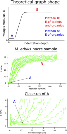
A new protocol has been devised for determining elastic properties of natural biocomposites in the form of bivalve shells under wet and dry conditions. Four-point bending on shell slices of Mytilus edulis, Ensis siliqua, and Pecten maximus give generally lower and more reliable values of Young’s modulus, E, than those in the literature from three-point bending, due to the more even distribution of strain. Finite element analysis of the prismatic microstructure of Pinna nobilis, obtained by X-ray tomography, shows that values of E ≈ 20 GPa can be understood in terms of the real microstructure containing a small proportion of organic matrix phase with E ≈ 1 GPa and a dominant proportion of calcite with E ≈ 90 GPa. Higher values of E obtained by nanoindentation give results which are biased toward the properties of the carbonate phase rather than of the biocomposite as a whole.

Type
Article
Copyright
Copyright © Materials Research Society 2019 

Access options

Get access to the full version of this content by using one of the access options below. (Log in options will check for institutional or personal access. Content may require purchase if you do not have access.)

References

Curry, J.R.: The design of mineralised hard tissues for their mechanical functions. J. Exp. Biol. 202, 3285 (1999).Google Scholar
Meyers, M.A., Lin, A.Y.M., Seki, Y., Chen, P-Y., Kad, B.K., and Bodde, S.: Structural biological composites: An overview. JOM 58, 35 (2006).CrossRefGoogle Scholar
Meyers, M.A., Chen, P-Y., Lin, A.Y.M., and Seki, Y.: Biological materials: Structure and mechanical properties. Prog. Mater. Sci. 53, 1 (2008).CrossRefGoogle Scholar
Chen, P-Y., Lin, A.Y.M., Lin, Y-S., Seki, Y., Stokes, A.G., Peyras, J., Olevsky, E.A., Meyers, M.A., and McKittrick, J.: Structure and mechanical properties of selected biological materials. J. Mech. Behav. Biomed. Mater. 1, 208 (2008).CrossRefGoogle ScholarPubMed
Barthelat, J., Rim, J.E., and Espinosa, H.D.: A review on the structure and mechanical properties of mollusk shells—Perspectives on synthetic biomimetic materials. In Applied Scanning Probe Methods XIII: Biomimetics and Industrial Applications, Bhushan, B., Fuchs, H. (eds.) (Springer, Switzerland, 2009); p. 1744.CrossRefGoogle Scholar
Dunlop, J.W.C. and Fratzl, P.: Biological composites. Annu. Rev. Mater. Res. 22, 1 (2010).CrossRefGoogle Scholar
Ehrlich, H., Simon, P., Carrillo-Cabrera, W., Bazhenov, V.V., Botting, J.P., Ilan, M., Ereskovsky, A.V., Muricy, G., Worch, H., Mensch, A., Born, R., Springer, A., Kummer, K., Vyalikh, D.V., Molodtsov, S.L., Kurek, D., Kammer, M., Paasch, S., and Brunner, E.: Insights into chemistry of biological materials: Newly discovered silica-aragonite-chitin biocomposites in demosponges. Chem. Mater. 22, 1462 (2010).CrossRefGoogle Scholar
Bieler, R., Mikkelsen, P.M., Collins, T.M., Glover, E.A., González, V.L., Graf, D.L., Harper, E.M., Healy, J., Kawauchi, G.Y., Sharma, P.P., Staubach, S., Strong, E.E., Taylor, J.D., Tëmkin, I., Zardus, J.D., Clark, S., Guzmán, A., McIntyre, E., Sharp, P., and Giribet, G.: Investigating the Bivalve Tree of Life—An exemplar-based approach combining molecular and novel morphological characters. Invertebr. Syst. 28, 32 (2014).CrossRefGoogle Scholar
Kaplan, D.: Mollusc shell structures: Novel design strategies for synthetic materials. Curr. Opin. Solid State Mater. Sci. 3, 232 (1998).CrossRefGoogle Scholar
Rousseau, M.: Nacre, a natural biomaterial. In Biomaterials Applications for Nanomedicine, Pignatello, R. (ed.) (InTech, Rijeka, Croatia, 2011); p. 281298.Google Scholar
Kim, Y-W., Kim, J.J., Kim, Y.H., and Rho, J.Y.: Effects of organic matrix proteins on the interfacial structure at the bone–biocompatible nacre interface in vitro. Biomaterials 23, 2089 (2002).CrossRefGoogle ScholarPubMed
Taylor, J.D., Kennedy, W.J., and Hall, A.: The shell structure and mineralogy of the Bivalvia. Introduction, nuculacea–trigonacea. Bull. Br. Mus. (Nat. Hist.) Zool. 3, 1 (1969).Google Scholar
Taylor, J.D., Kennedy, W.J., and Hall, A.: The shell structure and mineralogy of the Bivalvia. II. Lucinacea–Clavagellacea, conclusions. Bull. Br. Mus. (Nat. Hist.) Zool. Suppl. 22, 253 (1973).Google Scholar
Taylor, J. and Layman, M.: The mechanical properties of bivalve (Mollusca) shell structures. Palaeontology 15, 73 (1972).Google Scholar
Currey, J.D. and Taylor, J.D.: The mechanical behaviour of some molluscan hard tissues. J. Zool. 173, 395 (1974).CrossRefGoogle Scholar
Currey, J.D.: Further studies on the mechanical properties of mollusk shell material. J. Zool. 180, 445453 (1976).CrossRefGoogle Scholar
Currey, J.D.: Mechanical properties of mother of pearl in tension. Proc. R. Soc. London, Ser. B 196, 443 (1977).Google Scholar
Currey, J.D.: Mechanical properties of mollusc shell. Symp. Soc. Exp. Biol. 34, 75 (1980).Google ScholarPubMed
Jackson, A.P., Vincent, J.F.V., and Turner, R.M.: The mechanical design of nacre. Proc. R. Soc. London, Ser. B 234, 415 (1988).Google Scholar
Katti, D.R. and Katti, K.S.: Modeling microarchitecture and mechanical behavior of nacre using 3D finite element techniques part I elastic properties. J. Mater. Sci. 36, 1411 (2001).CrossRefGoogle Scholar
Katti, D.R., Katti, K.S., Sopp, J.M., and Sarikaya, M.: 3D finite element modeling of mechanical response in nacre-based hybrid nanocomposites. Comput. Theor. Polym. Sci. 11, 397 (2001).CrossRefGoogle Scholar
Katti, K., Katti, D.R., Tang, J., Pradhan, S., and Sarikaya, M.: Modeling mechanical responses in a laminated biocomposite part II nonlinear responses and nuances of nanostructure. J. Mater. Sci. 40, 1749 (2005).CrossRefGoogle Scholar
Katti, K.S., Mohanty, B., and Katti, D.R.: Nanomechanical properties of nacre. J. Mater. Res. 21, 1237 (2006).CrossRefGoogle Scholar
Tang, H., Barthelat, F., and Espinosa, H.D.: An elasto-viscoplastic interface model for investigating the constitutive behavior of nacre. J. Mech. Phys. Solids 55, 1410 (2007).CrossRefGoogle Scholar
Katti, K.S. and Katti, D.R.: Why is nacre so tough and strong? Mater. Sci. Eng., C 26, 1317 (2006).CrossRefGoogle Scholar
Balmain, J., Hannoyer, B., and Lopez, E.: Fourier transform infrared spectroscopy (FTIR) and X-ray diffraction analyses of mineral and organic matrix during heating of mother of pearl (nacre) from the shell of the mollusc Pinctada maxima. J. Biomed. Mater. Res. 48, 749 (1999).3.0.CO;2-P>CrossRefGoogle ScholarPubMed
Stempflé, P., Pantale, O., Rousseau, M., Lopez, E., and Bourrat, X.: Mechanical properties of the elemental nanocomponents of nacre structure. Mater. Sci. Eng., C 30, 715 (2010).CrossRefGoogle Scholar
Barthelat, F., Li, C.M., Comi, C., and Espinosa, H.D.: Mechanical properties of nacre constituents and their impact on mechanical performance. J. Mater. Res. 21, 1977 (2006).CrossRefGoogle Scholar
Bignardi, C., Petraroli, M., and Pugno, N.M.: Nanoindentations on conch shells of Gastropoda and Bivalvia molluscs reveal anisotropic evolution against external attacks. J. Nanosci. Nanotechnol. 10, 6453 (2010).CrossRefGoogle ScholarPubMed
Scurr, D. and Eichhorn, S.: Analysis of local deformation in indented Ensis siliqua mollusk shells using Raman spectroscopy. J. Mater. Res. 21, 3099 (2006).CrossRefGoogle Scholar
Li, X. and Nardi, P.: Micro/nanomechanical characterization of a natural nanocomposite material—The shell of Pectinidae. Nanotechnology 15, 211 (2004).CrossRefGoogle Scholar
Fleischli, F.D., Dietiker, M., Borgia, C., and Spolenak, R.: The influence of internal length scales on mechanical properties in natural nanocomposites: A comparative study on inner layers of seashells. Acta Biomater. 4, 1694 (2008).CrossRefGoogle ScholarPubMed
Wählisch, F., Peter, N.J., Abad, O.T., Oliveira, M.V., Schneider, A.S., Schmahl, W., Griesshaber, E., and Bennewitz, R.: Surviving the surf: The tribomechanical properties of the periostracum of Mytilus sp. Acta Biomater. 10, 3978 (2014).CrossRefGoogle ScholarPubMed
Ji, H-M. and Li, X-W.: Microstructural characteristic and its relation to mechanical properties of Clinocardium californiense shell. J. Am. Ceram. Soc. 97, 3991 (2014).CrossRefGoogle Scholar
Chen, C-C., Lin, C-C., Liu, L-G., Sinogeikin, S.V., and Bass, J.D.: Elasticity of single-crystal calcite and rhodochrosite by Brillouin spectroscopy. Am. Mineral. 86, 15251529 (2001).CrossRefGoogle Scholar
Liu, L-G., Chen, C-C., Lin, C-C., and Yang, Y-J.: Elasticity of single-crystal aragonite by Brillouin spectroscopy. Phys. Chem. Miner. 32, 97 (2005).CrossRefGoogle Scholar
Li, X., Chang, W-C., Chao, Y.J., Wang, R., and Chang, M.: Nanoscale structural and mechanical characterisation of a natural nanocomposite material: The shell of red abalone. Nano Lett. 4, 613 (2004).CrossRefGoogle Scholar
Rousseau, M., Lopez, E., Stempflé, P., Brendlé, M., Franke, L., Guette, A., Naslain, R., and Bourrat, X.: Multiscale structure of sheet nacre. Biomaterials 26, 6254 (2005).CrossRefGoogle ScholarPubMed
Li, X., Xu, Z-H., and Wang, R.: In situ observation of nanograin rotation and deformation in nacre. Nano Lett. 6, 23012304 (2006).CrossRefGoogle ScholarPubMed
Harper, E.M., Checa, A., and Rodríguez-Navarro, A.: Organization and mode of secretion of the granular prismatic microstructure of Entodesma navicula (Bivalvia: Mollusca). Acta Zool. 90, 132 (2009).CrossRefGoogle Scholar
Oliver, W. and Pharr, G.: An improved technique for determining hardness and elastic modulus using load and displacement sensing indentation experiments. J. Mater. Res. 7, 1564 (1992).CrossRefGoogle Scholar
Barthelat, F. and Espinosa, H.D.: Mechanical properties of nacre constituents: An inverse method approach. In Mechanical Properties of Bioinspired and Biological Materials, Viney, C. (ed.); Materials Research Society Symposium Proceedings, Vol. 844 (Warrendale, Pennsylvania, 2005); p. 6778.Google Scholar
Siu, K.W. and Ngan, A.H.W.: The continuous stiffness measurement technique in nanoindentation intrinsically modifies the strength of the sample. Philos. Mag. 93, 449 (2013).CrossRefGoogle Scholar
Fischer-Cripps, A.C.: Nanoindentation; Mechanical Engineering Series (Springer, New York, 2011); p 282.CrossRefGoogle Scholar
Menčík, J.: Uncertainties and errors in nanoindentation. In Nanoindentation in Materials Science, Nemecek, J. (ed.) (InTech, Rijeka, Croatia 2012); p. 5386.Google Scholar
Chen, X-Q., Niu, H., Li, D., and Li, Y.: Modeling hardness of polycrystalline materials and bulk metallic glasses. Intermetallics 19, 1275 (2011).CrossRefGoogle Scholar
Online source: Physics modules. Available at: https://www.synopsys.com/simpleware/products/software/physics-modules.html (accessed December, 2016).Google Scholar
Supplementary material: File

O’Toole-Howes et al. supplementary material

O’Toole-Howes et al. supplementary material
Download O’Toole-Howes et al. supplementary material(File)
File 4 MB