


No CrossRef data available.
Article contents
2201 – The Diagnosis Of Neurological Conditions Using Electrovestibulography (evestg)
Published online by Cambridge University Press: 15 April 2020
Abstract
Core share and HTML view are not available for this content. However, as you have access to this content, a full PDF is available via the ‘Save PDF’ action button.
Introduction
Dizziness is a defining condition of many pathologies within DSM4. There are many emotional and behavioural impacts on the balance system. EVestG is a purported test of the balance system that has been applied to the detection of schizophrenia. However, there is a need to show whether the EVestG recordings indeed contain vestibular signals.
Objective
To investigate a clear vestibular response in EVestG recordings by analysing the signals in response to whole body passive tilts.
Methods
EVestG signals were recorded in the ear canals of 5 healthy controls (50-69yrs) and 3 unmedicated Schizophrenics (29-53yrs) in response to whole body tilts. The signals were bandpass filtered (700-4000Hz) to remove muscle interference. The Root Mean Square (RMS) of the filtered signals was measured across 0.5 sec running windows (one sample at a time) and compared between the background and tilting responses.
Results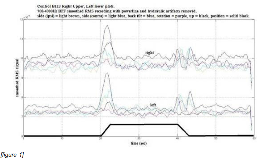
A typical example of the RMS signal for control subjects is shown in Fig 1. During the movement phase t=20-23 and 40-43 sec wherein the vestibular is active the RMS signal showed a marked increase for all signals of all subjects. The typical Schizophrenic response had the peaks seen at t=20-23 skewed to the left.
Fig. 1
[figure 1]
Conclusions
EVestG signals do show a vestibular component. When validated with larger sample size may be assistive in neurological disorder diagnosis.
- Type
- Abstract
- Information
- European Psychiatry , Volume 28 , Issue S1: Abstracts of the 21th European Congress of Psychiatry , 2013 , 28-E1362
- Copyright
- Copyright © European Psychiatric Association 2012
 You have
Access
You have
Access
Comments
No Comments have been published for this article.