


A 9-year-old female presented to neurology outpatient department of our hospital with complaints of recurrent generalized tonic–clonic seizures since birth and was being treated with anticonvulsants for the same. Patient also had complaints of giddiness and episodes of momentary loss of consciousness. There was history of twitching of left hemiface and eyelid during infancy, often associated with deviation of eyes to the left and groaning. The birth history was unremarkable. Family history revealed no known consanguinity. General examination revealed no dysmorphic features. Neurological examination revealed no cognitive deficits/signs to suggest cerebellar pathology. An electroencephalogram was done in view of her recurrent seizures, which was normal. Initial laboratory work-up was normal. The patient then underwent magnetic resonance imaging (MRI) brain, acquired with a 1.5-T unit (Siemens, Erlangen, Germany). MRI brain revealed hemihypertrophy of left cerebellar hemisphere with disorganized architecture, fissural malorientation with individual folia running vertically rather than horizontally with disorganized foliation, abnormal arborization of white matter predominantly involving mid and dorsal surface of left cerebellar hemisphere and a few suspicious areas of abnormal T2-hyperintense signal in subcortical white matter. Right cerebellar hemisphere and cerebellar vermis were normal. Corpus callosum was normal. Cerebral parenchyma was normal in signal intensity pattern with normal gray–white matter differentiation. Ventricular system was normal (Figures 1 and 2). Cerebellar malformations are uncommon and are usually associated with Dandy–Walker continuum, Joubert syndrome, rhombencephalosynapsis, lissencephaly, Fukuyama congenital muscular dystrophy, Walker–Warburg syndrome, muscle–eye–brain disease, congenital cytomegalovirus infection to name a few.Reference Patel and Barkovich1, Reference Demaerel2 Isolated unilateral cerebellar hemispheric dysplasia is exceedingly rare with only a few cases previously described in English literature. Cerebellar malformations are less adequately understood entity partly because of the complex cerebellar embryology and limited histologic studies of these disorders. Genes expressed in migration and maintenance of the Purkinje cells and/or in the generation and migration of granular cells when mutated will disrupt cerebellar migration and foliation and thus cause cerebellar malformation.Reference Soto-Ares, Delmaire, Deries, Vallee and Pruvo3–Reference Larroche5 Cerebellum is known to be a centre for motor learning, coordination, and higher cognitive functions. Clinical presentation of cerebellar malformations is highly variable and depends on the degree of cerebellar involvement, presence of associated cerebral involvement and the underlying disorders such as muscular dystrophy if any. Patel and Barkovich suggested an imaging-based classification of cerebellar malformations and classified the malformations broadly into two types, malformations with cerebellar hypoplasia and the ones with cerebellar dysplasia. Each of these was further classified into focal and diffuse.Reference Patel and Barkovich1 Demaerel gave a classification of abnormalities of cerebellar foliation and fissuration.Reference Demaerel2 Our index case with disorganized architecture, fissural malorientation and disorganized foliation of left cerebellar hemisphere associated with normal cerebellar vermis, corpus callosum, and absence of cerebral malformation falls into Type 2 category as per the classification by Demaerel.Reference Demaerel2 Treatment depends upon the severity of symptoms and the underlying disorder in case of syndromic malformations. Generally, treatment is symptomatic and supportive. Understanding of the basics of cerebellar embryology, knowledge of the imaging features, and clinical presentation aids in the precise diagnosis of this disorder and its optimal management.
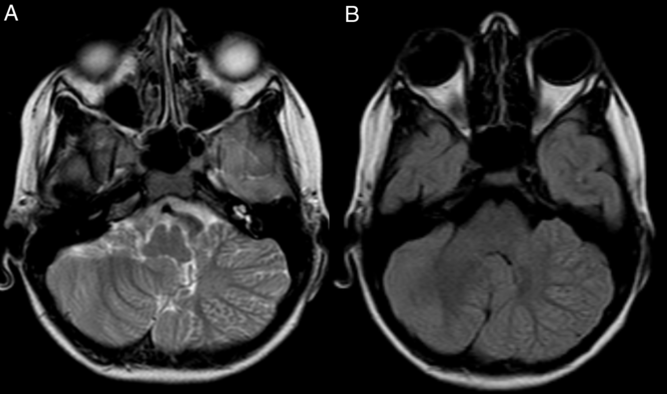
Figure 1: Axial T2 (A) and fluid-attenuated inversion recovery (B) sections of MRI brain of posterior fossa showing hemihypertrophy of left cerebellar hemisphere with disorganized architecture, fissural malorientation with individual folia running vertically rather than horizontally with disorganized foliation, abnormal arborization of white matter predominantly involving mid and dorsal surface of left cerebellar hemisphere.
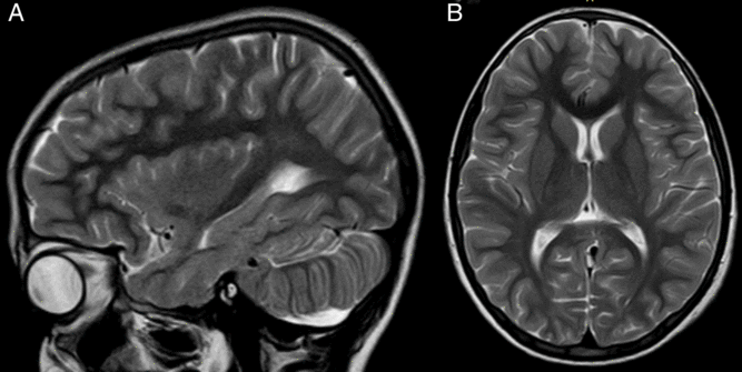
Figure 2: T2-weighted sagittal section (A) and axial section (B) of MRI brain shows disorganized foliation, with individual folia running vertically rather than horizontally (A). Cerebral parenchyma was normal in signal intensity with normal gray–white matter differentiation and ventricular system was normal (B).
Conflict of Interest
None.
Statement of Authorship
CC: Study concept and design, acquisition of data, drafting of manuscript.
AB: Acquisition of data, drafting of manuscript.
RV: Study supervision, revision of manuscript.
MGKM: Study supervision, revision of manuscript.
All authors have read and agreed to the content of the manuscript.


