Skip to main content Accessibility help
×
Hostname: page-component-848d4c4894-4rdrl Total loading time: 0 Render date: 2024-06-27T05:46:12.006Z Has data issue: false hasContentIssue false

Chapter 17 - Blood Flow Volume in Umbilical Vein in Fetal Growth Restriction

from Section 5 - Characteristics of Fetal Growth Restriction

Published online by Cambridge University Press:  23 July 2018

Christoph Lees
Affiliation:
Imperial College London
Gerard H. A. Visser
Affiliation:
Universiteit Utrecht, The Netherlands
Kurt Hecher
Affiliation:
University Medical Centre, Hamburg
Get access

Summary

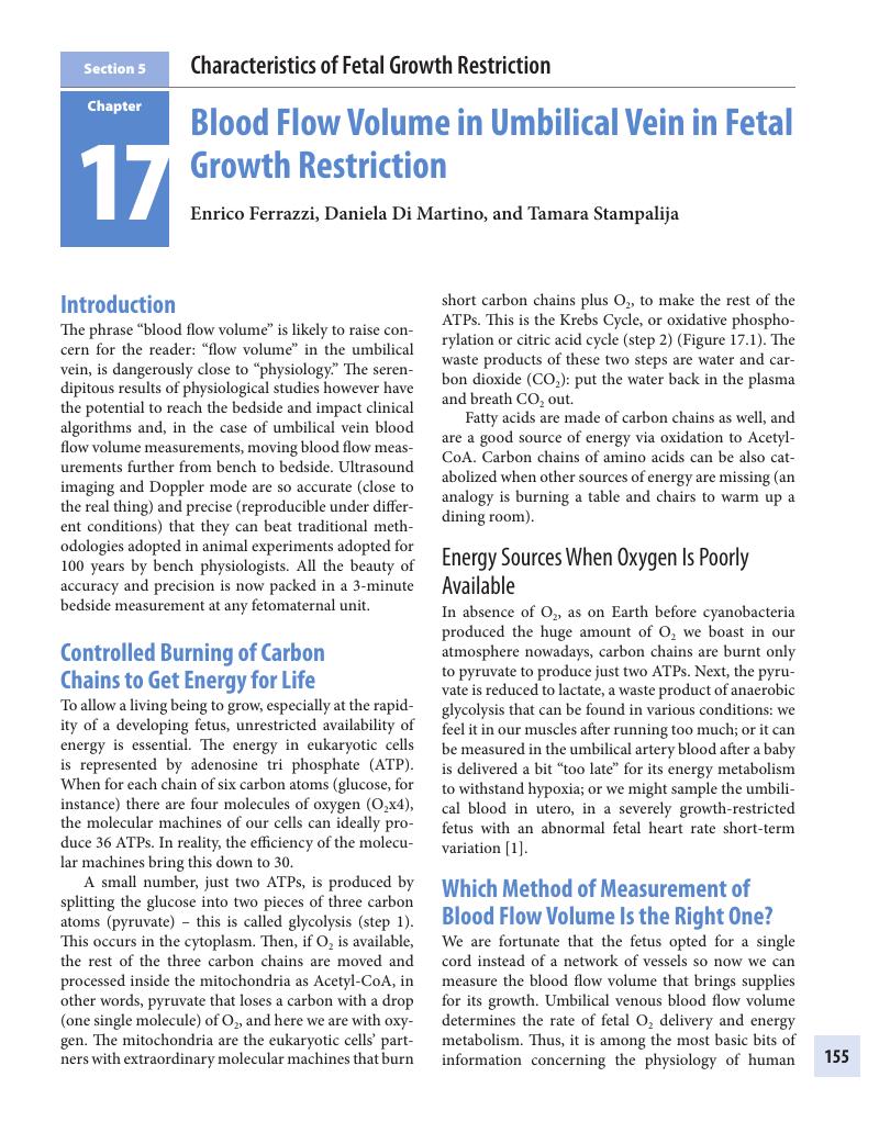
Image of the first page of this content. For PDF version, please use the ‘Save PDF’ preceeding this image.'
Type
Chapter
Information
Publisher: Cambridge University Press
Print publication year: 2018

Access options

Get access to the full version of this content by using one of the access options below. (Log in options will check for institutional or personal access. Content may require purchase if you do not have access.)

References

Pardi, G, Cetin, I, Marconi, AM, Lanfranchi, A, Bozzetti, P, Buscaglia, M, et al. Diagnostic value of blood sampling in fetuses with growth retardation. N Engl J Med 1993 Oct 2;328(10):692–6.Google Scholar
Galan, HL, Jozwik, M, Rigano, S, Regnault, TR, Hobbins, JC, Battaglia, FC, et al. Umbilical vein blood flow determination in the ovine fetus: Comparison of Doppler ultrasonographic and steady-state diffusion techniques. Am J Obstet Gynecol 1999 Nov 1;181(5 Pt 1):1149–53.CrossRefGoogle ScholarPubMed
Schmidt, KG, Di Tommaso, M, Silverman, NH, Rudolph, AM. Doppler echocardiographic assessment of fetal descending aortic and umbilical blood flows. Validation studies in fetal lambs. Circulation 1991 Apr 30;83(5):1731–7.CrossRefGoogle ScholarPubMed
Meschia, G, Cotter, JR, Makowski, EL, Burron, DH. Simultaneous measurement of uterine and umbilical blood flows and oxygen uptakes. Exp Physiol 1967 Aug 1;52:118.Google Scholar
Dawes, GS, Mott, JC. Changes on O2 distribution and consumption in foetal lambs with variations in umbilical blood flow. J Physiol 1964 Jan 25;170:524–40.CrossRefGoogle ScholarPubMed
Wallace, JM, Bourke, DA, Aitken, RP, Leitch, N, Hay, WW. Blood flows and nutrient uptakes in growth-restricted pregnancies induced by overnourishing adolescent sheep. Am J Physiol Regul Integr Comp Physiol 2002 Feb 25;282:1027–36.Google Scholar
Figueras, F, Fernández, S, Hernandez-Andrade, E, Gratacós, E. Umbilical venous blood flow measurement: Accuracy and reproducibility. Ultrasound Obstet Gynecol 2008 Sep;32(4):587–91.Google Scholar
Di Naro, E, Ghezzi, F, Raio, L, Franchi, M, D’Addario, V, Lanzillotti, G, et al. Umbilical vein blood flow in fetuses with normal and lean umbilical cord. Ultrasound Obstet Gynecol 2001 Feb 28;17(3):224–8.Google Scholar
Ferrazzi, E, Bellotti, M, Marconi, AM, Barbera, AF, Pardi, G. Peak velocity of the outflow of the aorta: Correlations with acid base status and oxygenation in growth retarded fetuses. 1965 Jan 4;85:663–7.Google Scholar
Meschia, G, Cotter, JR, Barron, DH. The hemoglobin, O2, CO2, and H+ concentrations in the umbilical bloods of sheep. Q J Exp Physiol Cogn Med Sci 1965 Aug 4;50:185–95.Google Scholar
Battaglia, FC, Meschia, G. Review of studies in human pregnancies of uterine and umbilical blood flows. Dev Period Med 2013 Feb 7;XVIII(4):287–92.Google Scholar
Pennati, G, Bellotti, M, De Gasperi, C, Rognoni, G. Spatial velocity profile changes along the cord in normal human fetuses: Can these affect Doppler measurements of venous umbilical blood flow? Ultrasound Obstet Gynecol 2004 Feb 4;23(2):131–7.CrossRefGoogle ScholarPubMed
Burton, GJ, Woods, AW, Jauniaux, E, Kingdom, JCP. Rheological and physiological consequences of conversion of the maternal spiral arteries for uteroplacental blood flow during human pregnancy. Placenta 2010 Nov 4;30(6):473–82.Google Scholar
Marconi, AM, Cetin, I, Ferrazzi, E, Ferrari, M, Pardi, G, Battaglia, FC. Lactate metabolism in normal and growth-retarded human fetuses. Pediatr Res 1990 Sep 28;28(6):652–6.Google Scholar
Alfirevic, Z, Neilson, JP. Doppler ultrasonography in high-risk pregnancies: Systematic review with meta-analysis. 1995 Jan 4;172:1379–87.CrossRefGoogle Scholar
Rigano, S, Bozzo, M, Padoan, A, Mustoni, P, Bellotti, M, Galan, HL, et al. Small size-specific umbilical vein diameter in severe growth restricted fetuses that die in utero. Prenat Diagn 2008 Oct;28(10):908–13.Google Scholar
Bellotti, M, Pennati, G, De, Gasperi, C, Battaglia, FC, Ferrazzi, E. Role of ductus venosus in distribution of umbilical blood flow in human fetuses during second half of pregnancy. Am J Physiol Heart Circ Physiol 2000 Aug 31;279(3):H1256–63.CrossRefGoogle ScholarPubMed
Kiserud, T. Physiology of the fetal circulation. Semin Fetal Neonatal Med 2005 Dec;10(6):493503.CrossRefGoogle ScholarPubMed
Kiserud, T, Ozaki, T, Nishina, H, Rodeck, C, Hanson, MA. Effect of NO, phenylephrine, and hypoxemia on ductus venosus diameter in fetal sheep. Am J Physiol Heart Circ Physiol 2000 Aug 31;279(3):H1166–71.Google Scholar
Bellotti, M, Pennati, G, Pardi, G, Fumero, R. Dilatation of the ductus venosus in human fetuses: Ultrasonographic evidence and mathematical modeling. Am J Physiol Heart Circ Physiol 1998;275(5):H1759–67.Google Scholar
Bellotti, M, Pennati, G, Gasperi, CD, Bozzo, M, Battaglia, FC, Ferrazzi, E. Simultaneous measurements of umbilical venous, fetal hepatic, and ductus venosus blood flow in growth-restricted human fetuses. Am J Obstet Gynecol 2004 May;190(5):1347–58.Google Scholar
Kiserud, T, Kessler, J, Ebbing, C, Rasmussen, S. Ductus venosus shunting in growth-restricted fetuses and the effect of umbilical circulatory compromise. Ultrasound Obstet Gynecol 2006;28(2):143–9.CrossRefGoogle ScholarPubMed
Regnault, TRH, Friedman, JE, Wilkening, RB, Anthony, RV, Hay, WW, Jr. Fetoplacental transport and utilization of amino acids in IUGR – a review. Placenta 2005 Apr;26:S52S62.CrossRefGoogle ScholarPubMed
Lees, C, Marlow, N, Arabin, B, Bilardo, CM, Brezinka, C, Derks, JB, et al. Perinatal morbidity and mortality in early-onset fetal growth restriction: Cohort outcomes of the trial of randomized umbilical and fetal flow in Europe (TRUFFLE). Ultrasound Obstet Gynecol 2013 Sep 23;42(4):400–8.Google Scholar
TRUFFLE Lancet Paper 2015.Google Scholar
Trudinger, B. Doppler: More or less? Ultrasound Obstet Gynecol 2007;29(3):243–6.Google Scholar
Figueras, F, Gardosi, J. Intrauterine growth restriction: New concepts in antenatal surveillance, diagnosis, and management. Am J Obstet Gynecol 2011 Apr 1;204(4):288300.Google Scholar
Cetin, I, Ronzoni, S, Marconi, AM, Perugino, G, Corbetta, C, Battaglia, FC, et al. Maternal concentrations and fetal-maternal concentration differences of plasma amino acids in normal and intrauterine growth-restricted pregnancies. Am J Obstet Gynecol 1996 May;174(5):1575–83.Google Scholar
Ronzoni, S, Marconi, AM, Paolini, CL, Teng, C, Pardi, G, Battaglia, FC. The effect of a maternal infusion of amino acids on umbilical uptake in pregnancies complicated by intrauterine growth restriction. Am J Obstet Gynecol 2002 Sep;187(3):741–6.Google Scholar
Ferrazzi, E, Rigano, S, Bozzo, M, Bellotti, M, Giovannini, N, Galan, H, et al. Umbilical vein blood flow in growth-restricted fetuses. Ultrasound Obstet Gynecol 2000 Oct 1;16(5):432–8.Google Scholar
Rigano, S, Bozzo, M, Ferrazzi, E, Bellotti, M, Battaglia, FC, Galan, HL. Early and persistent reduction in umbilical vein blood flow in the growth-restricted fetus: A longitudinal study. Am J Obstet Gynecol 2001 Oct 1;185(4):834–8.Google Scholar
Parra-Saavedra, M, Crovetto, F, Triunfo, S, Savchev, S, Parra, G, Sanz, M, et al. Added value of umbilical vein flow as a predictor of perinatal outcome in term small-for-gestational-age fetuses. Ultrasound Obstet Gynecol 2013 Jul 26;42(2):189–95.CrossRefGoogle ScholarPubMed
Sanz-Cortés, M, Figueras, F, Bargalló, N, Padilla, N, Amat-Roldan, I, Gratacós, E. Abnormal brain microstructure and metabolism in small-for-gestational-age term fetuses with normal umbilical artery Doppler. Ultrasound Obstet Gynecol 2010 Feb 3;36(2):159–65.CrossRefGoogle ScholarPubMed
Figueras, F, Oros, D, Cruz-Martinez, R, Padilla, N, Hernandez-Andrade, E, Botet, F, et al. Neurobehavior in term, small-for-gestational age infants with normal placental function. Pediatrics 2009 Oct 26;124(5):e934–41.Google Scholar
Mando, C, De Palma, C, Stampalija, T, Anelli, GM, Figus, M, Novielli, C, et al. Placental mitochondrial content and function in intrauterine growth restriction and preeclampsia. Am J Physiol Endocrinol Metab 2014 Feb 15;306(4):E404–13.Google Scholar
Barker, JA. Fetal origins of coronary heart disease. BMJ 1995;311:171–4.Google Scholar

Save book to Kindle

To save this book to your Kindle, first ensure coreplatform@cambridge.org is added to your Approved Personal Document E-mail List under your Personal Document Settings on the Manage Your Content and Devices page of your Amazon account. Then enter the ‘name’ part of your Kindle email address below. Find out more about saving to your Kindle.

Note you can select to save to either the @free.kindle.com or @kindle.com variations. ‘@free.kindle.com’ emails are free but can only be saved to your device when it is connected to wi-fi. ‘@kindle.com’ emails can be delivered even when you are not connected to wi-fi, but note that service fees apply.

Find out more about the Kindle Personal Document Service.

Available formats
×

Save book to Dropbox

To save content items to your account, please confirm that you agree to abide by our usage policies. If this is the first time you use this feature, you will be asked to authorise Cambridge Core to connect with your account. Find out more about saving content to Dropbox.

Available formats
×

Save book to Google Drive

To save content items to your account, please confirm that you agree to abide by our usage policies. If this is the first time you use this feature, you will be asked to authorise Cambridge Core to connect with your account. Find out more about saving content to Google Drive.

Available formats
×