Skip to main content Accessibility help
×
  • Cited by 4
Publisher:
Cambridge University Press
Online publication date:
March 2013
Print publication year:
2013
Online ISBN:
9781139088268

Book description

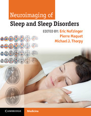
This up-to-date, superbly illustrated book is a practical guide to the effective use of neuroimaging in the patient with sleep disorders. There are detailed reviews of new neuroimaging techniques – including CT, MRI, advanced MR techniques, SPECT and PET – as well as image analysis methods, their roles and pitfalls. Neuroimaging of normal sleep and wake states is covered plus the role of neuroimaging in conjunction with tests of memory and how sleep influences memory consolidation. Each chapter carefully presents and analyzes the key findings in patients with sleep disorders indicating the clinical and imaging features of the various sleep disorders from clinical presentation to neuroimaging, aiding in establishing an accurate diagnosis. Written by neuroimaging experts from around the world, Neuroimaging of Sleep and Sleep Disorders is an invaluable resource for both researchers and clinicians including sleep specialists, neurologists, radiologists, psychiatrists, psychologists.

Reviews

“…Invaluable resource for researchers and clinicians in…sleep medicine, neurology, radiology, psychiatry, and psychology. A particular strength of the book is the incorporation of color figures and graphs of neuroimaging results.”

- Doody's Review Service

Refine List

Actions for selected content:

Select all | Deselect all
  • View selected items
  • Export citations
  • Download PDF (zip)
  • Save to Kindle
  • Save to Dropbox
  • Save to Google Drive

Save Search

You can save your searches here and later view and run them again in "My saved searches".

Please provide a title, maximum of 40 characters.
×

Contents


Page 2 of 3


  • Chapter 21 - Neuroimaging the effects of light on non-visual brain functions
    pp 171-178
  • View abstract

    Summary

    This chapter reviews the functional brain imaging studies, using positron emission tomography (PET) and functional magnetic resonance imaging (fMRI), that have examined neural activity patterns between non-rapid eye movement (NREM) sleep and wakefulness, and within NREM sleep in association with phasic neuronal oscillations. It explores recent fMRI data investigating the relationship between these rhythms and the processing of external stimulation during sleep. In order to further explore this relationship between external stimulation and NREM sleep phasic activity, brain responses to pure tones delivered during NREM sleep were evaluated in a recent event-related fMRI study. In the fMRI study, the relationship between auditory stimulation and slow waves was also explored. It is well known that external stimulation during NREM sleep can trigger a slow wave on electroencephalographic (EEG) recordings: such evoked slow waves, especially during stage N2, are also termed K-complexes.
  • Chapter 22 - Memory systems, sleep, and neuroimaging
    pp 179-186
  • View abstract

    Summary

    Although the neurophysiological origin of the functional magnetic resonance imaging (fMRI) blood oxygen level-dependent (BOLD) signal is still poorly understood, spontaneous fMRI signal fluctuations show consistent spatial correlations in functionally related networks. Large-scale functional brain networks as derived from fMRI time-series can be examined by graph theoretical analysis; such analysis has revealed a small-world organization of human functional brain networks during wakefulness, with high local clustering and short path length. A hierarchical cluster analysis indeed illustrated that frontoparietal clusters could be detected in wakefulness but not in deeper non-rapid eye movement (NREM) sleep stages. Functional connectivity of phasic events allows further spatial and temporal refinement of vigilance-state dependent connectivity patterns, and may be of special interest for phasic electroencephalography (EEG) events during sleep. Finally, although functional connectivity appears to overlap to a considerable extent with brain metabolism, these measures seem to represent correlated but different dimensions.
  • Chapter 24 - Functional neuroimaging of primary insomnia
    pp 197-208
  • View abstract

    Summary

    This chapter shows that the study of dreams provides meaningful and valuable information about cognitive and affective processes occurring during sleep. It demonstrates that typical features in large dream samples can be identified using statistical methods and that these features are in good correspondence with known patterns of brain activity during sleep, in particular rapid eye movement (REM) sleep. These analyses are based on the frequency of occurrence and degree of uniformity of dream contents, irrespective of whether the dreams mimicked real-life experiences or were extremely bizarre. The chapter also shows that bizarre but common aspects in dreams have much in common with known neuropsychological syndromes. Integrated approach to sleep and dreaming undoubtedly contribute to redefining the links between brain processes and the varieties of dream experiences, and lead to a more comprehensive model of human brain function during sleep.
  • Chapter 25 - Sleep neuroimaging in depression and schizophrenia
    pp 209-217
  • View abstract

    Summary

    This chapter reviews the techniques currently applied to study brain function during sleep deprivation (SD) as opposed to the consequence of SD. It provides a bird's eye view of functional imaging studies performed on healthy young adult volunteers to date and comment on how this research has evolved the conceptualization of how SD modulates behavior. The first functional imaging studies involving SD utilized positron emission tomography (PET). Based on the initial findings, cognitive domain and task difficulty was proposed as determinants of the neural response to SD. It was postulated that changes in dopamine signaling in the SD state contributed to the change in functional connectivity, an idea reprised when discussing risky decision making in SD. The interaction of SD and circadian effects, including the effects of chronotype, could be a further target of functional neuroimaging studies, including the effect of countermeasures such as naps and stimulants.
  • Chapter 27 - Functional neuroimaging of narcolepsy
    pp 223-227
  • View abstract

    Summary

    Deficits in attention are a central mechanism through which performance in higher cognitive domains such as memory may be affected in sleep deprivation (SD). This chapter reviews how different facets of attention and information processing are compromised in sleep-deprived persons. It discusses the link between behavioral alterations and concurrently observed shifts in task-related functional magnetic resonance imaging (fMRI) signal as well as how imaging can reveal alterations in processing not evident in overt behavior. Complimenting attention's enhancement effects is its ability to suppress irrelevant distractors. This ability is impaired by SD. The perceptual load theory of attention provides a useful framework for evaluating SD-induced change in visual information processing. Reduced engagement of frontoparietal regions that mediate top-down control of attention has been demonstrated in experiments evaluating visual short-term memory (VSTM), preparatory attention, and selective attention.
  • Chapter 28 - Neuroimaging of treatment response in narcolepsy
    pp 228-230
  • View abstract

    Summary

    An improved understanding of risky decision-making in sleep-deprived persons could have important real-world consequences. Traditional expectation-based models assume that individuals integrate information across outcomes and probabilities. Neuroeconomic studies that seek to understand the neural mechanisms underlying economic decision-making often target a particular decision variable, incorporate that variable into a model function, manipulate the level of that variable across a range of stimuli, and then identify aspects of brain function that track changes in that variable. Separating decision and outcome phases in the imaging analysis could also be important as sleep deprivation (SD) might interact with task context and feedback to influence neural responses and behavior. The shifts in economic preferences in the multiple outcome gambling experiment as well as relative valuation for social and monetary stimuli were independent of the effects of SD on psychomotor vigilance, consistent with the suggestion that effects of SD vary according to cognitive domain.
  • Chapter 29 - Modafinil effects in narcolepsy
    pp 231-239
  • View abstract

    Summary

    Functional MRI provides a reproducible, non-invasive, and flexible means to study inter-individual variation in performance impairment in the setting of sleep deprivation (SD). An important long-term goal of studying inter-individual differences in responses to SD is to elucidate phenotypes that predict how an individual perform in an operational setting after being sleep deprived. The seemingly contradictory findings regarding the benefit of greater activation in areas of the brain led one to examine whether evaluating shifts in activation across states would prove a reliable marker of inter-individual variability in behavior. The thalamus plays an important part in mediating arousal and attention, which in turn have substantial effects on behavioral performance. Thalamic activation is less reliably reproducible across scan sessions than frontoparietal activation. More complex decision making tasks, where different strategies may be engaged, allow some latitude regarding which neural circuits are engaged and are potentially less at risk of use-dependent effects.
  • Chapter 30 - Neuroimaging of Kleine–Levin syndrome
    pp 240-245
  • View abstract

    Summary

    This chapter focuses on two recent studies capitalizing on inter-individual differences in response to increased sleep homeostasis and misalignment between the circadian signal and sleep to better characterize the brain mechanisms involved in the maintenance of wakefulness and associated cognitive processes. Extreme chronotypes are characterized by marked differences in their preferred timing for sleep and wakefulness, as well as optimal times of day to perform cognitively demanding tasks. A study detailed in the chapter used a genetic trait as a means to characterize the regulation of cognitive performance throughout a normal waking day and following total sleep deprivation. A variable-number-of-tandem-repeat (VNTR) polymorphism in the coding region of the clock gene PERIOD3 (PER3) was reported to present a weak association with chronotype. According to a recent model of cognition, the VLPFC plays a key role in higher cognitive control and is involved in complex neurobehavioral processes.
  • Chapter 31 - Neuroimaging of cataplexy
    pp 246-247
  • View abstract

    Summary

    This chapter describes the physiological bases of the impact of light on non-image-forming functions before summarizing recent neuroimaging investigations that substantially increased the understanding of the brain mechanisms involved. In accordance with animal research, the neuroimaging results presented in the chapter are compatible with a scenario in which light would influence subcortical structures involved in arousal regulation before significantly affecting the cortical areas involved in the ongoing non-image-forming process. Several factors modulate the effects described in this scenario including light intensity, duration, and wavelength, and possibly task difficulty. The interaction between circadian and sleep homeostasis signals and PERIOD3 (PER3) genotype can amplify or diminish the non-image-forming impact of light on cognitive brain function. For tasks triggering emotional responses, effects within cortical areas specially devoted to decoding the emotional content of a particular type of stimulus appear to be quick and mediated through a network based on hypothalamus and amygdala interactions.
  • Chapter 32 - Structural brain neuroimaging changes in obstructive sleep apnea
    pp 248-255
  • View abstract

    Summary

    This chapter looks at how different memory systems are influenced by sleep. It describes the currently most-widely accepted model of consolidation of hippocampus-dependent memory. The chapter also looks at human functional magnetic resonance imaging (fMRI) studies which provide evidence that, in fact, memories are re-activated, re-organized, and re-processed during sleep. Reactivation occurs during post-learning sleep, and it seems to be an important component of memory consolidation. In general, it has been found that it occurs in those brain regions most strongly related to the specific learning task. Re-activation could therefore support synaptic consolidation of memory traces. However, recent studies also provide more and more evidence for systems memory consolidation. Looking for signs of re-processing during sleep is the most difficult to do, because based on imaging data alone it is hard to distinguish from re-organization, and there are only few behavioral tasks that are designed to examine such changes.
  • Chapter 34 - Neuroimaging of cognitive effects in obstructive sleep apnea
    pp 264-274
  • View abstract

    Summary

    This chapter discusses imaging studies in insomnia and in association with insomnia complaints in people not diagnosed with insomnia. This review includes studies applying structural and functional MRI, magnetic resonance spectroscopy (MRS), high-density electroencephalography, and transcranial magnetic stimulation (TMS). The studies reviewed have reported almost exclusively on regions of the temporal lobe, frontal lobe, and parietal lobe. These cortical regions are of interest because of their key involvement in the cognitive domains that are most affected in insomnia and after sleep deprivation. For each lobe, the chapter systematically addresses differences between insomniacs and controls and correlations of insomnia symptom severity with brain changes in both insomniacs and people not diagnosed with insomnia. Subsequently, the findings are summarized and interpreted with respect to functional relevance, pitfalls, and conclusions on cause, risk factor, or consequence. Neuroimaging has a high promise to reveal insights into the causes and consequences of insomnia.
  • Chapter 36 - Neuroimaging of treatment effects in obstructive sleep apnea
    pp 284-292
  • View abstract

    Summary

    This chapter reviews brain imaging studies that comment on whether or not there is abnormal brain function in insomnia patients that may in some way relate to their difficulty in sleeping. It provides a systems neuroscience view of a hierarchical arousal network in the central nervous system. Human sleep neuroimaging studies in healthy subjects support the involvement of these basic arousal networks in non-rapid eye movement (NREM) sleep. Blood flow has been shown to correlate negatively with the presence of NREM sleep in the pontine reticular formation, and in the basal forebrain/hypothalamus. Pharmacotherapy for insomnia may have some of its mechanism of action on the limbic and paralimbic structures, especially the antidepressant medications. Traditional sedative-hypnotic approaches appear to target brainstem and hypothalamic arousal networks in insomnia patients while behavioral treatments and frontal cerebral hypothermia appear to target frontal hyperarousal in insomnia patients.
  • Chapter 37 - Structural and functional neuroimaging of congenital central hypoventilation syndrome
    pp 293-300
  • View abstract

    Summary

    Extensive electroencephalographic (EEG) sleep studies have demonstrated increases in rapid eye movement (REM) sleep and changes in non-rapid eye movement (NREM) sleep in depression. Preclinical evidence shows that REM sleep is generated in the brainstem. It also shows that NREM sleep is characterized by slower frequency, higher amplitude thalamocortical electrical oscillations. The alterations in NREM sleep in depressed patients may lead to impaired restoration of prefrontal cortex function during NREM sleep. Functional neuroimaging studies of sleep extend the preclinical understanding of the mechanisms of sleep/wake regulation by providing potential links between neural systems involved in emotional behavior and those involved in sleep. The notion of hyperarousal in paralimbic structures in depressed patients has received further support from an extensive literature describing the functional neuroanatomical correlates of the antidepressant response to sleep deprivation in depressed patients. Patients with schizophrenia are known to have severely disturbed subjective sleep.
  • Chapter 38 - Neuroimaging of disorders of arousal and other parasomnias
    pp 301-304
  • View abstract

    Summary

    The neuroimaging studies have provided new information about brain abnormalities in narcolepsy patients. Differences in brain morphology that are not identifiable by routine visual inspection of individual brain magnetic resonance imaging (MRI) can be investigated using voxel-based morphometry (VBM). The VBM method has some limitations in representing gray matter morphology, and localization in the sulcal regions where the fine details of the anatomy are often obscured by a partial volume effect. On the other hand, the thickness of the cerebral cortex reflects the density and arrangement of cells. Measuring cortical thickness using the cortical surface method has been suggested in studies of gray matter morphometry as a strategy for overcoming the limitation of volumetric analyses. Higher tesla MRI scanners and further development of analysis software of brain MR images are able to better characterize the structural changes in narcoleptic brains.
  • Chapter 39 - Neuroimaging of Parkinson’s disease and multiple system atrophy in patients with sleep disturbance
    pp 305-315
  • View abstract

    Summary

    This chapter reviews functional brain imaging studies conducted in narcoleptic patients. Several functional neuroimaging studies were conducted to evaluate the distribution of brain activity during wakefulness and sleep in narcolepsy. Three of them described CMRglu and rCBF patterns during resting wakefulness. Processing of emotional information potentially plays an important role in narcolepsy-cataplexy. Brain responses to unpleasant stimuli were investigated in nine unmedicated narcoleptic patients with cataplexy and nine matched controls. Anticipation of reward constitutes a particular emotional experience prone to trigger cataplexy in humans, which suggests a potential involvement of the hypocretin system in reward brain circuits, and possible alterations of these circuits in narcolepsy with cataplexy. In the narcolepsy group, significant positive correlations were found between disease duration and functional magnetic resonance imaging (fMRI) responses to high motivational cues in the nucleus accumbens and ventromedial prefrontal cortex.
  • Chapter 40 - Neuroimaging of idiopathic REM sleep behavior disorder
    pp 316-332
  • View abstract

    Summary

    This chapter reviews the neuroimaging findings devoted to the effects of treatment in narcolepsy. These studies involved functional neuroimaging methods such as single photon emission computed tomography (SPECT), positron emission tomography (PET), and functional magnetic resonance imaging (fMRI). The majority of them focused on modafinil, with also early reports on amphetamines and methylphenidate. In narcoleptic patients single-dose treatment effects were also studied with fMRI. PET and SPECT were used to investigate brain activity changes induced by a prolonged administration of modafinil in narcoleptic patients. An 18F-fluorodeoxyglucose (18F-FDG) PET study evaluated brain glucose metabolism (CMRglu) during baseline wakefulness in eight patients and eight controls before and after two weeks of treatment with modafinil. Decreased CMRglu was observed in the brainstem, hypothalamus, thalamus, and mesiotemporal areas in narcoleptics compared to controls, both before and after treatment.
  • Chapter 41 - Cardiac123I-MIBG scintigraphic findings and REM sleep behavior disorder
    pp 333-339
  • View abstract

    Summary

    A recent double-blind, placebo-controlled study investigated the effect of modafinil on cortical excitability in narcolepsy by means of transcranial magnetic stimulation (TMS) and explored the relation between these TMS measures and conventional measures of sleepiness. Electroencephalographic (EEG) low-resolution brain electromagnetic tomography (LORETA) was developed in order to identify brain regions that are involved in neuropsychiatric disorders and are the targets of therapeutic drug action. Modafinil did not influence thymopsychic variables in narcolepsy, but it significantly improved cognitive performance, which may be related to medial prefrontal activity processes identified by LORETA. To bridge the currently wide gap between the neurotransmitter and the behavioral level and to improve temporal resolution up to the millisecond level, it may be useful to study the individual components of event-related brain potentials (ERPs). The ERP study revealed significantly shortened N2 and P300 latencies under modafinil compared with placebo, which reflects an improvement of information processing speed.
  • Chapter 42 - Neuroimaging and posttraumatic stress disorder
    pp 340-346
  • View abstract

    Summary

    This chapter reviews the spectrum of neuroimaging in Kleine-Levin syndrome (KLS). Although structural neuroimaging in KLS is within the normal range in most cases, the vast majority of studies report hypoperfusion in several brain regions. Cognitive impairment and altered perception occur in all KLS patients during sleep episodes according to the review by Arnulf. It is thus pertinent to investigate the neural concomitants to cognitive function in KLS by functional neuroimaging. N-Acetylasparate (NAA) is a biomarker for neuronal health, associated with either neuronal loss or neuronal dysfunction. Hypothalamic pathology constitutes a main hypothesis for KLS etiology. This hypothesis is based on the important function of the hypothalmus in sleep and appetite regulation as well as regarding sexual activity. The sparsity of neuroimaging findings in the hypothalamus might be a result of the detection limit for functional imaging in this region of the brain.
  • Chapter 43 - Neuroimaging of sleepwalking
    pp 347-348
  • View abstract

    Summary

    This chapter focuses on the neuroimaging of cataplexy using a case of a 68-year-old woman, who had suffered from narcolepsy since she was 15 years old, as an example. Her mean sleep latency during a multiple sleep latency test was 0.5 minutes, with three sleep onset rapid eye movement (REM) periods among the four naps. The patient underwent two 99mTc-ethylcysteinate dimer brain single-photon emission computed tomography (SPECT) studies during symptomatic and asymptomatic periods of cataplexy on two non-consecutive days. Symptomatic SPECT images were coregistered with asymptomatic images and both images were then co-registered with 3-dimensional magnetic resonance imaging (MRI). The normalized subtracted SPECT and MRI volumes were merged for visual analysis. A characteristic of human REM sleep is right-hemisphere activation, as shown by SPECT imaging and spectral electroencephalographic (EEG) analysis. The right hemisphere is also more activated during cataplexy than the left hemisphere.
  • Chapter 44 - Neuroimaging of the brainstem in parasomnia overlap disorder
    pp 349-351
  • View abstract

    Summary

    Numerous findings of brain structural changes in obstructive sleep apnea (OSA) give strong support to the notion that the disorder does cause brain injury. This chapter describes findings by technique, influences of factors other than the sleep disordered breathing on structural changes in OSA, and a summary of the brain regions shown across multiple studies to be affected in the disorder. Psychological symptoms of depression and anxiety are associated with neural changes in non-OSA populations, so one can hypothesize that the structural changes in OSA would be exacerbated in the presence of these symptoms. Many areas in the brain show structural impairments in OSA, including cortical, limbic, brainstem and cerebellar regions. Neuroimaging methods give numerical measures that are associated with a variety of biological pathologies, and technical limitations due to scanning and analysis issues limit the interpretability of the data.
  • Chapter 45 - Nocturnal wanderings and an arachnoid cyst
    pp 352-354
  • View abstract

    Summary

    This chapter focuses on upper airway imaging, and examines risk factors for obstructive sleep apnea (OSA) (both static and dynamic) and the anatomical factors that are changed when successful treatment modalities are applied. OSA is known to have a significant familial component. Volumetric magnetic resonance imaging (MRI) has been employed to investigate whether or not oropharyngeal soft tissue structures segregate in a familial fashion. These data implicate that changes in airway conformation and the size of structures surrounding the oropharynx are important identifiable risk factors for the development of OSA. Interventions which successfully improve sleep disordered breathing often have positive impacts on these same factors, which may contribute to the reduction in airway collapsibility. Imaging can be used to classify patients for the purpose of targeting treatment modalities in order to enhance the success in treating sleep disordered breathing especially in patients undergoing upper airway surgery.
  • Chapter 46 - Structural and functional neuroimaging of restless legs syndrome and periodic limb movements in sleep
    pp 355-362
  • View abstract

    Summary

    Sleep apnea is an important cause in sleep medicine for a direct sustained assault on the brain, and also indirectly through a range of mechanisms. This chapter summarizes some of these mechanisms, as they are all active in the obstructive sleep apnea (OSA) patient. Sleep deprivation/fragmentation, and sleep apnea, alters task-related activation in the executive network. Brain-derived neurotrophic factor (BDNF) may be a special link between sleep, cognition, and brain health. Supportive evidence of a central role for BDNF in sleep homeostasis has emerged, particularly providing a link between wake and use-dependent synaptic plasticity and subsequent slow-wave sleep. Structural and functional imaging can establish if genomic modifiers such as these modify the impact of sleep hypoxia or sleep fragmentation on the adult or pediatric brain structure and function, including cognition. Such imaging genomics has shown some utility in assessing the effects of stimulants in relation to catecholamine metabolizing pathway polymorphisms.
  • Chapter 47 - Functional neuroimaging of dopamine, iron, and opiates in restless legs syndrome
    pp 363-374
  • View abstract

    Summary

    This chapter summarizes functional neuroimaging findings from a variety of autonomic and respiratory challenges the author's group has performed in people with obstructive sleep apnea (OSA). A cold pressor challenge involves exposing a body region to a cold stimulus, which elicits a sympathetic activation that leads to a vasoconstriction and a blood pressure increase. The Valsalva maneuver is an autonomic challenge involving straining by forceful expiration against a closed glottis, and the tasks elicit a sequence of blood pressure and heart rate responses mediated through a coordination of autonomic regulatory activity. The inspiratory and expiratory loading tasks led to a degree of dyspnea in most subjects, as did the Valsalva maneuver to a lesser extent. An abnormal pattern in OSA is altered insular functional neuroanatomy in response to autonomic stimuli, as seen with higher-resolution functional magnetic resonance imaging (fMRI) in response to the Valsalva maneuver.
  • Chapter 48 - Neuroimaging and fatal familial insomnia
    pp 375-383
  • View abstract

    Summary

    A previous review of the neuroimaging studies in obstructive sleep apnea (OSA) called for specific attention to longitudinal studies of the treatment effects of OSA on neuroimaging. This chapter focuses on those studies where treatment effects were considered. The structural studies suggest that there are some notable changes in the structure of the human brain when continuous positive airway pressure (CPAP) is used to correct OSA. Some of these changes are even associated with cognitive changes in the expected cognitive domains and are seen with as little as only three months of treatment. The functional imaging studies together suggest that changes in brain function associated with working memory are evident when comparing treatment with no-treatment conditions in patients with OSA. Specifically, treatment often results in the recruitment of fewer cognitive resources to perform at the same level or better.

Page 2 of 3


Metrics

Full text views

Total number of HTML views: 0
Total number of PDF views: 0 *
Loading metrics...

Book summary page views

Total views: 0 *
Loading metrics...

* Views captured on Cambridge Core between #date#. This data will be updated every 24 hours.

Usage data cannot currently be displayed.