
Book contents
- Frontmatter
- Contents
- List of Contributors
- Foreword
- Preface
- List of Abbreviations
- Section 1 Techniques
- Section 2 Clinical applications
- 8 MR perfusion imaging in neurovascular disease
- 9 MR perfusion imaging in neurodegenerative disease
- 10 MR perfusion imaging in clinical neuroradiology
- 11 MR perfusion imaging in oncology: neuro applications
- 12 MR perfusion imaging in oncology: applications outside the brain
- 13 MR perfusion imaging in breast cancer
- 14 MR perfusion imaging in the body: kidney, liver, and lung
- 15 MR perfusion imaging in cardiac diseases
- 16 MR perfusion imaging in pediatrics
- Index
- References
15 - MR perfusion imaging in cardiac diseases
from Section 2 - Clinical applications
Published online by Cambridge University Press: 05 May 2013

Book contents
- Frontmatter
- Contents
- List of Contributors
- Foreword
- Preface
- List of Abbreviations
- Section 1 Techniques
- Section 2 Clinical applications
- 8 MR perfusion imaging in neurovascular disease
- 9 MR perfusion imaging in neurodegenerative disease
- 10 MR perfusion imaging in clinical neuroradiology
- 11 MR perfusion imaging in oncology: neuro applications
- 12 MR perfusion imaging in oncology: applications outside the brain
- 13 MR perfusion imaging in breast cancer
- 14 MR perfusion imaging in the body: kidney, liver, and lung
- 15 MR perfusion imaging in cardiac diseases
- 16 MR perfusion imaging in pediatrics
- Index
- References
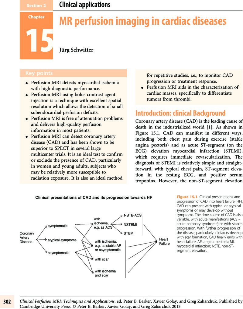
Summary
A summary is not available for this content so a preview has been provided. Please use the Get access link above for information on how to access this content.
- Type
- Chapter
- Information
- Clinical Perfusion MRITechniques and Applications, pp. 302 - 325Publisher: Cambridge University PressPrint publication year: 2013
References
, , , et al. American Heart Association Statistics Committee and Stroke Statistics Subcommittee. Heart disease and stroke statistics – 2009 update: report from the American Heart Association Statistics Committee and Stroke Statistics Subcommittee. Circulation 2009;119.
, , , et al. Coronary event and case fatality rates in an English population: results of the Oxford myocardial infarction incidence study. The Oxford Myocardial Infarction Incidence Study Group. Heart 1998;80:40–4.CrossRefGoogle Scholar
, , , et al. Prognostic value of the admission electrocardiogram in acute coronary syndromes. JAMA 1999;281:707–13.CrossRefGoogle ScholarPubMed
, , , et al. Impact of ischaemia and scar on the therapeutic benefit derived from myocardial revascularization vs. medical therapy among patients undergoing stress-rest myocardial perfusion scintigraphy. Eur Heart J 2011;32:1012–24.CrossRefGoogle ScholarPubMed
Future strategies in the management of coronary artery disease. Fut Cardiol 2006;2(5):555–65.CrossRefGoogle ScholarPubMed
, , , et al. Impact of unrecognized myocardial scar detected by cardiac magnetic resonance imaging on event-free survival in patients presenting with signs or symptoms of coronary artery disease. Circulation 2006;113(23):2733–43.CrossRefGoogle ScholarPubMed
, , , Myocardial viability testing and impact of revascularization on prognosis in patients with coronary artery disease and left ventricular dysfunction: a meta-analysis. J Am Coll Cardiol 2002;39:1151–8.CrossRefGoogle ScholarPubMed
, , , et al. Positron emission tomography and recovery following revascularization (PARR-1): the importance of scar and the development of a prediction rule for the degree of recovery of left ventricular function. J Am Coll Cardiol 2002;40(10):1735–43.CrossRefGoogle ScholarPubMed
, , , et al. Myocardial viability and survival in ischemic left ventricular dysfunction. N Engl J Med 2011;364(17):1617–25.CrossRefGoogle ScholarPubMed
, , , et al. Contrast-enhanced MRI and routine single photon emission computed tomography (SPECT) perfusion imaging for detection of subendocardial myocardial infarcts: an imaging study. Lancet 2003;361(9355):374–9.CrossRefGoogle ScholarPubMed
, Risk assessment using single-photon emission computed tomographic technetium-99m sestamibi imaging. J Am Coll Cardiol 1998;32(1):57–62.CrossRefGoogle ScholarPubMed
, , , et al. Prognostic value of cardiac magnetic resonance stress tests: adenosine stress perfusion and dobutamine stress wall motion imaging. Circulation 2007;115:1769–76.CrossRefGoogle ScholarPubMed
, , , et al. Complementary prognostic values of stress myocardial perfusion and late gadolinium enhancement imaging by cardiac magnetic resonance in patients with known or suspected coronary artery disease. Circulation 2009;120:1390–400.CrossRefGoogle ScholarPubMed
, , , et al. Magnetic resonance imaging determination of cardiac prognosis. Circulation 2002;106(18):2328–33.CrossRefGoogle ScholarPubMed
, Pressure-flow relations in coronary circulation. Physiol Rev 1990;70:331–90.CrossRefGoogle ScholarPubMed
, , , Control of coronary blood flow by an autoregulatory mechanism. Circ Res 1964;14:250–9.CrossRefGoogle ScholarPubMed
, , , et al. Magnetic resonance-based assessment of global coronary flow and flow reserve and its relation to left ventricular functional parameters: a comparison with positron emission tomography. Circulation 2000;101(23):2696–702.CrossRefGoogle ScholarPubMed
, , , et al. Morphometry of canine coronary arteries, arterioles and capillaries during hypertension and left ventricular hypertrophy. Circ Res 1986;58:38–46.CrossRefGoogle ScholarPubMed
, , , et al. Relation between myocardial blood flow and the severity of coronary artery stenosis. N Engl J Med 1994;330:1782–8.CrossRefGoogle ScholarPubMed
, , , et al. Assessment of anatomic and physiological severity of single-vessel coronary artery lesions by dipyridamole echocardiography. Comparison with positron emission tomography and quantitative arteriography. Circulation 1994;89:753–61.CrossRefGoogle ScholarPubMed
, , , et al. Relation among stenosis severity, myocardial blood flow, and flow reserve in patients with coronary artery disease. Circulation 1995;91:1944–51.CrossRefGoogle ScholarPubMed
, , , et al. Alterations in regulation of myocardial blood flow in one-vessel coronary artery disease determined by positron emission tomography. Am J Cardiol 1993;72:538–43.CrossRefGoogle Scholar
, , , et al. Assessment of myocardial perfusion in coronary artery disease by magnetic resonance: a comparison with positron emission tomography and coronary angiography. Circulation 2001;103(18):2230–5.CrossRefGoogle ScholarPubMed
, , , et al. Magnetic resonance myocardial first-pass perfusion imaging: parameter optimization for signal response and cardiac coverage. J Magn Reson Imaging 2001;14(5):556–62.CrossRefGoogle ScholarPubMed
, , , et al. Detection of coronary artery disease by magnetic resonance myocardial perfusion imaging with various contrast medium doses: first European multicenter experience. Eur Heart J 2004;25:1657–65.CrossRefGoogle Scholar
, , , et al. MR-IMPACT: comparison of perfusion-cardiac magnetic resonance with single-photon emission computed tomography for the detection of coronary artery disease in a multicentre, multivendor, randomized trial. Eur Heart J 2008;29:480–9.CrossRefGoogle Scholar
, , , et al. High spatial resolution myocardial perfusion cardiac magnetic resonance for the detection of coronary artery disease. Eur Heart J 2008;29:2148–55.CrossRefGoogle ScholarPubMed
CMR Update, 2nd edn. Lausanne: Schwitter, J. 2012; Available from (accessed November 19, 2012).
, Imaging: assessment of cardiac ischaemia and viability: role of cardiovascular magnetic resonance. Eur Heart J 2011;32:799–809.CrossRefGoogle Scholar
, , , et al. EuroCMR (European Cardiovascular Magnetic Resonance) Registry. J Am Coll Cardiol 2009;54:1457–66.CrossRefGoogle ScholarPubMed
, , , et al. Superior diagnostic performance of perfusion-cardiovascular magnetic resonance versus SPECT to detect coronary artery disease: the secondary endpoints of the multicenter multivendor MR-IMPACT II (Magnetic Resonance Imaging for Myocardial Perfusion Assessment in Coronary Artery Disease Trial). J. Cardiovasc Magn Reson 2012;14:61–71.CrossRefGoogle Scholar
, , , et al. Magnetic resonance perfusion measurements for the noninvasive detection of coronary artery disease. Circulation 2003;108(4):432–7.CrossRefGoogle ScholarPubMed
, , , et al. Coronary artery disease: myocardial perfusion MR imaging with sensitivity encoding versus conventional angiography. Radiology 2005;235(2):423–30.CrossRefGoogle ScholarPubMed
, , , et al. Comparison of dobutamine stress magnetic resonance, adenosine stress magnetic resonance, and adenosine stress magnetic resonance perfusion. Circulation 2004;110(7):835–42.CrossRefGoogle ScholarPubMed
, , , et al. Detecting acute coronary syndrome in the emergency department with cardiac magnetic resonance imaging. Circulation 2003;107:531–7.CrossRefGoogle ScholarPubMed
, , , et al. Prognosis of negative adenosine stress magnetic resonance in patients presenting to an emergency department with chest pain. J Am Coll Cardiol 2006;47(7):1427–32.CrossRefGoogle Scholar
, , , et al. Assessment of non-ST-segment elevation acute coronary syndromes with cardiac magnetic resonance imaging. J Am Coll Cardiol 2004;44:2173–81.CrossRefGoogle ScholarPubMed
, , , et al. Usefulness of magnetic resonance imaging of cardiac and paracardiac masses. Am J Cardiol 2003;92(7):890–5.CrossRefGoogle ScholarPubMed
, , , et al. MRI of intimal sarcoma of the pulmonary arteries. Circ Cardiovasc Imaging 2009;2:e37–9.CrossRefGoogle ScholarPubMed
, , , et al. Cardiovascular magnetic resonance imaging for diagnosis and clinical management of suspected cardiac masses and tumours. Eur Heart J 2011;32(12):1551–60.CrossRefGoogle ScholarPubMed
, , , et al. Clinical, imaging, and pathological characteristics of left ventricular thrombus: a comparison of contrast-enhanced magnetic resonance imaging, transthoracic echocardiography, and transesophageal echocardiography with surgical or pathological validation. Am Heart J 2006;152(1):75–84.CrossRefGoogle ScholarPubMed
, , , et al. Characterization of dysfunctional myocardium by positron emission tomography and magnetic resonance: relation to functional outcome after revascularization. Circulation 2003;108(9):1095–100.CrossRefGoogle ScholarPubMed
, , , et al. Absolute myocardial perfusion in canines measured by using dual-bolus first-pass MR imaging. Radiology 2004;232(3):677–84.CrossRefGoogle ScholarPubMed
, , , et al. Accurate assessment of the arterial input function during high-dose myocardial perfusion cardiovascular magnetic resonance. J Magn Reson Imaging 2004;20(1):39–45.CrossRefGoogle ScholarPubMed
Biological effects of ionizing radiation (BEIR) reports VII-Phase 2. National Research Council. Available from: (accessed November 19, 2012).
, , , et al. Cancer risk related to low-dose ionizing radiation from cardiac imaging in patients after acute myocardial infarction. CMAJ 2011;183:430–6.CrossRefGoogle ScholarPubMed
, , , et al. Risk of cancer after low doses of ionising radiation: retrospective cohort study in 15 countries. Br Med J 2005;331:77–82.CrossRefGoogle ScholarPubMed
, , , , Feasibility of perfusion cardiovascular magnetic resonance in paediatric patients. J Cardiovasc Magn Reson 2009;11:51.CrossRefGoogle ScholarPubMed
, , , et al. Healed plaque ruptures and sudden coronary death: evidence that subclinical rupture has a role in plaque progression. Circulation 2001;103:934–40.CrossRefGoogle Scholar
, , , et al. 2D-spatially-selective real-time magnetic resonance imaging for the assessment of microvascular function and its relation to the cardiovascular risk profile. J Cardiovasc Magn Reson 2006;8(5):759–69.CrossRefGoogle ScholarPubMed
, , , et al. Oral administration of 17beta-estradiol over 3 months without progestin co-administration does not improve coronary flow reserve in post-menopausal women: a randomized placebo-controlled cross-over CMR study. J Cardiovasc Magn Reson 2007;9:665–72.CrossRefGoogle Scholar
, , , et al. Assessing coronary sinus blood flow in patients with coronary artery diseaseAJR Am J Roentgenol 2001;177:1161–6.CrossRefGoogle ScholarPubMed
, , , et al. Hypertrophic cardiomyopathy: MR measurement of coronary blood flow and vasodilator flow reserve in patients and healthy subjects. Radiology 1999;211:129–35.CrossRefGoogle ScholarPubMed
, , , et al. Noninvasive visualization of coronary artery endothelial function in healthy subjects and in patients with coronary artery disease. J Am Coll Cardiol 2010;56:1657–65.CrossRefGoogle ScholarPubMed
, , , et al. Myocardial intensity changes associated with flow stimulation in blood oxygenation sensitive magnetic resonance imaging. Magn Reson Med 1996;36:78–82.CrossRefGoogle ScholarPubMed
, , , et al. Myocardial perfusion imaging based on the blood oxygen level-dependent effect using T2-prepared steady-state free-precession magnetic resonance imaging. Circulation 2004;110:1284–90.CrossRefGoogle ScholarPubMed
, , , et al. Assessment of regional differences in myocardial blood flow using T2-weighted 3D BOLD imaging. Magn Reson Med 2001;46:573–8.CrossRefGoogle ScholarPubMed
, , , , Quantitative cardiac perfusion: a noninvasive spin-labeling method that exploits coronary vessel geometry. Radiology 1996;200:177–84.CrossRefGoogle ScholarPubMed
, , , et al. In vivo quantitative mapping of cardiac perfusion in rats using a noninvasive MR spin-labeling method. J Magn Reson Imaging 1998;8:1240–5.CrossRefGoogle ScholarPubMed
, , , , Feasibility study of myocardial perfusion and oxygenation by noncontrast MRI: comparison with PET study in a canine model. Magn Reson Imaging 2008;26:11–19.CrossRefGoogle Scholar
, , , , Molecular imaging with endogenous substances. Proc Natl Acad Sci U S A 2003;100(18):10435–9.CrossRefGoogle ScholarPubMed
Myocardial perfusion imaging by cardiac magnetic resonance. J Nucl Cardiol 2006;13(6):841–54.CrossRefGoogle ScholarPubMed
Extending the frontiers of cardiac magnetic resonance. Circulation 2008;118:109–12.CrossRefGoogle ScholarPubMed
, , , et al. Safety and feasibility of high-dose dobutamine-atropine stress cardiovascular magnetic resonance for diagnosis of myocardial ischaemia: experience in 1000 consecutive cases. Eur Heart J 2004;25(14):1230–6.CrossRefGoogle ScholarPubMed
, , , et al. Utility of fast cine magnetic resonance imaging and display for the detection of myocardial ischemia in patients not well suited for second harmonic stress echocardiography. Circulation 1999;100(16):1697–702.CrossRefGoogle Scholar
, , , et al. Magnetic resonance imaging in patients with a pacemaker system designed for the magnetic resonance environment. Heart Rhythm 2011;8(1):65–73.CrossRefGoogle ScholarPubMed
, , Long-term safety of cardiac magnetic resonance imaging performed in the first few days after bare-metal stent implantation. J Magn Reson Imaging 2006;24(5):1056–61.CrossRefGoogle ScholarPubMed
, , , , Extended coverage first-pass perfusion imaging using slice-interleaved TSENSE. Magn Reson Med 2004;51:200–4.CrossRefGoogle ScholarPubMed
, , , et al. Myocardial perfusion imaging with 99mTc tetrofosmin. Comparison to 201TI imaging and coronary angiography in a phase III multicenter trial. Tetrofosmin International Trial Study Group. Circulation 1995;91:313–19.CrossRefGoogle Scholar
, , , et al. Multicenter trial validation for quantitative analysis of same-day, rest-stress technetium-99m-sestamibi myocardial tomograms. J Nucl Med 1994;35:609–18.Google ScholarPubMed
, , , et al. Multicenter clinical trial to evaluate the efficacy of correction for photon attenuation and scatter in SPECT myocardial perfusion imaging. Circulation 1999;99:2742–9.CrossRefGoogle ScholarPubMed