Hostname: page-component-76fb5796d-25wd4 Total loading time: 0 Render date: 2024-04-26T00:52:35.708Z Has data issue: false hasContentIssue false

Using Xe Plasma FIB for High-Quality TEM Sample Preparation

Published online by Cambridge University Press:  15 March 2022

Suzy M. Vitale*
Affiliation:
Earth and Planets Laboratory, Carnegie Institution for Science, 5241 Broad Branch Rd NW, Washington, DC 20015, USA
Joshua D. Sugar
Affiliation:
Sandia National Laboratories, Livermore, CA 94550, USA
*
*Corresponding author: Suzy M. Vitale, E-mail: svitale@carnegiescience.edu
Get access

Abstract

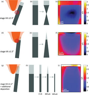
A direct comparison between electron transparent transmission electron microscope (TEM) samples prepared with gallium (Ga) and xenon (Xe) focused ion beams (FIBs) is performed to determine if equivalent quality samples can be prepared with both ion species. We prepared samples using Ga FIB and Xe plasma focused ion beam (PFIB) while altering a variety of different deposition and milling parameters. The samples’ final thicknesses were evaluated using STEM-EELS t/λ data. Using the Ga FIB sample as a standard, we compared the Xe PFIB samples to the standard and to each other. We show that although the Xe PFIB sample preparation technique is quite different from the Ga FIB technique, it is possible to produce high-quality, large area TEM samples with Xe PFIB. We also describe best practices for a Xe PFIB TEM sample preparation workflow to enable consistent success for any thoughtful FIB operator. For Xe PFIB, we show that a decision must be made between the ultimate sample thickness and the size of the electron transparent region.

Type
Software and Instrumentation
Copyright
Copyright © The Author(s), 2022. Published by Cambridge University Press on behalf of the Microscopy Society of America

Access options

Get access to the full version of this content by using one of the access options below. (Log in options will check for institutional or personal access. Content may require purchase if you do not have access.)

References

Assaf, H, Ntsoenzok, E, Leoni, E, Barthe, MF, Ruault, MO, Kaitasov, O & Ashok, S (2007). Nanocavity generation in SiO2 by Kr and Xe ion implantation. Electrochem Solid-State Lett 10(10), G72G75.CrossRefGoogle Scholar
Belianinov, A, Burch, MJ, Kim, S, Tan, S, Hlawacek, G & Ovchinnikova, O (2017). Noble gas ion beams in materials science for future applications and devices. MRS Bull 42, 660666.CrossRefGoogle Scholar
Brogden, V, Johnson, C, Rue, C, Graham, J, Langworthy, K, Golledge, S, McMorran, B & Dinda, GP (2021). Material sputtering with a multi-ion species plasma focused ion beam. Adv Mater Sci Eng 2021, 19.CrossRefGoogle Scholar
Burnett, TL, Kelley, R, Winiarski, B, Contreras, L, Daly, M, Gholinia, A, Burke, MG & Withers, PJ (2016). Large volume serial section tomography by Xe plasma FIB dual beam microscopy. Ultramicroscopy 161, 119129.CrossRefGoogle ScholarPubMed
Egerton, RF (2011). Electron Energy-Loss Spectroscopy in the Electron Microscope. New York: Springer.CrossRefGoogle Scholar
Giannuzzi, L & Smith, N (2017). TEM specimen preparation with plasma FIB Xe+ ions. Microsc Microanal 17(S2), 646647.CrossRefGoogle Scholar
Giannuzzi, LA & Stevie, FA (1999). A review of focused ion beam milling techniques for TEM specimen preparation. Micron 30, 197204.CrossRefGoogle Scholar
Gonzalez, CM, Timilsina, R, Li, G, Duscher, G, Rack, PD, Slingenbergh, W, van Dorp, WF, De Hosson, JTM, Klein, KL, Wu, HM & Stern, LA (2014). Focused helium and neon ion beam induced etching for advanced extreme ultraviolet lithography mask repair. J Vac Sci Technol B, Nanotechnol Microelectron 32(2), 02160210216029.Google Scholar
Greenzweig, Y, Drezner, Y, Tan, S, Livengood, RH & Raveh, A (2016). Current density profile characterization and analysis method for focused ion beam. Microelectron Eng 155, 1924.CrossRefGoogle Scholar
Harriott, LR (1990). Beam-size measurements in focused ion beam systems. J Vac Sci Technol A 8(2), 899901.CrossRefGoogle Scholar
Hugo, RC & Hoagland, RG (1999). Gallium penetration of aluminum: In-situ TEM observations at the penetration front. Scr Mater 41, 13411346.CrossRefGoogle Scholar
Ishii, Y, Isoya, A, Kojima, T & Arakawa, K (2003). Estimation of keV submicron ion beam width using a knife-edge method. Nucl Instrum Methods Phys Res Sect B 211(3), 415424.CrossRefGoogle Scholar
Ishitani, T & Yaguchi, T (1996). Cross-sectional sample preparation by focused ion beam: A review of ion-sample interaction. Microsc Res Tech 35, 320333.3.0.CO;2-Q>CrossRefGoogle ScholarPubMed
Kelley, RD, Song, K, Van Leer, B, Wall, D & Kwakman, L (2013). Xe+ FIB milling and measurement of amorphous silicon damage. Microsc Microanal 19(S2), 862863.CrossRefGoogle Scholar
Livengood, RH, Greenzweig, Y, Liang, T & Grumski, M (2007). Helium ion microscope invasiveness and imaging study for semiconductor applications. J Vac Sci Technol B 25(6), 25472552.CrossRefGoogle Scholar
MacLaren, I, Nord, M, Jiao, C & Yucelen, E (2019). Liftout of high-quality thin sections of a perovskite oxide thin film using a xenon plasma focused ion beam microscope. Microsc Microanal 25(1), 115118.CrossRefGoogle ScholarPubMed
Malis, T, Cheng, SC & Egerton, RF (1988). EELS log-ratio technique for specimen-thickness measurement in the TEM. J Electron Microsc Tech 8, 193200.CrossRefGoogle ScholarPubMed
Nicholas, MG & Old, CF (1979). Liquid metal embrittlement. J Mater Sci 14, 118.CrossRefGoogle Scholar
Pekin, TC, Allen, FI & Minor, AM (2016). Evaluation of neon focused ion beam milling for TEM sample preparation. J Microsc 264, 5963.CrossRefGoogle ScholarPubMed
Postek, MT, Orloff, J, Newbury, DE, Platek, SF & Joy, DC (2010). Measuring the beam size of a focused ion beam (FIB) system. Scanning Microscopy 7729, 77290C1–77290C9.Google Scholar
Rishton, SA (1984). Measurement of the profile of finely focused electron beams in a scanning electron microscope. J Phys E: Sci Instrum 17, 296303.CrossRefGoogle Scholar
Senel, E, Walmsley, JC, Diplas, S & Nisancioglu, K (2014). Liquid metal embrittlement of aluminum by segregation of trace element gallium. Corros Sci 85, 167173.CrossRefGoogle Scholar
Smith, NS, Notte, JA & Steele, AV (2014). Advances in source technology for focused ion beam instruments. MRS Bull 39(4), 329335.CrossRefGoogle Scholar
Smith, NS, Skoczylas, WP, Kellog, SM, Kinion, DE & Tesch, PP (2006). High brightness inductively coupled plasma source for high current focused ion beam applications. J Vac Sci Technol B 24(6), 29022906.CrossRefGoogle Scholar
Tan, S, Livengood, R, Greenzweig, Y, Drezner, Y & Shima, D (2012). Probe current distribution characterization technique for focused ion beam. J Vac Sci Technol B, Nanotechnol Microelectron 30(6), 06F606106F6066.Google Scholar
Tan, S, Livengood, R, Shima, D, Notte, J & McVey, S (2010). Gas field ion source and liquid metal ion source charged particle material interaction study for semiconductor nanomachining applications. J Vac Sci Technol B, Nanotechnol Microelectron 28(6), C6F15C16F21.CrossRefGoogle Scholar
Templier, C, Gaboriaud, RJ & Garem, H (1985). Precipitation of implanted xenon in aluminium. Mater Sci Eng 69, 6366.CrossRefGoogle Scholar
Unocic, KA, Mills, MJ & Daehn, GS (2010). Effect of gallium focused ion beam milling on preparation of aluminium thin foils. J Microsc 240(3), 227238.CrossRefGoogle ScholarPubMed
Xia, D, Jiang, Y-B, Notte, J & Runt, D (2021). Gaas milling with neon focused ion beam: Comparison with gallium focused ion beam milling and subsurface damage analysis. Appl Surf Sci 538, 14792211479228.CrossRefGoogle Scholar
Zhong, X, Wade, CA, Withers, PJ, Zhou, X, Cai, C, Haigh, SJ & Burke, MG (2021). Comparing Xe(+) pFIB and Ga(+) FIB for TEM sample preparation of Al alloys: Minimising FIB-induced artefacts. J Microsc 282(2), 101112.CrossRefGoogle ScholarPubMed