


A previously healthy 59-year-old female presented to the emergency department (ED) with a 1-month history of progressive shortness of breath on exertion rapidly resolving with rest. She had no chest pain nausea or diaphoresis. An exercise stress test was positive for ischemia but a follow-up sestamibi scan was normal. A subsequent computed tomography coronary angiogram was suspicious for an atrial myxoma.
In the ED, her vital signs were within normal limits, and her physical exam was unremarkable. An electrocardiogram revealed normal sinus rhythm, and her troponin was negative. A point-of-care ultrasound demonstrated a 67-mm×35-mm hyperechoic lesion in the left atrium extruding into the left ventricle during atrial systole, resulting in moderate mitral regurgitation (Figures 1–3, Supplemental Videos 1–3). Both atria were dilated, and the left ventricular ejection fraction was normal. The mass was subsequently resected and diagnosed via histopathology as an atrial myxoma.
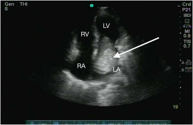
Figure 1 Apical four-chamber view of a left atrial myxoma (arrow) extruding into the left ventricle during atrial systole. LA=left atrium; LV=left ventricle; RA=right atrium; RV=right ventricle.

Figure 2 Parasternal long-axis view of a left atrial myxoma (arrow) extruding into the left ventricle during atrial systole. LA=left atrium; LV=left ventricle; LVOT=left ventricular outflow tract; RV=right ventricle.
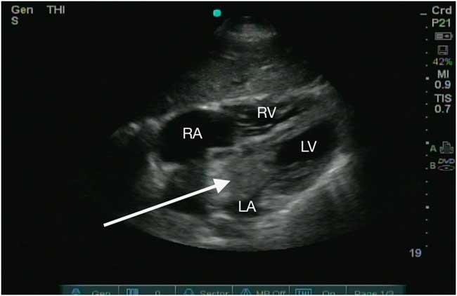
Figure 3 Subxiphoid view of a left atrial myxoma (arrow) extruding into the left ventricle during atrial systole. LA=left atrium; LV=left ventricle; RA=right atrium; RV=right ventricle.
Myxomas are the most common primary benign cardiac neoplasm, with 80% arising from the left atrium.Reference Vander Salm 1 The main alternative consideration for this type of lesion on echocardiography is an atrial thrombus. A mobile hyperechoic lesion with a stalk arising from the interatrial septum favours myxoma.Reference Dhawan and Tak 2 On physical examination, one-third of patients will have an early diastolic murmur after the second heart sound, called a “tumor plop.”Reference Reynen 3 They present on a wide clinical spectrum from asymptomatic to acute heart failure and malignant arrhythmias.Reference Pinede, Duhaut and Loire 4 They can also present with neurologic symptoms secondary to embolization or constitutional symptoms secondary to tumour release of growth factor and cytokines.Reference Wang, Chen and Zhu 5 - Reference Seino, Ikeda and Shimada 7 These tumours require surgical excision due to the risk of embolization and cardiac complications, including arrhythmias and sudden death.Reference Keeling, Oberwalder and Anelli-Monti 8 - Reference Cina, Smialek and Burke 10
Competing interests: None declared.
Supplementary Material
To view supplementary material for this article, please visit https://doi.org/10.1017/cem.2017.351


