CASE HISTORY
A fully vaccinated 7-month-old male with no significant past medical history presented to the emergency department (ED) with his mother complaining of 3 days of worsening cough and tachypnea. His mother stated that he has begun belly-breathing and had significantly decreased oral intake over the past day. She denied fever, vomiting, diarrhea, rashes, lethargy, rhinorrhea, and wheezing.
His vital signs were temperature of 36.1°C, heart rate (HR) of 130 beats/minute, respiratory rate (RR) of 60 breaths/minute, oxygen saturation of 94% on room air, and weight of 7.28 kg. He appeared to be in moderate distress with accessory muscle use. His head and neck examination was normal without stridor. For his age, he had a normal HR and rhythm without a murmur. He was tachypneic, but his lung sounds were clear of wheezes, rhonchi, and rales. His abdomen was soft without tenderness or masses. He had strong distal pulses, and his capillary refill was less than 2 seconds.
His initial lab results were notable for a white blood cell count of 21.1 × 10⁹/L and platelet count of 622×109/L. A chest radiograph was obtained (Figure 1) and read by the radiologist as, “Perihilar haziness. Question bronchial infection. Correlate clinically.” The patient was diagnosed with sepsis secondary to pneumonia, given a 20-ml/kg normal saline bolus, intravenous ceftriaxone, and admitted to the pediatrics floor.

Figure 1 Patient’s chest radiograph.
Several hours later, a rapid response was called to the patient’s bedside for tachycardia. The patient’s HR had risen to 300 beats/minute without significant variability. His other vital signs were RR 40, oxygen saturation of 92%, and blood pressure of 62/23 mm Hg. Despite the tachycardia, the patient’s cardiopulmonary examination was normal, including capillary refill, but hepatomegaly was now noted. An electrocardiogram (ECG) was obtained showing a narrow complex tachycardia with P waves, QRS alternans, and a ventricular rate of 298 beats/minute (Figure 2).

Figure 2 Patient’s ECG at the rapid response.
QUESTION
The most likely cause of the infant’s presenting dyspnea was:
-
A) Pneumonia
-
B) Structural heart disease
-
C) Paroxysmal supraventricular tachycardia (SVT) with acute congestive heart failure
-
D) Salicylate toxicity
-
E) Diabetic ketoacidosis
ANSWER
The correct answer is C (paroxysmal SVT with acute congestive heart failure). Vagal maneuvers, including ice to the face and gently flipping him upside down were attempted without success. Because of the alternans, a limited bedside echocardiogram was performed showing no pericardial effusion or gross structural abnormalities. The patient was then given 0.7 mg or roughly 0.1 mg/kg of adenosine, and the dysrhythmia converted to a normal sinus rhythm at 149 beats/minute without evidence of a delta wave or short PR interval for his age (normal: 82-141 msec) on a repeat ECG (Figure 3).Reference Rijnbeek, Witsenburg and Schrama 1 No flutter waves or other ectopic beats were observed during the pause. The patient was transferred to the nearest pediatric hospital.

Figure 3 Patient’s ECG after chemical cardioversion.
The patient was later diagnosed with SVT by his pediatric cardiologist and placed on propranolol prophylaxis. About 6 months later, he was seen again in the ED for tachycardia. His mother stated that she forgot to give him his propranolol the day prior and noticed that he appeared sleepy. When she checked his pulse, it was >200 beats/minute. She stated that she flipped him upside down, and the tachycardia resolved. On examination at the repeat ED visit, he was afebrile with normal vital signs and no clinical signs of infection. His ECG (Figure 4) showed a normal sinus rhythm at 121 beats/minute without evidence of a delta wave or short PR interval for his age (normal: 86-151 msec).Reference Rijnbeek, Witsenburg and Schrama 1 In retrospect, the patient’s initial dyspnea and “perihilar haziness” on chest radiograph (see Figure 1) at the first ED visit is likely more consistent with acute congestive heart failure rather than pneumonia, because of his lack of fever on presentation and knowledge of this second visit for recurrent SVT without signs of infection.

Figure 4 Patient’s ECG at his repeat ED visit.
SVT is defined as a sustained, narrow complex tachycardia involving conduction through the atrial tissue or the atrioventricular node, otherwise originating above the atrioventricular junction.Reference Salerno and Seslar 2 , Reference Ashok, Sharma and Jain 3 SVT occurs in as many as 1 in every 250 to 500 children but can easily be missed in infants who present with vague symptoms and when the dysrhythmia has temporarily subsided.Reference Salerno and Seslar 2 , Reference Kantoch 4
Infants with SVT and resultant acute congestive heart failure can often have initial signs, symptoms, and findings suggestive of other pathology such as pneumonia, as in this case. Whereas older children may be able to relay symptoms of presyncope, syncope, palpitations, or chest discomfort, an infant cannot relay these specific symptoms concerning a dysrhythmia. Therefore, the diagnosis of paroxysmal SVT is difficult to make in infants, especially if the dysrhythmia has temporarily subsided. Symptoms of rapid breathing and poor feeding, described by the mother of the patient, can easily lead the clinician down an alternative diagnostic pathway away from SVT.Reference Ashok, Sharma and Jain 3 Although the patient’s hepatomegaly was a critical indication of his acute congestive heart failure secondary to SVT,Reference Macicek, Macias and Jefferies 5 it was not present on abdominal examination in the ED. It is also likely that the administration of an intravenous fluid bolus and several hours of maintenance fluids contributed to the hepatomegaly, making it more prominent later at the rapid response.
Laboratory and imaging studies can also be misleading. The systemic stress caused by SVT can create a leukocytosis leading to a false positive indication of infection. Also, with SVT at rates greater than 250 beats/minutes, the dysrhythmia can lead to severe congestive heart failure,Reference Kantoch 4 producing vascular congestion on the chest X-ray, which can be mistaken for a pneumonia. Therefore, it is critical for the emergency provider to consider paroxysmal SVT and other cardiac pathology in infants with pulmonary signs and symptoms.
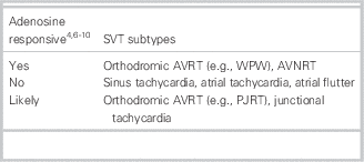
Similarly, specific subtypes of SVT can be difficult to elucidate during the acute dysrhythmia. Our initial interpretation of the rapid response ECG was a 1:1 atrial flutter because of the P waves seen in V2-3 and the fast, constant rate of roughly 300 beats/minute (see Figure 2). However, atrial flutter should not have converted with adenosine, making other subtypes of SVT more likely (Table 1).Reference Kantoch 4 , Reference Marx, Hockberger and Walls 6
Table 1 Responsiveness to adenosine of selected subtypes of regular narrow complex SVT

AVRT = atrioventricular reentry tachycardia; AVNRT = atrioventricular node reentry tachycardia; PJRT = permanent junctional reciprocating tachycardia; SVT = supraventricular tachycardia; WPW = Wolff-Parkinson-White syndrome.
This ambiguity in the rhythm is created by several factors. First, infants with SVT often have faster HRs than older children and adults. Rates can reach 220 to 320 beats/minute, making conditions, such as atrioventricular node reentry tachycardia (AVNRT), more difficult to distinguish from a 1:1 atrial flutter based off of the rate alone.Reference Salerno and Seslar 2 , Reference Kantoch 4 Also, with elevated rates, it can be challenging to differentiate between anterograde and retrograde P waves, which help delineate the subtypes of SVT. Recording a rhythm strip immediately after vagal maneuvers or the administration of adenosine to look at P-wave location and morphology can help elucidate the diagnosis of specific SVT subtypes.Reference Kantoch 4
Although adenosine is the most widely used abortive therapy for infantile SVT, vagal maneuvers, including carotid massage, facial cooling, ice water immersion, direct eyeball pressure, and the Valsalva maneuver for older children, are considered first-line if the infant is hemodynamically stable.Reference Ashok, Sharma and Jain 3 , Reference Chu, Hill and Clark 11 - Reference de Caen, Berg and Chameides 13 If vagal maneuvers fail, adenosine is considered second-line, but it is contraindicated for irregular or polymorphic wide complex tachycardias because of the risk of conversion to ventricular fibrillation.Reference Chu, Hill and Clark 11 , Reference de Caen, Berg and Chameides 13 , Reference Link, Berkow and Kudenchuk 14 Last, if the infant is hemodynamically unstable, electrical cardioversion is recommended.Reference Kantoch 4 , Reference de Caen, Berg and Chameides 13
Paroxysmal SVT can be difficult to diagnose in infants, especially when the dysrhythmia has temporarily subsided. Vague symptoms and misleading laboratory and imaging findings can easily deceive providers. In addition, subtypes of SVT can be difficult to differentiate from one another in the acute setting; therefore, a systematic approach using vagal maneuvers and adenosine, if necessary, with a continuous rhythm strip, is essential to obtain the correct diagnosis.
Acknowledgments
Both authors have made substantial contributions to the concept and design of the manuscript, have partaken in the drafting and revising of the manuscript, and have approved the final version.
Competing interests: None declared.


