


The prevalence of conjoined twins is reported in 1 of every 75,000 live births (Castilla et al., Reference Castilla, Lopez-Camelo, Orioli, Sanchez and Paz1988); these events are classified in terms of symmetry, place of fusion, and grade of duplication (Potter & Craig, Reference Potter and Craig1975). The most frequent type is thoracopagus (32.7%), and the least frequent is diprosopus (0.4%). The latter are Siamese twins characterized by having two faces and one single body (Machin, Reference Machin1993). This craniofacial duplication is an extremely rare event, with very few reports in the medical literature (Molina et al., Reference Molina, Duque, Motta, Alvarado and Torres2008; Okazaki et al., Reference Okazaki, Wilson, Holmes and Vandermark1987); its estimated prevalence is 1 of every 180,000 to 15 million live births (Al Muti Zaitoun et al., Reference Al Muti Zaitoun, Chang and Booker1999). Here is presented a case of craniofacial duplication with bilateral cleft palate in a pre-Hispanic ceramic representation of the Chimú culture from Peru, and a comparative analysis of this case with a current one.
Materials and Methods
From 2011 to 2013, a multidisciplinary research project on health and diseases in prehispanic art from the Pacific coast of South America was conducted by the Universidad Icesi and Universidad del Valle in Cali, Colombia. A hallmark of this project was a rare anthropomorphic ceramic crock identified by the Brünnin National Archeological Museum (Lambayeque, Peru). This crock, of exceptional iconography, belonged to the Chimú culture (Peru, 900–1470 A.D.) and appears to be the representation of Siamese twins with craniofacial duplication plus bilateral cleft lip and palate. A review of the literature was made from searches in Pubmed, Scopus, Embase and DOAJ databases using as keywords the terms ‘diprosopus’, ‘conjoined twins’ and ‘Siamese’; however, there was no evidence of a similar case reported in the medical literature.
Shortly after, a similar case of a 28 week-old fetus was presented during a meeting from the Latin-American Collaborative Study of Congenital Malformations (ECLAMC), which was held in Caxias do Sul, Brazil, in November 2013. The report and presentation of this case led us to review and write about this subject from a multidisciplinary point of view, taking into account iconographic, archeological, medical and genetic data.
Results
Anthropological Analysis
The anthropological analysis suggests an anthropomorphic representation made in the top of an effigy jar or crock, where a pair of conjoined twins with bilateral cleft palate can be distinguished (see Figures 1 and 2).

FIGURE 1 Effigy crock from the Chimú culture. Possibly representing conjoined twins with craniofacial duplication and cleft lip/palate.
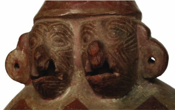
FIGURE 2 Detail of bilateral cleft lip/palate on the possible diprosopus twins represented on the Chimú crock.
The ceramic piece is representative of the early Chimú culture, which existed from 900 to 1470 A.D. in the north coast of Peru. This culture was the continuation of a previous civilization, the Moche. The capital, or political-administrative centre, of this civilization, the city of Chan-Chan, was the largest city in the world at that time; it was remarkable for being built entirely from adobe, and until today it is still recognized as one of the most splendorous cities from the pre-Hispanic culture.
Case Report Analysis
This analysis reports on the case of a fetus from the third pregnancy of a 37-year-old woman with a 55-year-old man. She was admitted from perinatology at the 28th week of pregnancy after detailed ultrasound findings revealed a fetus with two heads conjoined by the neck, sharing a single trunk and extremities (two arms and two legs). Each head had developed an individual spine in the most cephalic portion, which fused with each other at the thoracolumbar level where a defect of the normal curvature was evident. Both heads presented an acrania/anencephaly sequence, with the encephalic mass exposed to the amniotic cavity, and bilateral cleft lip/palate. The fetus was identified as female by the characteristics of the external genitals. The placenta was a grade 1, located in anterior position, and augmented amniotic fluid (over 8 cm) was noticed. There was no evidence of a gastric chamber and an analysis of the cardiac structure was not possible because of the fetal position. The ultrasound conclusion was ‘25-week-old monochorionic, monoamniotic conjoined-twins, with dicephaly and multiple structural defects not compatible with life’, suggesting the indication of pregnancy termination. Following counseling, the mother agreed to schedule a C-section, which was done without any complications.
The fetus presented the characteristics previously described by the perinatologist; a muscle biopsy was taken to determine the karyotype, giving a normal result, 46,XX. The fetus was sent to anatomical pathology for postmortem examination. The subsequent report stated it was a body corresponding to female conjoined twins with two heads and a single body; both heads with anencephaly, encephalocele and bilateral cleft lip/palate (see Figures 3 and 4). A bilateral cranial rachischisis down to the mid thoracic spine was also evident; and after dissection, a defect in cardiac structure with a big interatrial communication was documented.

FIGURE 3 28 week-old fetus with craniofacial duplication and bilateral cleft lip/palate.
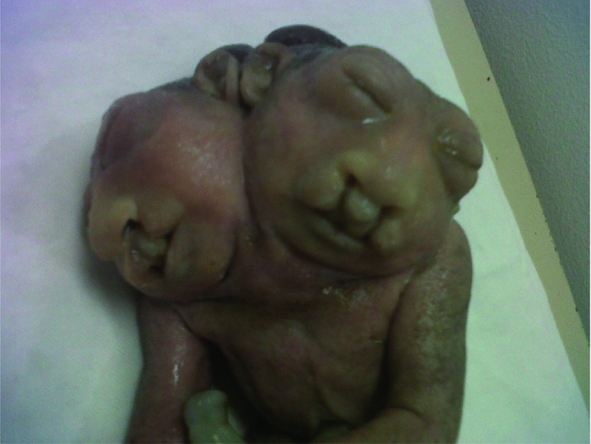
FIGURE 4 Detail of midline defect: bilateral cleft lip/palate in both heads.
Discussion
Many anthropomorphic malformations can be seen in artistic representations of different Peruvian cultures; most of these belong to the Moche civilization, which later gave rise to the Chimú culture. Some of these malformations include dwarfism, achondroplasia, flat feet and facial cleft (Guerra & Sanchez, Reference Guerra and Sanchez1990). Conjoined twins of the diprosopus type have been represented in various sculptures from different cultures, such as the Tlatilco (Bendersky, Reference Bendersky2000), and have been described in European literature from the 16th century (Bates, Reference Bates2002). The crock presented in this article is probably one of the first representations of this type of Siamese twins, and likely to be the only one to document the presence of bilateral cleft palate in addition to the abnormal fusion. Nevertheless, it is also possible that this representation may be the one of classical, non-conjoined twins with bilateral lip/palate defects.
Diprosopus malformation is an entity with a variable spectrum in terms of clinical presentation, being possible to find rather partial or complete duplication of the different facial structures (Brodsky, Reference Brodsky1939; Castilla et al., Reference Castilla, Lopez-Camelo, Orioli, Sanchez and Paz1988; Machin, Reference Machin1993; Okazaki et al., Reference Okazaki, Wilson, Holmes and Vandermark1987; Pachajoa & Rodriguez, Reference Pachajoa and Rodriguez2013). Although craniofacial duplication has been historically considered as a type of conjoined twins (Al Muti Zaitoun et al., Reference Al Muti Zaitoun, Chang and Booker1999; Bates, Reference Bates2002; Bendersky, Reference Bendersky2000; Hahnel et al., Reference Hahnel, Shramm, Hassfeld, Steiner and Seitz2003; Okazaki et al., Reference Okazaki, Wilson, Holmes and Vandermark1987), it has also been suggested that it could be due to a duplication of the rostral aspect of the notochord (Hahnel et al., Reference Hahnel, Shramm, Hassfeld, Steiner and Seitz2003). This defect could lead to the formation of two spinal axis and two neural plates, which finally turns into the duplication of the different derivates from the neural crest (Kotrikova et al., Reference Kotrikova, Hassfeld, Steiner, Hahnel, Krempien and Muhling2007; Maruotti et al., Reference Maruotti, Paladini, Napolitano, Mazzarelli, Russo, Quarantelli and Martinelli2009).
The congenital defects associated to diprosopus malformations include other different systems, such as the cardiovascular, gastrointestinal, respiratory, central nervous system; and also neural tube defects, the most frequent being the presentation of anencephaly, cranial rachischisis, spina bifida, duplication of the brain hemispheres and fusion of the structures in the posterior fossa (Chen, Reference Chen2008; Kotrikova et al., Reference Kotrikova, Hassfeld, Steiner, Hahnel, Krempien and Muhling2007; Molina et al., Reference Molina, Duque, Motta, Alvarado and Torres2008; Wu et al., Reference Wu, Staffenberg, Mulliken and Shanske2002). In the reviewed literature there were very few reports of cleft lip/palate in both twins (Potter & Craig, Reference Potter and Craig1975). Gorlin (Reference Gorlin1988) and Bulbul (Reference Bulbul, Drummond, Hillion, Bidat and Vile2004) reported unilateral cleft lip/palate in both heads, but no reports of bilateral cleft lip/palate were found, leaving only with the two cases reported here. Although we recognize it is possible that this old artistic crock could be the representation of a mid-line defect in otherwise normal twins, we would still like to think it is actual evidence of a much rarer event.
Acknowledgments
The authors are very grateful to Dr Carlos Wester La Torre, director of the Brunning National Archeological Museum, in Lambayeque, Peru, for his collaboration and for allowing us to work with the ceramic collection of the museum.


