Hostname: page-component-84b7d79bbc-4hvwz Total loading time: 0 Render date: 2024-07-27T23:21:49.056Z Has data issue: false hasContentIssue false

Structure and Immune Recognition of the Porcine Epidemic Diarrhea Virus Spike Protein

Published online by Cambridge University Press:  30 July 2020

Robert Kirchdoerfer*
Affiliation:
University of Wisconsin-Madison, Madison, Wisconsin, United States

Abstract

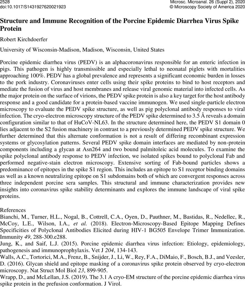
Image of the first page of this content. For PDF version, please use the ‘Save PDF’ preceeding this image.'
Type
Microbes in Focus
Copyright
Copyright © Microscopy Society of America 2020

References

Bianchi, M., Turner, H.L., Nogal, B., Cottrell, C.A., Oyen, D., Pauthner, M., Bastidas, R., Nedellec, R., McCoy, L.E., Wilson, I.A., et al. . (2018). Electron-Microscopy-Based Epitope Mapping Defines Specificities of Polyclonal Antibodies Elicited during HIV-1 BG505 Envelope Trimer Immunization. Immunity 49, 288-300.e288.10.1016/j.immuni.2018.07.009CrossRefGoogle ScholarPubMed
Jung, K., and Saif, L.J. (2015). Porcine epidemic diarrhea virus infection: Etiology, epidemiology, pathogenesis and immunoprophylaxis. Vet J 204, 134-143.10.1016/j.tvjl.2015.02.017CrossRefGoogle ScholarPubMed
Walls, A.C., Tortorici, M.A., Frenz, B., Snijder, J., Li, W., Rey, F.A., DiMaio, F., Bosch, B.J., and Veesler, D. (2016). Glycan shield and epitope masking of a coronavirus spike protein observed by cryo-electron microscopy. Nat Struct Mol Biol 23, 899-905.10.1038/nsmb.3293CrossRefGoogle ScholarPubMed
Wrapp, D., and McLellan, J.S. (2019). The 3.1 A cryo-EM structure of the porcine epidemic diarrhea virus spike protein in the prefusion conformation. J Virol.Google Scholar