Hostname: page-component-8448b6f56d-sxzjt Total loading time: 0 Render date: 2024-04-18T22:17:04.466Z Has data issue: false hasContentIssue false

Quantification of Nanoparticles in Dispersions Using Transmission Electron Microscopy

Published online by Cambridge University Press:  11 May 2021

Ralf Kaegi*
Affiliation:
Eawag, Swiss Federal Institute of Aquatic Science and Technology, 8600Dübendorf, Switzerland
Martin Fierz
Affiliation:
naneos particle solutions GmbH, Dorfstr. 69, 5210Windisch, Switzerland
Bodo Hattendorf
Affiliation:
Department of Chemistry and Applied Biosciences, ETH Zurich, Zurich, Switzerland
*
*Author for correspondence: Ralf Kaegi, E-mail: ralf.kaegi@eawag.ch
Get access

Abstract

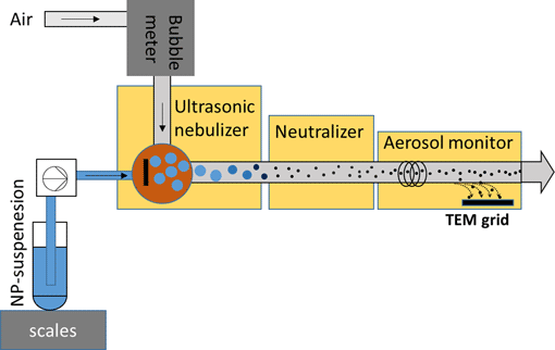
The quantification of the particle size and the number concentration (PNC) of nanoparticles (NPs) is key for the characterization of nanomaterials. Transmission electron microscopy (TEM) is often considered as the gold standard for assessing the size of NPs; however, the TEM sample preparation suitable for estimating the PNC based on deposited NPs is challenging. Here, we use an ultrasonic nebulizer (USN) to transfer NPs from aqueous suspensions into dried aerosols which are deposited on TEM grids in an electrostatic precipitator of an aerosol monitor. The deposition efficiency of the electrostatic precipitator was ≈2%, and the transport efficiency of the USN was ≈7%. Experiments using SiO2 NPs (50–200 nm) confirmed an even deposition of the nebulized particles in the center of the TEM grids. PNCs of the SiO2 NPs derived from TEM images underestimated the expected PNCs of the suspensions by a factor of up to three, most likely resulting from droplet coagulation and NP aggregation in the USN. Nevertheless, single particles still dominated the PNC. Our approach results in reproducible and even deposition of particles on TEM grids suitable for morphological analysis and allows an estimation of the PNC in the suspensions based on the number of particles detected by TEM.

Type
Software and Instrumentation
Copyright
Copyright © The Author(s), 2021. Published by Cambridge University Press on behalf of the Microscopy Society of America

Access options

Get access to the full version of this content by using one of the access options below. (Log in options will check for institutional or personal access. Content may require purchase if you do not have access.)

References

Baalousha, M & Lead, JR (2013). Characterization of natural and manufactured nanoparticles by atomic force microscopy: Effect of analysis mode, environment and sample preparation. Colloids Surf, A 419, 238247.CrossRefGoogle Scholar
Babick, F, Mielke, J, Wohlleben, W, Weigel, S & Hodoroaba, V-D (2016). How reliably can a material be classified as a nanomaterial? Available particle-sizing techniques at work. J Nanopart Res 18, 158.CrossRefGoogle ScholarPubMed
Boverhof, DR, Bramante, CM, Butala, JH, Clancy, SF, Lafranconi, M, West, J & Gordon, SC (2015). Comparative assessment of nanomaterial definitions and safety evaluation considerations. Regul Toxicol Pharmacol 73, 137150.CrossRefGoogle ScholarPubMed
De Temmerman, P-J, Van Doren, E, Verleysen, E, Van der Stede, Y, Francisco, MAD & Mast, J (2012). Quantitative characterization of agglomerates and aggregates of pyrogenic and precipitated amorphous silica nanomaterials by transmission electron microscopy. J Nanobiotechnol 10, 24.CrossRefGoogle ScholarPubMed
De Temmerman, P-J, Verleysen, E, Lammertyn, J & Mast, J (2014). Semi-automatic size measurement of primary particles in aggregated nanomaterials by transmission electron microscopy. Powder Technol 261, 191200.CrossRefGoogle Scholar
Domingos, RF, Baalousha, MA, Ju-Nam, Y, Reid, MM, Tufenkji, N, Lead, JR, Leppard, GG & Wilkinson, KJ (2009). Characterizing manufactured nanoparticles in the environment: Multimethod determination of particle sizes. Environ Sci Technol 43, 72777284.CrossRefGoogle ScholarPubMed
European Commission (2011). Commission recommendation of 18 October 2011 on the definition of nanomaterial. Off J Eur Union L 275, 3840.Google Scholar
Farre, M, Sanchis, J & Barcelo, D (2011). Analysis and assessment of the occurrence, the fate and the behavior of nanomaterials in the environment. TrAC Trend Anal Chem 30, 517527.CrossRefGoogle Scholar
Fierz, M, Kaegi, R & Burtscher, H. (2007). Theoretical and experimental evaluation of a portable electrostatic TEM sampler. Aerosol Sci Technol 41, 520528.CrossRefGoogle Scholar
Hassellöv, M & Kaegi, R (2009). Analysis and characterization of manufactured nanoparticles in aquatic environments. In Environmental and Human Health Impacts of Nanotechnology, Lead, JR & Smith, E (Eds.), pp. 211266. Chichester, West Sussex, UK: John Wiley & Sons, Ltd.CrossRefGoogle Scholar
Hassellöv, M, Readman, JW, Ranville, JF & Tiede, K (2008). Nanoparticle analysis and characterization methodologies in environmental risk assessment of engineered nanoparticles. Ecotoxicology 17, 344361.CrossRefGoogle ScholarPubMed
Hodoroaba, V-D, Unger, WES & Shard, AG (2020). Characterization of Nanoparticles, 1st ed. Elsevier. Available at https://linkinghub.elsevier.com/retrieve/pii/C20170003129 (retrieved November 14, 2020).Google Scholar
International Organization for Standardization (2020). ISO 21363:2020(en), Nanotechnologies — Measurements of Particle Size and Shape Distributions by Transmission Electron Microscopy. Available at https://www.iso.org/obp/ui/#iso:std:iso:21363:ed-1:v1:en (retrieved February 2, 2021).Google Scholar
Khadangi, A, Boudier, T & Rajagopal, V (2020). EM-net: Deep learning for electron microscopy image segmentation. bioRxiv 2020.02.03.933127.Google Scholar
Kim, BH, Yang, J, Lee, D, Choi, BK, Hyeon, T & Park, J (2018). Liquid-phase transmission electron microscopy for studying colloidal inorganic nanoparticles. Adv Mater 30, 1703316.CrossRefGoogle ScholarPubMed
Laborda, F, Bolea, E, Cepriá, G, Gómez, MT, Jiménez, MS, Pérez-Arantegui, J & Castillo, JR (2016). Detection, characterization and quantification of inorganic engineered nanomaterials: A review of techniques and methodological approaches for the analysis of complex samples. Anal Chim Acta 904, 1032.CrossRefGoogle ScholarPubMed
Lespes, G & Gigault, J (2011). Hyphenated analytical techniques for multidimensional characterisation of submicron particles: A review. Anal Chim Acta 692, 2641.CrossRefGoogle ScholarPubMed
Linsinger, TPJ, Roebben, G, Gilliland, D, Calzolai, L, Rossi, F, Gibson, N & Klein, C (2012). Requirements on Measurements for the Implementation of the European Commission Definition of the Term ‘Nanomaterial’. EUR 25404 EN,Publications Office of the European Union, Luxembourg.Google Scholar
Lorenzo, R, Kaegi, R, Gehrig, R, Scherrer, L, Grobéty, B & Burtscher, H (2007). A thermophoretic precipitator for the representative collection of atmospheric ultrafine particles for microscopic analysis. Aerosol Sci Technol 41, 934943.CrossRefGoogle Scholar
Mast, J & Demeestere, L (2009). Electron tomography of negatively stained complex viruses: Application in their diagnosis. Diagn Pathol 4, 5.CrossRefGoogle ScholarPubMed
Mast, J, Verleysen, E, Hodoroaba, V-D & Kaegi, R (2020). Characterization of Nanomaterials by Transmission Electron Microscopy: Measurement Procedures. In Characterization of Nanoparticles. Hodoroaba, VD, Unger, WES & Shard, AG (Eds.), 1st ed., pp. 2948. Amsterdam: Elsevier Science Bv.CrossRefGoogle Scholar
Mavrocordatos, D & Perret, D (1995). Non-artifacted specimen preparation for transmission electron-microscopy. Commun Soil Sci Plant Anal 26, 25932602.CrossRefGoogle Scholar
Michen, B, Geers, C, Vanhecke, D, Endes, C, Rothen-Rutishauser, B, Balog, S & Petri-Fink, A (2015). Avoiding drying-artifacts in transmission electron microscopy: Characterizing the size and colloidal state of nanoparticles. Sci Rep 5, 9793.CrossRefGoogle ScholarPubMed
Mielke, J, Dohanyosova, P, Mueller, P, Lopez-Vidal, S & Hodoroaba, V-D (2017). Evaluation of electrospray as a sample preparation tool for electron microscopic investigations: Toward quantitative evaluation of nanoparticles. Microsc Microanal 23, 163172.CrossRefGoogle ScholarPubMed
Nomizu, T & Mizuike, A (1986). Electron-microscopy of submicron particles in natural-waters–specimen preparation by centrifugation. Mikrochim Acta 1, 6572.CrossRefGoogle Scholar
Pace, HE, Rogers, NJ, Jarolimek, C, Coleman, VA, Higgins, CP & Ranville, JF (2011). Determining transport efficiency for the purpose of counting and sizing nanoparticles via single particle inductively coupled plasma mass spectrometry. Anal Chem 83, 93619369.CrossRefGoogle ScholarPubMed
Prasad, A, Lead, JR & Baalousha, M (2015). An electron microscopy based method for the detection and quantification of nanomaterial number concentration in environmentally relevant media. Sci Total Environ 537, 479486.CrossRefGoogle ScholarPubMed
Roebben, G, Rauscher, H, Amenta, V, Aschberger, K, Boix-Sanfeliu, A, Calzolai, L, Emons, H, Gaillard, C, Gibson, P, Holzwarth, U, Koeber, R, Linsinger, T, Rasmussen, K, Sokull-Kluettgen, B & Stamm, H (2014). Towards a review of the EC recommendation for a definition of the term ‘nanomaterial’ part 2: Assessment of collected information concerning the experience with the defintion. EU Science Hub – European Commission. p. 83. Available at https://ec.europa.eu/jrc/en/publication/eur-scientific-and-technical-research-reports/towards-review-ec-recommendation-definition-term-nanomaterial-part-2-assessment-collected (retrieved July 17, 2020).Google Scholar
Schmid, O & Stoeger, T (2016). Surface area is the biologically most effective dose metric for acute nanoparticle toxicity in the lung. J Aerosol Sci 99, 133143.CrossRefGoogle Scholar
Simonet, BM & Valcarcel, M (2009). Monitoring nanoparticles in the environment. Anal Bioanal Chem 393, 1721.CrossRefGoogle ScholarPubMed
Sorzano, COS, Recarte, E, Alcorlo, M, Bilbao-Castro, JR, San-Martín, C, Marabini, R & Carazo, JM (2009). Automatic particle selection from electron micrographs using machine learning techniques. J Struct Biol 167, 252260.CrossRefGoogle ScholarPubMed
Tiede, K, Boxall, ABA, Tear, SP, Lewis, J, David, H & Hassellov, M (2008). Detection and characterization of engineered nanoparticles in food and the environment. Food Addit Contam A 25, 795821.CrossRefGoogle ScholarPubMed
Uusimaeki, T, Wagner, T, Lipinski, H-G & Kaegi, R (2019). AutoEM: A software for automated acquisition and analysis of nanoparticles. J Nanopart Res 21, 122.CrossRefGoogle Scholar
Verleysen, E, De Temmerman, P-J, Van Doren, E, Francisco, MAD & Mast, J (2014). Quantitative characterization of aggregated and agglomerated titanium dioxide nanomaterials by transmission electron microscopy. Powder Technol 258, 180188.CrossRefGoogle Scholar
Verleysen, E, Wagner, T, Lipinski, H-G, Kagi, R, Koeber, R, Boix-Sanfeliu, A, De Temmerman, P-J & Mast, J (2019). Evaluation of a TEM based approach for size measurement of particulate (nano)materials. Materials 12, 2274.CrossRefGoogle ScholarPubMed
von der Kammer, F, Ferguson, PL, Holden, PA, Masion, A, Rogers, KR, Klaine, SJ, Koelmans, AA, Horne, N & Unrine, JM (2012). Analysis of engineered nanomaterials in complex matrices (environment and biota): General considerations and conceptual case studies. Environ Toxicol Chem 31, 3249.CrossRefGoogle ScholarPubMed
Wagner, T (2016). ParticleSizer. ij-particlesizer: ParticleSizer 1.0.1. Zenodo. 10.5281/zenodo.56457.Google Scholar
Wang, Y, Kalinina, A, Sun, T & Nowack, B (2016). Probabilistic modeling of the flows and environmental risks of nano-silica. Sci Total Environ 545, 6776.CrossRefGoogle ScholarPubMed
Zänker, H & Schierz, A (2012). Engineered nanoparticles and their identification Among natural nanoparticles. Annu Rev Anal Chem 5, 107132.CrossRefGoogle ScholarPubMed
Zhu, Y, Ouyang, Q & Mao, Y (2017). A deep convolutional neural network approach to single-particle recognition in cryo-electron microscopy. BMC Bioinformatics 18, 348.CrossRefGoogle ScholarPubMed
Supplementary material: File

Kaegi et al. supplementary material

Kaegi et al. supplementary material

Download Kaegi et al. supplementary material(File)
File 1 MB