


No CrossRef data available.
Article contents
Electrostatic Contribution to the Photo-Assisted Piezoresponse Force Microscopy by Photo-Induced Surface Charge
Published online by Cambridge University Press: 26 May 2022
Abstract
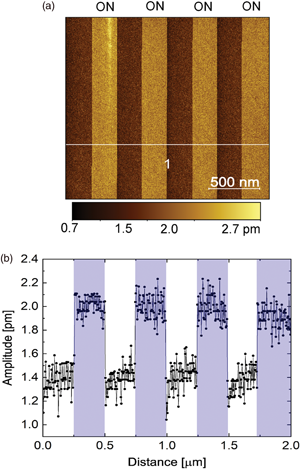
The surging interest in manipulating the polarization of piezo/ferroelectric materials by means of light has driven an increasing number of studies toward their light-polarization interaction. One way to investigate such interaction is by performing piezoresponse force microscopy (PFM) while/after the sample is exposed to light illumination. However, caution must be exercised when analyzing and interpreting the data, as demonstrated in this paper, because sizeable photo-response observed in the PFM amplitude image of the sample is shown to be caused by the electrostatic interaction between the photo-induced surface charge and tip. Through photo-assisted Kelvin probe force microscopy (KPFM), positive surface potential is found to be developed near the sample's surface under 405 nm light illumination, whose effects on the measured PFM signal is revealed by the comparative studies on its amplitude curves that are obtained using PFM spectroscopy mode with/without illumination. This work exemplifies the need for complementary use of KPFM, PFM imaging mode, and PFM spectroscopy mode in order to distinguish real behavior from artifacts.
Keywords
- Type
- Software and Instrumentation
- Information
- Copyright
- Copyright © The Author(s), 2022. Published by Cambridge University Press on behalf of the Microscopy Society of America
References
Ali Deeb, M, Ledig, J, Wei, J, Wang, X, Wehmann, H-H & Waag, A (2017). Photo-assisted Kelvin probe force microscopy investigation of three dimensional GaN structures with various crystal facets, doping types, and wavelengths of illumination. J Appl Phys 122(8), 085307.CrossRefGoogle Scholar
Balke, N, Bdikin, I, Kalinin, SV & Kholkin, AL (2009). Electromechanical imaging and spectroscopy of ferroelectric and piezoelectric materials: State of the art and prospects for the future. J Am Ceram Soc 92(8), 1629–1647.CrossRefGoogle Scholar
Balke, N, Jesse, S, Li, Q, Maksymovych, P, Baris Okatan, M, Strelcov, E, Tselev, A & Kalinin, SV (2015). Current and surface charge modified hysteresis loops in ferroelectric thin films. J Appl Phys 118(7), 072013.CrossRefGoogle Scholar
Balke, N, Jesse, S, Yu, P, Ben, C, Kalinin, SV & Tselev, A (2016). Quantification of surface displacements and electromechanical phenomena via dynamic atomic force microscopy. Nanotechnology 27(42), 425707.CrossRefGoogle ScholarPubMed
Balke, N, Maksymovych, P, Jesse, S, Kravchenko, II, Li, Q & Kalinin, SV (2014). Exploring local electrostatic effects with scanning probe microscopy: Implications for piezoresponse force microscopy and triboelectricity. ACS Nano 8(10), 10229–10236.CrossRefGoogle ScholarPubMed
Coll, M, Gomez, A, Mas-Marza, E, Almora, O, Garcia-Belmonte, G, Campoy-Quiles, M & Bisquert, J (2015). Polarization switching and light-enhanced piezoelectricity in lead halide perovskites. J Phys Chem Lett 6(8), 1408–1413.CrossRefGoogle ScholarPubMed
Collins, L, Liu, Y, Ovchinnikova, OS & Proksch, R (2019). Quantitative electromechanical atomic force microscopy. ACS Nano 13(7), 8055–8066.CrossRefGoogle ScholarPubMed
Dufrêne, YF, Ando, T, Garcia, R, Alsteens, D, Martinez-Martin, D, Engel, A, Gerber, C & Müller, DJ (2017). Imaging modes of atomic force microscopy for application in molecular and cell biology. Nat Nanotechnol 12(4), 295–307.CrossRefGoogle ScholarPubMed
Gomez, A, Puig, T & Obradors, X (2018). Diminish electrostatic in piezoresponse force microscopy through longer or ultra-stiff tips. Appl Surf Sci 439, 577–582.CrossRefGoogle Scholar
Gruverman, A, Alexe, M & Meier, D (2019). Piezoresponse force microscopy and nanoferroic phenomena. Nat Commun 10(1), 1–9.CrossRefGoogle ScholarPubMed
Harnagea, C, Alexe, M, Hesse, D & Pignolet, A (2003). Contact resonances in voltage-modulated force microscopy. Appl Phys Lett 83(2), 338–340.CrossRefGoogle Scholar
Hong, S, Woo, J, Shin, H, Jeon, JU, Pak, YE, Colla, EL, Setter, N, Kim, E & No, K (2001). Principle of ferroelectric domain imaging using atomic force microscope. J Appl Phys 89(2), 1377–1386.CrossRefGoogle Scholar
Jesse, S, Baddorf, AP & Kalinin, SV (2006 a). Switching spectroscopy piezoresponse force microscopy of ferroelectric materials. Appl Phys Lett 88(6), 062908.CrossRefGoogle Scholar
Jesse, S, Kumar, A, Arruda, TM, Kim, Y, Kalinin, SV & Ciucci, F (2012). Electrochemical strain microscopy: Probing ionic and electrochemical phenomena in solids at the nanometer level. MRS Bulletin 37(7), 651–658.CrossRefGoogle Scholar
Jesse, S, Lee, HN & Kalinin, SV (2006 b). Quantitative mapping of switching behavior in piezoresponse force microscopy. Rev Sci Instrum 77(7), 073702.CrossRefGoogle Scholar
Kalinin, SV, Rodriguez, BJ, Jesse, S, Shin, J, Baddorf, AP, Gupta, P, Jain, H, Williams, DB & Gruverman, A (2006). Vector piezoresponse force microscopy. Microsc Microanal 12(3), 206–220.CrossRefGoogle ScholarPubMed
Kazakova, O, Puttock, R, Barton, C, Corte-León, H, Jaafar, M, Neu, V & Asenjo, A (2019). Frontiers of magnetic force microscopy. J Appl Phys 125(6), 060901.CrossRefGoogle Scholar
Killgore, JP, Robins, L & Collins, L (2021). A universal approach to electrostatically-blind quantitative piezoresponse force microscopy. arXiv preprint arXiv:2112.09665.Google Scholar
Kim, B, Seol, D, Lee, S, Lee, HN & Kim, Y (2016). Ferroelectric-like hysteresis loop originated from non-ferroelectric effects. Appl Phys Lett 109(10), 102901.CrossRefGoogle Scholar
Kim, H-S, Kim, SK, Kim, BJ, Shin, K-S, Gupta, MK, Jung, HS, Kim, S-W & Park, N-G (2015). Ferroelectric polarization in CH3NH3PbI3 perovskite. J Phys Chem Lett 6(9), 1729–1735.CrossRefGoogle ScholarPubMed
Kim, S, Seol, D, Lu, X, Alexe, M & Kim, Y (2017). Electrostatic-free piezoresponse force microscopy. Sci Rep 7(1), 1–8.Google ScholarPubMed
Kim, Y, Bühlmann, S, Hong, S, Kim, S-H & No, K (2007). Injection charge assisted polarization reversal in ferroelectric thin films. Appl Phys Lett 90(7), 072910.CrossRefGoogle Scholar
Kim, Y, Kumar, A, Tselev, A, Kravchenko, II, Han, H, Vrejoiu, I, Lee, W, Hesse, D, Alexe, M & Kalinin, SV (2011). Nonlinear phenomena in multiferroic nanocapacitors: Joule heating and electromechanical effects. ACS Nano 5(11), 9104–9112.CrossRefGoogle ScholarPubMed
Kim, Y, Morozovska, AN, Kumar, A, Jesse, S, Eliseev, EA, Alibart, F, Strukov, D & Kalinin, SV (2012). Ionically-mediated electromechanical hysteresis in transition metal oxides. ACS Nano 6(8), 7026–7033.CrossRefGoogle ScholarPubMed
Krieg, M, Fläschner, G, Alsteens, D, Gaub, BM, Roos, WH, Wuite, GJL, Gaub, HE, Gerber, C, Dufrêne, YF & Müller, DJ (2019). Atomic force microscopy-based mechanobiology. Nat Rev Phys 1(1), 41–57.CrossRefGoogle Scholar
Li, T, Lipatov, A, Lu, H, Lee, H, Lee, J-W, Torun, E, Wirtz, L, Eom, C-B, Íñiguez, J & Sinitskii, A (2018). Optical control of polarization in ferroelectric heterostructures. Nat Commun 9(1), 1–8.Google ScholarPubMed
Loo, CC, Ng, SS & Chang, WS (2021). Photostrictive behavior as the piezo-phototronic effect in InGaN/GaN multiple quantum wells. Nano Energy 86, 106085.CrossRefGoogle Scholar
MacDonald, GA, DelRio, FW & Killgore, JP (2018). Higher-eigenmode piezoresponse force microscopy: A path towards increased sensitivity and the elimination of electrostatic artifacts. Nano Futures 2(1), 015005.CrossRefGoogle Scholar
Melitz, W, Shen, J, Kummel, AC & Lee, S (2011). Kelvin probe force microscopy and its application. Surf Sci Rep 66(1), 1–27.CrossRefGoogle Scholar
Pawlak, R, Kisiel, M, Klinovaja, J, Meier, T, Kawai, S, Glatzel, T, Loss, D & Meyer, E (2016). Probing atomic structure and Majorana wavefunctions in mono-atomic Fe chains on superconducting Pb surface. npj Quantum Inf 2(1), 1–5.CrossRefGoogle Scholar
Reshchikov, MA, Foussekis, M & Baski, AA (2010). Surface photovoltage in undoped n-type GaN. J Appl Phys 107(11), 113535.CrossRefGoogle Scholar
Si, H, Zhang, S, Ma, S, Xiong, Z, Kausar, A, Liao, Q, Zhang, Z, Sattar, A, Kang, Z & Zhang, Y (2020). Emerging conductive atomic force microscopy for metal halide perovskite materials and solar cells. Adv Energy Mater 10(10), 1903922.CrossRefGoogle Scholar
Stan, G & King, SW (2020). Atomic force microscopy for nanoscale mechanical property characterization. J Vac Sci Technol B Nanotechnol Microelectron Mater Process Meas Phenom 38(6), 060801.Google Scholar
Vasudevan, RK, Balke, N, Maksymovych, P, Jesse, S & Kalinin, SV (2017). Ferroelectric or non-ferroelectric: Why so many materials exhibit “ferroelectricity” on the nanoscale. Appl Phys Rev 4(2), 021302.CrossRefGoogle Scholar
Wang, P, Zhao, J, Wei, L, Zhu, Q, Xie, S, Liu, J, Meng, X & Li, J (2017). Photo-induced ferroelectric switching in perovskite CH3NH3PbI3 films. Nanoscale 9(11), 3806–3817.CrossRefGoogle ScholarPubMed
Winnerl, A, Pereira, RN & Stutzmann, M (2015). Kinetics of optically excited charge carriers at the GaN surface. Phys Rev B 91(7), 075316.CrossRefGoogle Scholar
Yang, MM & Alexe, M (2018). Light-induced reversible control of ferroelectric polarization in BiFeO3. Adv Mater 30(14), 1704908.CrossRefGoogle ScholarPubMed

Loo et al. supplementary material
Figures S1-S2

File
295.2 KB