Hostname: page-component-76fb5796d-x4r87 Total loading time: 0 Render date: 2024-04-26T17:29:36.206Z Has data issue: false hasContentIssue false

Direct Visualization of the Earliest Stages of Crystallization

Published online by Cambridge University Press:  11 May 2021

Manish Kumar Singh*
Affiliation:
Department of Materials Science and Engineering, University of Connecticut, Storrs, CT06269, USA
Chanchal Ghosh*
Affiliation:
Department of Materials Science and Engineering, University of Connecticut, Storrs, CT06269, USA
Benjamin Miller*
Affiliation:
Gatan Inc., 5794 W. Las Positas Blvd, Pleasanton, CA94588, USA
C. Barry Carter
Affiliation:
Department of Chemical and Biomolecular Engineering, University of Connecticut, Storrs, CT06269, USA Center for Integrated Nanotechnologies, Sandia National Laboratories, Albuquerque, NM87185, USA
*
*Authors for correspondence: Manish Kumar Singh, E-mail: manish.singh@uconn.edu; Chanchal Ghosh, E-mail: chanchal.ghosh@uconn.edu and Benjamin Miller, E-mail: Benjamin.Miller@ametek.com
Get access

Abstract

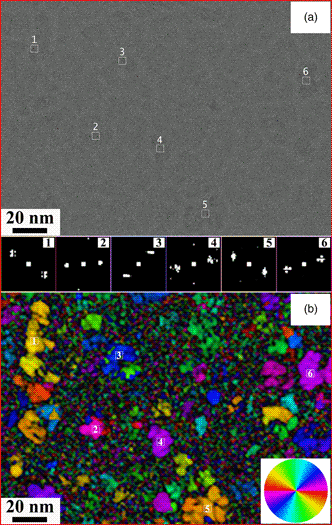
Investigating the earliest stages of crystallization requires the transmission electron microscope (TEM) and is particularly challenging for materials which can be affected by the electron beam. Typically, when imaging at magnifications high enough to observe local crystallinity, the electron beam's current density must be high to produce adequate image contrast. Yet, minimizing the electron dose is necessary to reduce the changes caused by the beam. With the advent of a sensitive, high-speed, direct-detection camera for a TEM that is corrected for spherical aberration, it is possible to probe the early stages of crystallization at the atomic scale. High-quality images with low contrast can now be analyzed using new computing methods. In the present paper, this approach is illustrated for crystallization in a Ge2Sb2Te5 (GST-225) phase-change material which can undergo particularly rapid phase transformations and is sensitive to the electron beam. A thin (20 nm) film of GST-225 has been directly imaged in the TEM and the low-dose images processed using Python scripting to extract details of the nanoscale nuclei. Quantitative analysis of the processed images in a video sequence also allows the growth of such nuclei to be followed.

Type
Materials Science Applications
Copyright
Copyright © The Author(s), 2021. Published by Cambridge University Press on behalf of the Microscopy Society of America

Access options

Get access to the full version of this content by using one of the access options below. (Log in options will check for institutional or personal access. Content may require purchase if you do not have access.)

Footnotes

a

Manish Kumar Singh, Chanchal Ghosh, and Benjamin Miller equally contributed to the present study and can be regarded, therefore, as being main authors.

References

Bustillo, KC, Panova, O, Gammer, C, Trigg, EB, Chen, XC, Yan, L, Balsara, NP, Winey, KI & Minor, AM (2016). Development of diffraction scanning techniques for beam sensitive polymers. Microsc Microanal 22(S3), 492493.CrossRefGoogle Scholar
Carter, CB & Williams, DB (2016). Transmission Electron Microscopy: Diffraction, Imaging, and Spectrometry. Switzerland: Springer International Publishing.CrossRefGoogle Scholar
Choi, Y, Jung, M & Lee, Y-K (2009). Effect of heating rate on the activation energy for crystallization of amorphous Ge2Sb2Te5 thin film. Electrochem Solid State Lett 12(7), F17.CrossRefGoogle Scholar
DeRocher, KA, Smeets, PJ, Goodge, BH, Zachman, MJ, Balachandran, PV, Stegbauer, L, Cohen, MJ, Gordon, LM, Rondinelli, JM & Kourkoutis, LF (2020). Chemical gradients in human enamel crystallites. Nature 583(7814), 6671.CrossRefGoogle ScholarPubMed
Gao, WP, Tieu, P, Addiego, C, Ma, YL, Wu, JB & Pan, XQ (2019). Probing the dynamics of nanoparticle formation from a precursor at atomic resolution. Sci Adv 5, 1.CrossRefGoogle ScholarPubMed
Jiang, T-T, Wang, J-J, Lu, L, Ma, C-S, Zhang, D-L, Rao, F, Jia, C-L & Zhang, W (2019). Progressive amorphization of GeSbTe phase-change material under electron beam irradiation. APL Mater 7(8), 081121.CrossRefGoogle Scholar
Jiang, T-T, Wang, X-D, Wang, J-J, Zhou, Y-X, Zhang, D-L, Lu, L, Jia, C-L, Wuttig, M, Mazzarello, R & Zhang, W (2020). In situ study of vacancy disordering in crystalline phase-change materials under electron beam irradiation. Acta Mater 187, 103111.CrossRefGoogle Scholar
Kooi, B, Groot, W & De Hosson, JTM (2004). In situ transmission electron microscopy study of the crystallization of Ge2Sb2Te5. J Appl Phys 95(3), 924932.CrossRefGoogle Scholar
Kwon, MH, Lee, BS, Bogle, SN, Nittala, LN, Bishop, SG, Abelson, JR, Raoux, S, Cheong, BK & Kim, KB (2007). Nanometer-scale order in amorphous Ge2Sb2Te5 analyzed by fluctuation electron microscopy. Appl Phys Lett 90, 2.CrossRefGoogle Scholar
Lee, B-S, Shelby, RM, Raoux, S, Retter, CT, Burr, GW, Bogle, SN, Darmawikarta, K, Bishop, SG & Abelson, JR (2014). Nanoscale nuclei in phase change materials: Origin of different crystallization mechanisms of Ge2Sb2Te5 and AgInSbTe. J Appl Phys 115, 6.CrossRefGoogle Scholar
Li, JJ, Li, YP, Li, Q, Wang, ZC & Deepak, FL (2019). Atomic-scale dynamic observation reveals temperature-dependent multistep nucleation pathways in crystallization. Nanoscale Horiz 4(6), 13021309.CrossRefGoogle Scholar
McMullan, G, Clark, AT, Turchetta, R & Faruqi, AR (2009). Enhanced imaging in low dose electron microscopy using electron counting. Ultramicroscopy 109(12), 14111416.CrossRefGoogle ScholarPubMed
Miller, B, Pakzad, A & Mick, S (2019). Real-time electron counting for continuous TEM imaging of sensitive samples. Microsc Microanal 25(S2), 17181719.CrossRefGoogle Scholar
Murthy, AA, Stanev, TK, dos Reis, R, Hao, SQ, Wolverton, C, Stern, NP & Dravid, VP (2020). Direct visualization of electric-field-induced structural dynamics in monolayer transition metal dichalcogenides. ACS Nano 14(2), 15691576.CrossRefGoogle ScholarPubMed
Nam, KW, Park, SS, dos Reis, R, Dravid, VP, Kim, H, Mirkin, CA & Stoddart, JF (2019). Conductive 2D metal-organic framework for high-performance cathodes in aqueous rechargeable zinc batteries. Nat Commun 10 (1), 4948.CrossRefGoogle ScholarPubMed
Noé, P, Sabbione, C, Bernier, N, Castellani, N, Fillot, F & Hippert, F (2016). Impact of interfaces on scenario of crystallization of phase change materials. Acta Mater 110, 142148.CrossRefGoogle Scholar
Ogata, AF, Rakowski, AM, Carpenter, BP, Fishman, DA, Merham, JG, Hurst, PJ & Patterson, JP (2020). Direct observation of amorphous precursor phases in the nucleation of protein-metal-organic frameworks. J Am Chem Soc 142(3), 14331442.CrossRefGoogle ScholarPubMed
Ophus, C (2019). Four-dimensional scanning transmission electron microscopy (4D-STEM): From scanning nanodiffraction to ptychography and beyond. Microsc Microanal 25(3), 563582.CrossRefGoogle ScholarPubMed
Orava, J & Greer, AL (2017). Classical-nucleation-theory analysis of priming in chalcogenide phase-change memory. Acta Mater 139, 226235.CrossRefGoogle Scholar
Panova, O, Ophus, C, Takacs, CJ, Bustillo, KC, Balhorn, L, Salleo, A, Balsara, N & Minor, AM (2019). Diffraction imaging of nanocrystalline structures in organic semiconductor molecular thin films. Nat Mater 18(8), 860865.CrossRefGoogle ScholarPubMed
Pekin, TC, Ding, J, Gammer, C, Ozdol, B, Ophus, C, Asta, M, Ritchie, RO & Minor, AM (2019). Direct measurement of nanostructural change during in situ deformation of a bulk metallic glass. Nat Commun 10(1), 17.CrossRefGoogle ScholarPubMed
Raoux, S (2009). Phase change materials. Annu Rev Mater Res 39, 2548.CrossRefGoogle Scholar
Rezikyan, A & Moore, GG (2020). Fluctuation electron microscopy study of crystal nucleation in TiO2-SiO2 glass with heat treatment. J Phys Condens Matter 32(48), 485402.CrossRefGoogle ScholarPubMed
Simpson, R, Krbal, M, Fons, P, Kolobov, A, Tominaga, J, Uruga, T & Tanida, H (2010). Toward the ultimate limit of phase change in Ge2Sb2Te5. Nano Lett 10(2), 414419.CrossRefGoogle Scholar
Song, SA, Zhang, W, Sik Jeong, H, Kim, JG & Kim, YJ (2008). In situ dynamic HR-TEM and EELS study on phase transitions of Ge2Sb2Te5 chalcogenides. Ultramicroscopy 108(11), 14081419.CrossRefGoogle Scholar
Treacy, M & Gibson, J (1996). Variable coherence microscopy: A rich source of structural information from disordered materials. Acta Crystallogr Sect A 52(2), 212220.CrossRefGoogle Scholar
Tripathi, S, Janish, M, Dirisaglik, F, Cywar, A, Zhu, Y, Jungjohann, K, Silva, H & Carter, CB (2018). Phase-change materials; the challenges for TEM. Microsc Microanal 24(S1), 19041905.CrossRefGoogle Scholar
Tripathi, S, Kotula, P, Singh, MK, Ghosh, C, Bakan, G, Silva, H & Carter, CB (2020). Role of oxygen on chemical segregation in uncapped Ge2Sb2Te5 thin films on silicon nitride. ECS J Solid State Sci Technol 9(5), 054007.CrossRefGoogle Scholar
Voyles, P & Muller, D (2002). Fluctuation microscopy in the STEM. Ultramicroscopy 93(2), 147159.CrossRefGoogle ScholarPubMed
Wuttig, M & Yamada, N (2007). Phase-change materials for rewriteable data storage. Nat Mater 6(11), 824832.CrossRefGoogle ScholarPubMed
Yamada, N, Ohno, E, Nishiuchi, K, Akahira, N & Takao, M (1991). Rapid-phase transitions of GeTe-Sb2Te3 pseudobinary amorphous thin films for an optical disk memory. J Appl Phys 69(5), 28492856.CrossRefGoogle Scholar
Zhang, DL, Zhu, YH, Liu, LM, Ying, XR, Hsiung, CE, Sougrat, R, Li, K & Han, Y (2018). Atomic-resolution transmission electron microscopy of electron beam-sensitive crystalline materials. Science 359(6376), 675679.CrossRefGoogle ScholarPubMed
Zhu, YH, Ciston, J, Zheng, B, Miao, XH, Czarnik, C, Pan, YC, Sougrat, R, Lai, ZP, Hsiung, CE, Yao, KX, Pinnau, I, Pan, M & Han, Y (2017). Unravelling surface and interfacial structures of a metal-organic framework by transmission electron microscopy. Nat Mater 16(5), 532536.CrossRefGoogle ScholarPubMed