


No CrossRef data available.
Article contents
Dimensional and Angular Measurements from Microvascular Corrosion Casts: 2D vs. 3D
Published online by Cambridge University Press: 02 July 2020
Extract
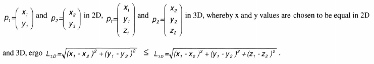
In microvascular network analysis on vascular corrosion casts (VCC) detection of vascular relations between distant regions within vascular territories deserves to operate the SEM at low magnification (< 500x). As a result in low powered SEM micrographs of VCCs vessels may remain in the wide zone of focal depth (Dƒ), but can approach upper and lower focal planes of this zone (Fig.l). Their lengths, thus may severely be underestimated when measuring in 2D. The differences in lengths in 2D (L3D) and in 3D (L3D) of a distance L between points p1 and p2 (Fig.3) is obvious, denoting
While data from 2D-morphometry (1, 2, 3) have to be corrected for dimensional measurements in the direction of tilt by expanding the dimensions by [l/cos(tilt angle)] (4), modern 3D-morphometry techniques (5, 6, 7) consider the perspective projections by calculating the parallax in their working algorithms.
- Type
- Applications and Advances in Vascular Corrosion Casting in Microvascular Research
- Information
- Microscopy and Microanalysis , Volume 6 , Issue S2: Proceedings: Microscopy & Microanalysis 2000, Microscopy Society of America 58th Annual Meeting, Microbeam Analysis Society 34th Annual Meeting, Microscopical Society of Canada/Societe de Microscopie de Canada 27th Annual Meeting, Philadelphia, Pennsylvania August 13-17, 2000 , August 2000 , pp. 564 - 565
- Copyright
- Copyright © Microscopy Society of America