Hostname: page-component-76fb5796d-zzh7m Total loading time: 0 Render date: 2024-04-26T01:41:46.278Z Has data issue: false hasContentIssue false

Developing Atom Probe Tomography to Characterize Sr-Loaded Bioactive Glass for Bone Scaffolding

Published online by Cambridge University Press:  25 October 2021

Yanru Ren*
Affiliation:
Department of Materials, University of Oxford, Oxford OX1 3PH, UK
Hélène Autefage
Affiliation:
Department of Materials, Imperial College London, London SW7 2AZ, UK Department of Bioengineering, Imperial College London, London SW7 2AZ, UK
Julian R. Jones
Affiliation:
Department of Materials, Imperial College London, London SW7 2AZ, UK Department of Bioengineering, Imperial College London, London SW7 2AZ, UK
Molly M. Stevens
Affiliation:
Department of Materials, Imperial College London, London SW7 2AZ, UK Department of Bioengineering, Imperial College London, London SW7 2AZ, UK
Paul A.J. Bagot
Affiliation:
Department of Materials, University of Oxford, Oxford OX1 3PH, UK
Michael P. Moody
Affiliation:
Department of Materials, University of Oxford, Oxford OX1 3PH, UK
*
*Corresponding author: Yanru Ren, E-mail: yanru.ren@materials.ox.ac.uk
Get access

Abstract

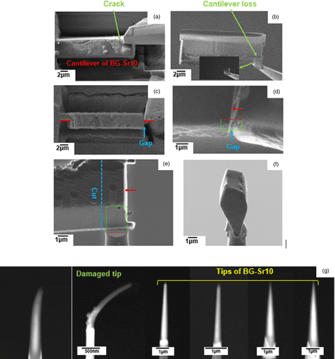
In this study, atom probe tomography (APT) was used to investigate strontium-containing bioactive glass particles (BG-Sr10) and strontium-releasing bioactive glass-based scaffolds (pSrBG), both of which are attractive biomaterials with applications in critical bone damage repair. We outline the challenges and corresponding countermeasures of this nonconductive biomaterial for APT sample preparation and experiments, such as avoiding direct contact between focussed ion beam micromanipulators and the extracted cantilever to reduce damage during liftout. Using a low imaging voltage (≤3 kV) and current (≤500 pA) in the scanning electron microscope and a low acceleration voltage (≤2 kV) and current (≤200 pA) in the focussed ion beam prevents tip bending in the final stages of annular milling. To optimize the atom probe experiment, we considered five factors: total detected hits, multiple hits, the background level, the charge-state ratio, and the accuracy of the measured compositions, to explore the optimal laser pulse for BG-Sr10 bioactive glass. We show that a stage temperature of 30 K, 200–250 pJ laser pulse energy, 0.3% detection rate, and 200 kHz pulse rate are optimized experimental parameters for bioactive glass. The use of improved experimental preparation methods and optimized parameters resulted in a 90% successful yield of pSrBG samples by APT.

Type
Applications in Biology
Copyright
Copyright © The Author(s), 2021. Published by Cambridge University Press on behalf of the Microscopy Society of America

Access options

Get access to the full version of this content by using one of the access options below. (Log in options will check for institutional or personal access. Content may require purchase if you do not have access.)

References

Andrew, JL (2020). AtomProbeLab. Available at https://sourceforge.net/projects/atomprobelab/.Google Scholar
Arepalli, SK, Tripathi, H, Hira, SK, Manna, PP, Pyare, R & Singh, S (2016). Enhanced bioactivity, biocompatibility and mechanical behavior of strontium substituted bioactive glasses. Mater Sci Eng C 69, 108116.CrossRefGoogle ScholarPubMed
Autefage, H, Allen, F, Tang, H, Kallepitis, C, Gentleman, E, Reznikov, N, Nitiputri, K, Nommeots-Nomm, A, O'Donnell, M & Lange, C (2019). Multiscale analyses reveal native-like lamellar bone repair and near perfect bone-contact with porous strontium-loaded bioactive glass. Biomaterials 209, 152162.CrossRefGoogle ScholarPubMed
Bassim, ND, De Gregorio, BT, Kilcoyne, ALD, Scott, K, Chou, T, Wirick, S & Stroud, RM (2012). Minimizing damage during FIB sample preparation of soft materials. J Microsc 245(3), 288301.CrossRefGoogle Scholar
Bunton, J, Olson, J, Lenz, D, Larson, D & Kelly, T (2010). Optimized laser thermal pulsing of atom probe tomography: LEAP 4000x™. Microsc Microanal 16(S2), 1011.CrossRefGoogle Scholar
Chandran, S, Shenoy, SJ, Nair, RP, Varma, H & John, A (2018). Strontium hydroxyapatite scaffolds engineered with stem cells aid osteointegration and osteogenesis in osteoporotic sheep model. Colloids Surf B Biointerfaces 163, 346354.CrossRefGoogle ScholarPubMed
Eder, K, Otter, LM, Yang, L, Jacob, DE & Cairney, JM (2019). Overcoming challenges associated with the analysis of nacre in the atom probe. Geostand Geoanalytical Res 43(3), 385395.CrossRefGoogle Scholar
Fredholm, YC, Karpukhina, N, Brauer, DS, Jones, JR, Law, RV & Hill, RG (2011). Influence of strontium for calcium substitution in bioactive glasses on degradation, ion release and apatite formation. J R Soc Interface 9(70), 880889.CrossRefGoogle ScholarPubMed
Gault, B, Moody, MP, Cairney, JM & Ringer, SP (2012). Atom Probe Microscopy. (Vol. 160). New York: Springer Science & Business Media.CrossRefGoogle Scholar
Gault, B, Saxey, DW, Ashton, MW, Sinnott, SB, Chiaramonti, AN, Moody, MP & Schreiber, DK (2016). Behavior of molecules and molecular ions near a field emitter. New J Phys 18(3), 033031.CrossRefGoogle Scholar
Geiser, BP, Schneir, J, Roberts, J, Wiener, S, Larson, D & Kelly, T (2006). Spatial distribution maps for atom probe tomography. In 2006 19th International Vacuum Nanoelectronics Conference. IEEE.CrossRefGoogle Scholar
Gentleman, E, Fredholm, YC, Jell, G, Lotfibakhshaiesh, N, O'Donnell, MD, Hill, RG & Stevens, MM (2010). The effects of strontium-substituted bioactive glasses on osteoblasts and osteoclasts in vitro. Biomaterials 31(14), 39493956.CrossRefGoogle ScholarPubMed
Gordon, LM, Cohen, MJ, MacRenaris, KW, Pasteris, JD, Seda, T & Joester, D (2015). Amorphous intergranular phases control the properties of rodent tooth enamel. Science 347(6223), 746750.CrossRefGoogle ScholarPubMed
Gordon, LM & Joester, D (2015). Mapping residual organics and carbonate at grain boundaries and the amorphous interphase in mouse incisor enamel. Front Physiol 6, 57.CrossRefGoogle ScholarPubMed
Gordon, LM, Tran, L & Joester, D (2012). Atom probe tomography of apatites and bone-type mineralized tissues. ACS Nano 6(12), 1066710675.CrossRefGoogle ScholarPubMed
Hyde, JM & English, CA (2015). Microstructural characterisation techniques for the study of reactor pressure vessel (RPV) embrittlement. In Embrittlement of Reactor Pressure Vessels (RPVs) in Nuclear Power Plants, Naoki, S (Ed.), pp. 211294. Elsevier: Woodhead Publishing.Google Scholar
Joester, D & Gordon, LM (2011). Nanoscale chemical tomography of buried organic–inorganic interfaces in the chiton tooth. Nature 469(7329), 194197.Google Scholar
Jones, JR (2013). Review of bioactive glass: From Hench to hybrids. Acta Biomater 9(1), 44574486.CrossRefGoogle ScholarPubMed
Karlsson, J, Sundell, G, Thuvander, M & Andersson, M (2014). Atomically resolved tissue integration. Nano Lett 14(8), 42204223.CrossRefGoogle ScholarPubMed
La Fontaine, A, Zavgorodniy, A, Liu, H, Zheng, R, Swain, M & Cairney, J (2016). Atomic-scale compositional mapping reveals Mg-rich amorphous calcium phosphate in human dental enamel. Sci Adv 2(9), e1601145.CrossRefGoogle ScholarPubMed
Langelier, B, Wang, X & Grandfield, K (2017). Atomic scale chemical tomography of human bone. Sci Rep 7(1), 19.CrossRefGoogle ScholarPubMed
Lee, BE, Langelier, B & Grandfield, K (2020). Visualization of Collagen–Mineral Arrangement using Atom Probe Tomography. bioRxiv.Google Scholar
Leu, A & Leach, JK (2008). Proangiogenic potential of a collagen/bioactive glass substrate. Pharm Res 25(5), 12221229.CrossRefGoogle ScholarPubMed
Lis, GDA, Hitchcock, AP, Berejnov, V, Susac, D, Stumper, J & Botton, GA (2016). Evaluating focused ion beam and ultramicrotome sample preparation for analytical microscopies of the cathode layer of a polymer electrolyte membrane fuel cell. J Power Sources 312, 2335.Google Scholar
Liu, J, Hu, H, Li, P, Shuai, C & Peng, S (2013). Fabrication and characterization of porous 45S5 glass scaffolds via direct selective laser sintering. Mater Manuf Processes 28(6), 610615.Google Scholar
Marquis, EA & Gault, B (2008). Determination of the tip temperature in laser assisted atom-probe tomography using charge state distributions. J Appl Phys 104(8), 084914.CrossRefGoogle Scholar
Meunier, PJ, Roux, C, Seeman, E, Ortolani, S, Badurski, JE, Spector, TD, Cannata, J, Balogh, A, Lemmel, E-M & Pors-Nielsen, S (2004). The effects of strontium ranelate on the risk of vertebral fracture in women with postmenopausal osteoporosis. N Engl J Med 350(5), 459468.CrossRefGoogle ScholarPubMed
Miller, M, Russell, K & Thompson, GJU (2005). Strategies for fabricating atom probe specimens with a dual beam FIB. Ultramicroscopy 102(4), 287298.CrossRefGoogle ScholarPubMed
Mosiman, DS, Chen, YS, Yang, L, Hawkett, B, Ringer, SP, Mariñas, BJ & Cairney, JM (2020). Atom probe tomography of encapsulated hydroxyapatite nanoparticles. Small Methods 5(2), 2000692.CrossRefGoogle ScholarPubMed
Neves, N, Linhares, D, Costa, G, Ribeiro, C & Barbosa, M (2017). In vivo and clinical application of strontium-enriched biomaterials for bone regeneration: A systematic review. Bone Joint Res 6(6), 366375.CrossRefGoogle ScholarPubMed
Querido, W, Rossi, AL & Farina, MJM (2016). The effects of strontium on bone mineral: A review on current knowledge and microanalytical approaches. Micron 80, 122134.CrossRefGoogle Scholar
Rahaman, MN, Day, DE, Bal, BS, Fu, Q, Jung, SB, Bonewald, LF & Tomsia, AP (2011). Bioactive glass in tissue engineering. Acta Biomaterialia 7(6), 23552373.CrossRefGoogle ScholarPubMed
Reginster, J-Y, Seeman, E, De Vernejoul, M, Adami, S, Compston, J, Phenekos, C, Devogelaer, J-P, Curiel, MD, Sawicki, A & Goemaere, S (2005). Strontium ranelate reduces the risk of nonvertebral fractures in postmenopausal women with osteoporosis: Treatment of Peripheral Osteoporosis (TROPOS) Study. J Clin Endocrinol Metab 90(5), 28162822.CrossRefGoogle ScholarPubMed
Reitmaier, S, Kovtun, A, Schuelke, J, Kanter, B, Lemm, M, Hoess, A, Heinemann, S, Nies, B & Ignatius, A (2018). Strontium (II) and mechanical loading additively augment bone formation in calcium phosphate scaffolds. J Orthop Res 36(1), 106117.Google ScholarPubMed
Schrof, S, Varga, P, Galvis, L, Raum, K & Masic, A (2014). 3D Raman mapping of the collagen fibril orientation in human osteonal lamellae. J Struct Biol 187(3), 266275.CrossRefGoogle ScholarPubMed
Thavornyutikarn, B, Tesavibul, P, Sitthiseripratip, K, Chatarapanich, N, Feltis, B, Wright, PF & Turney, TW (2017). Porous 45S5 bioglass®-based scaffolds using stereolithography: Effect of partial pre-sintering on structural and mechanical properties of scaffolds. Mater Sci Eng C 75, 12811288.CrossRefGoogle ScholarPubMed
Thompson, K, Lawrence, D, Larson, D, Olson, J, Kelly, T & Gorman, BJU (2007). In situ site-specific specimen preparation for atom probe tomography. Ultramicroscopy 107(2–3), 131139.CrossRefGoogle ScholarPubMed
Vogel, M, Voigt, C, Knabe, C, Radlanski, RJ, Gross, UM & Müller-Mai, CM (2004). Development of multinuclear giant cells during the degradation of bioglass® particles in rabbits. J Biomed Mater Res Part A 70(3), 370379.CrossRefGoogle Scholar
Wang, W & Yeung, KWK (2017). Bone grafts and biomaterials substitutes for bone defect repair: A review. Bioact Mater 2(4), 224247.CrossRefGoogle ScholarPubMed
Xynos, ID, Edgar, AJ, Buttery, LD, Hench, LL & Polak, JM (2001). Gene-expression profiling of human osteoblasts following treatment with the ionic products of bioglass® 45S5 dissolution. J Biomed Mater Res 55(2), 151157.3.0.CO;2-D>CrossRefGoogle Scholar