Hostname: page-component-76fb5796d-vfjqv Total loading time: 0 Render date: 2024-04-26T22:38:29.025Z Has data issue: false hasContentIssue false

Contamination of TEM Holders Quantified and Mitigated With the Open-Hardware, High-Vacuum Bakeout System

Published online by Cambridge University Press:  14 July 2020

Yin Min Goh
Affiliation:
Department of Physics, University of Michigan, Ann Arbor, MI48109, USA
Jonathan Schwartz
Affiliation:
Department of Materials Science and Engineering, University of Michigan, Ann Arbor, MI 48109, USA
Emily Rennich
Affiliation:
Department of Mechanical Engineering, University of Michigan, Ann Arbor, MI48109, USA
Tao Ma
Affiliation:
Michigan Center for Materials Characterization, University of Michigan, Ann Arbor, MI48109, USA
Bobby Kerns
Affiliation:
Applied Physics Program, University of Michigan, Ann Arbor, MI48109, USA
Robert Hovden*
Affiliation:
Department of Materials Science and Engineering, University of Michigan, Ann Arbor, MI 48109, USA Applied Physics Program, University of Michigan, Ann Arbor, MI48109, USA
*
*Author for correspondence: Robert Hovden, E-mail: hovden@umich.edu
Get access

Abstract

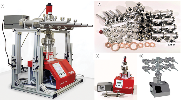
Hydrocarbon contamination plagues high-resolution and analytical electron microscopy by depositing carbonaceous layers onto surfaces during electron irradiation, which can render carefully prepared specimens useless. Increased specimen thickness degrades resolution with beam broadening alongside loss of contrast. The large inelastic cross-section of carbon hampers accurate atomic species detection. Oxygen and water molecules pose problems of lattice damage by chemically etching the specimen during imaging. These constraints on high-resolution and spectroscopic imaging demand clean, high-vacuum microscopes with dry pumps. Here, we present an open-hardware design of a high-vacuum manifold for transmission electron microscopy (TEM) holders to mitigate hydrocarbon and residual species exposure. We quantitatively show that TEM holders are inherently dirty and introduce a range of unwanted chemical species. Overnight storage in our manifold reduces contaminants by one to two orders of magnitude and promotes two to four times faster vacuum recovery. A built-in bakeout system further reduces contaminants partial pressure to below 10−10 hPa (Torr) (approximately four orders of magnitude down from ambient storage) and alleviates monolayer adsorption during a typical TEM experiment. We determine that bakeout of TEM holder with specimen held therein is the optimal cleaning method. Our high-vacuum manifold design is published with open-source blueprints, parts, and cost list.

Type
Software and Instrumentation
Copyright
Copyright © Microscopy Society of America 2020

Access options

Get access to the full version of this content by using one of the access options below. (Log in options will check for institutional or personal access. Content may require purchase if you do not have access.)

References

Bance, UR, Drummond, IW, Finbow, D, Harden, EH & Kenway, P (1978). Hydrocarbon contamination in vacuum dependent scientific instruments. Vacuum 28, 1011. https://doi.org/10.1016/S0042-207X(78)80029-3.CrossRefGoogle Scholar
Conru, HW & Laberge, PC (1975). Oil contamination with the SEM operated in the spot scanning mode. J Phys E: Sci Instrum 8, 136.CrossRefGoogle Scholar
Coyne, D (Ed.) (2013). LIGO Vacuum Compatible List. LIGO Science Collaboration. Available at https://dcc.ligo.org/public/0003/E960050/012/E960050-v12%20Vacuum%20Compatible%20Materials%20List.pdf.Google Scholar
Dayton, BB (1961). Outgassing rate of contaminated metal surfaces. Trans. 8th National Vacuum Symp. Am. Vacuum Soc., 42–57.Google Scholar
de Jonge, N, Houben, L, Dunin-Borkowski, RE & Ross, FM (2019). Resolution and aberration correction in liquid cell transmission electron microscopy. Nat Rev Mater 4, 6178. https://doi.org/10.1038/s41578-018-0071-2.CrossRefGoogle Scholar
Egerton, RF (2011). Electron Energy-Loss Spectroscopy in the Electron Microscope. Boston, MA: Springer.CrossRefGoogle Scholar
Ennos, AE (1953). The origin of specimen contamination in the electron microscope. J Appl Phys 4, 101106. https://doi.org/10.1088/0508-3443/4/4/302.Google Scholar
Fraser, HL (1978). Elemental analysis of second-phase carbides using electron energy-loss spectroscopy. In Scanning Electron Microscopy, Johari, O (Ed.), Part 1, pp. 627632. Chicago, IL: SEM Inc., A. M. F. O'Hare.Google Scholar
Griffiths, AJV & Walther, T (2010). Quantification of carbon contamination under electron beam irradiation in a scanning transmission electron microscope and its suppression by plasma cleaning. J Phys: Conf Ser 241, 012017. https://doi.org/10.1088/1742-6596/241/1/012017.Google Scholar
Grinham, R & Chew, A (2017). A review of outgassing and methods for its reduction. Appl Sci Converg Technol 26, 95109. https://doi.org/10.5757/ASCT.2017.26.5.95.CrossRefGoogle Scholar
Hettler, S, Dries, M, Hermann, P, Obermair, M, Gerthsen, D & Malac, M (2017). Carbon contamination in scanning transmission electron microscopy and its impact on phase-plate applications. Micron 96, 3847. https://doi.org/10.1016/j.micron.2017.02.002.CrossRefGoogle ScholarPubMed
Isabell, TC, Fischione, PE, O'Keefe, C, Guruz, MU & Dravid, VP (1999). Plasma cleaning and its applications for electron microscopy. Microsc Microanal 5, 126135. https://doi.org/10.1017/S1431927699000094.CrossRefGoogle ScholarPubMed
Jenninger, B & Chiggiato, P (2017). CAS tutorial on RGA Interpretation of RGA spectra. In CERN Accelerator School: Vacuum for Particle Accelerators, Glumslov, Sweden, 6 to 16 June 2017. Available at https://indico.cern.ch/event/565314/contributions/2285748/attachments/1467497/2273709/RGA_tutorial-interpretation.pdf.Google Scholar
Jousten, K (1998). Dependence of the outgassing rate of a “vacuum fired” 316N stainless steel chamber on bake-out-temperature. Vacuum 49, 359360. https://doi.org/10.1016/S0042-207X(98)00002-5.CrossRefGoogle Scholar
Jousten, K (1999). Thermal outgassing. In CERN Accelerator School: Vacuum Technology, Turner, S (Ed.), pp. 111124. Geneva: CERN Accelerator School.Google Scholar
Krivanek, OL, Corbin, GJ, Delby, N, Elston, BF, Keyse, RJ, Murfitt, MF, Own, CS, Szilagyi, ZS & Woodruff, JW (2008). An electron microscope for the aberration-corrected era. Ultramicroscopy 108, 179195. https://doi.org/10.1016/j.ultramic.2007.07.010.CrossRefGoogle ScholarPubMed
Leuthner, GT, Hummel, S, Mangler, C, Pennycook, TJ, Susi, T, Meyer, JC & Kotakoski, J (2019). Scanning transmission electron microscopy under controlled low-pressure atmospheres. Ultramicroscopy 203, 7681. https://doi.org/10.1016/j.ultramic.2019.02.002.CrossRefGoogle ScholarPubMed
Love, G, Scott, VD, Dennis, NMT & Laurenson, L (1981). Sources of contamination in electron optical equipment. Scanning 4, 3239. https://doi.org/10.1002/sca.4950040105.CrossRefGoogle Scholar
Matthewson, AG & Gröbner, O (1999). Thermal outgassing and beam induced desorption. In Handbook of Accelerator Physics and Engineering, Chao, AW & Tigner, M (Eds.), pp. 227230. Singapore: World Scientific.Google Scholar
McGilvery, CM, Goode, AE, Shaffer, MSP & McComb, DW (2012). Contamination of holey/lacey carbon films in STEM. Micron 43, 450455. https://doi.org/10.1016/j.micron.2011.10.026.CrossRefGoogle ScholarPubMed
Mitchell, DRG (2015). Contamination mitigation strategies for scanning transmission electron microscopy. Micron 73, 3646. https://doi.org/10.1016/j.micron.2015.03.013.CrossRefGoogle ScholarPubMed
Nerl, HC, Winther, KT, Hage, FS, Thygesen, KS, Houben, L, Backes, C, Coleman, JN, Ramasse, QM & Nicolosi, V (2017). Probing the local nature of excitons and plasmons in few-layer MoS2. npj 2D Mater Appl 1, 2. https://doi-org.proxy.lib.umich.edu/10.1038/s41699-017-0003-9.CrossRefGoogle Scholar
Postek, MT (1996). An approach to the reduction of hydrocarbon contamination in the scanning electron microscope. Scanning 18, 269274. https://doi.org/10.1002/sca.1996.4950180402.CrossRefGoogle Scholar
Reimer, L & Wächter, M (1978). Contribution to the contamination problem in transmission electron microscopy. Ultramicroscopy 3, 169174. https://doi.org/10.1016/S0304-3991(78)80023-0.CrossRefGoogle ScholarPubMed
Soong, C, Woo, P & Hoyle, D (2012). Contamination cleaning of TEM/SEM samples with the ZONE cleaner. Microsc Today 20, 4448. https://doi.org/10.1017/S1551929512000752.CrossRefGoogle Scholar
Stanford Research Systems, Inc. (2009). Models RGA100, RGA200, and RGA300 Residual Gas Analyzer: Operating Manual and Programming Reference. SRS Residual Gas Analyzer. Available at https://www.thinksrs.com/downloads/pdfs/manuals/RGAm.pdf .Google Scholar
Stewart, RL (1934). Insulating films formed under electron and ion bombardment. Phys Rev 45, 488490. https://doi.org/10.1103/PhysRev.45.488.CrossRefGoogle Scholar
Thompson, RF, Walker, M, Siebert, CA, Muench, SP & Ranson, NA (2016). An introduction to sample preparation and imaging by cryo-electron microscopy for structural biology. Methods 100, 315. https://doi.org/10.1016/j.ymeth.2016.02.017.CrossRefGoogle ScholarPubMed
Watson, JHL (1947). An effect of electron bombardment upon carbon black. J Appl Phys 18, 153161. https://doi.org/10.1063/1.1697597.CrossRefGoogle Scholar
Yoshimura, N, Hirano, H & Etoh, T (1983). Mechanism of contamination build-up induced by fine electron probe irradiation. Vacuum 33, 391395. https://doi.org/10.1016/0042-207X(83)90658-9.CrossRefGoogle Scholar
Supplementary material: File

Goh et al. supplementary material

Goh et al. supplementary material 1

Download Goh  et al. supplementary material(File)
File 968.3 KB
Supplementary material: PDF

Goh et al. supplementary material

Goh et al. supplementary material 2

Download Goh  et al. supplementary material(PDF)
PDF 357.9 KB