


No CrossRef data available.
Article contents
Age-Related Changes in Calcitonin-Producing Thyroid C-Cells of Male Wistar Rats
Published online by Cambridge University Press: 20 May 2022
Abstract
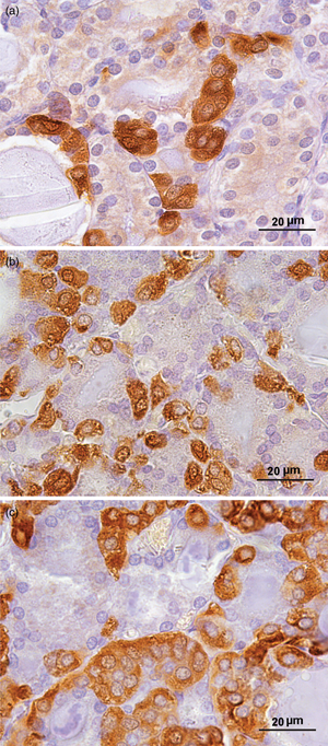
Thyroid C-cells secrete the hormone calcitonin (CT) which acts as an inhibitor of bone resorption. Our aim was to examine the age-related changes in the structure and function of CT-producing C-cells, using histomorphometric, ultrastructural, and biochemical analyses. We used young adult (3-months-old), middle-aged (16-months-old), and old (24-months-old) male rats. The peroxidase-antiperoxidase method was applied for localization of CT. Stereological analysis was performed using the newCAST stereological software package. Serum samples were analyzed for the determination of CT, testosterone (T), calcium (Ca2+), and phosphorus (P). We found a significant increase in the volume density (Vv) of C-cells in both older groups (p < 0.05). The percentage of smaller volume range C-cells increased (p < 0.0001), while the proportion of greater volume range C-cells decreased (p < 0.05) with ageing. Ultrastructural analysis revealed a larger number of secretory granules in older rats. Serum CT increased (p < 0.001), while serum T and P were reduced (p < 0.01) in older rats. Serum Ca2+ was lower (p < 0.0001) in middle-aged rats compared to young adults. We revealed a 20% incidence of C-cell hyperplasia in older rats and one case of medullary thyroid carcinoma in an old rat. Our findings indicate that the ageing process causes significant histomorphometric changes at the thyroid C-cell level.
- Type
- Biological Applications
- Information
- Copyright
- Copyright © The Author(s), 2022. Published by Cambridge University Press on behalf of the Microscopy Society of America
References
Albores-Saavedra, JA & Krueger, JE (2001). C-cell hyperplasia and medullary thyroid microcarcinoma. Endocr Pathol 12, 365–377.CrossRefGoogle ScholarPubMed
Barbagallo, M, Dominguez, LJ, Licata, G & Resnick, LM (1999). Effects of aging on serum ionized and cytosolic free calcium: Relation to hypertension and diabetes. Hypertension 34, 902–906.Google ScholarPubMed
Barlet, JP, Gaumet, N, Coxam, V & Davicco, MJ (1998). Calcitonin and stanniocalcin. Particular aspects of the endocrine regulation of phospho-calcium metabolism in mammals and fish. Ann Endocrinol (Paris) 59, 281–290.Google Scholar
Caverzasio, J, Bonjour, JP & Fleisch, H (1982). Tubular handling of Pi in young growing and adult rats. Am J Physiol 242, F705–F710.Google ScholarPubMed
Conde, E, Martín-Lacave, I, Utrilla, JC, González-Cámpora, R & Galera-Davidson, H (1995). Postnatal variations in the number and size of C-cells in the rat thyroid gland. Cell Tissue Res 280, 659–663.CrossRefGoogle ScholarPubMed
Diaz-Cano, SJ, de Miguel, M, Blanes, A, Tashjian, R & Wolfe, HJ (2001). Germline RET 634 mutation positive MEN 2A-related C-cell hyperplasias have genetic features consistent with intraepithelial neoplasia. J Clin Endocrinol Metab 86, 3948–3957.Google ScholarPubMed
Filipović, B, Šošić-Jurjević, B, Ajdžanović, V, Pantelić, J, Nestorović, N, Milošević, V & Sekulić, M (2013). The effects of sex steroids on thyroid C cells and trabecular bone structure in the rat model of male osteoporosis. J Anat 222, 313–320.CrossRefGoogle ScholarPubMed
Foresta, C, Zanatta, GP, Busnardo, B, Scanelli, G & Scandellari, C (1985). Testosterone and calcitonin plasma levels in hypogonadal osteoporotic young men. J Endocrinol Invest 8, 377–379.Google ScholarPubMed
Gibson, WG, Peng, TC & Croker, BP (1982). Age-associated C-cell hyperplasia in the human thyroid. Am J Pathol 106, 388–393.Google ScholarPubMed
Greenberg, C, Kukreja, SC, Bowser, EN, Hargis, GK, Henderson, WJ & Williams, GA (1986). Effects of estradiol and progesterone on calcitonin secretion. Endocrinology 118, 2594–2598.CrossRefGoogle ScholarPubMed
Guyétant, S, Bléchet, C & Saint-André, JP (2006). C-cell hyperplasia. Ann Endocrinol (Paris) 67, 190–197.CrossRefGoogle ScholarPubMed
Guyétant, S, Rousselet, MC, Durigon, M, Chappard, D, Franc, B, Guerin, O & Saint-André, JP (1997). Sex-related C cell hyperplasia in the normal human thyroid: A quantitative autopsy study. J Clin Endocrinol Metab 82, 42–47.CrossRefGoogle ScholarPubMed
Haschek, WM, Rousseaux, CG, Wallig, MA, Bolon, B & Ochoa, R (2013). Haschek Rousseaux's Handbook of Toxicologic Pathology, 3rd ed. New York: Academic Press.Google Scholar
Kalu, DN, Herbert, DC, Hardin, RR, Yu, BP, Kaplan, G & Jacobs, JW (1988). Mechanism of dietary modulation of calcitonin levels in Fischer rats. J Gerontol 43, 125–131.Google ScholarPubMed
Kaserer, K, Scheuba, C, Neuhold, N, Weinhäusel, A, Haas, OA, Vierhapper, H & Niederle, B (2001). Sporadic versus familial medullary thyroid microcarcinoma: A histopathologic study of 50 consecutive patients. Am J Surg Pathol 25, 1245–1251.CrossRefGoogle ScholarPubMed
Kiebzak, GM & Sacktor, B (1986). Effect of age on renal conservation of phosphate in the rat. Am J Physiol 251, F399–F407.Google ScholarPubMed
LiVolsi, VA (1997). C cell hyperplasia/neoplasia. J Clin Endocrinol Metab 82, 39–41.CrossRefGoogle ScholarPubMed
LiVolsi, V, DeLellis, R & Komminoth, P (2017). Multiple endocrine neoplasia type 2. In WHO Classification of Tumours of Endocrine Organs, 4th edn. Lloyd, RV, Osamura, RY, Kloppel, G & Rosai, J (Eds.), pp. 248–250. Lyon: IARC Press.Google Scholar
Lu, CC, Tsai, SC, Chien, EJ, Tsai, CL & Wang, PS (2000). Age-related differences in the secretion of calcitonin in male rats. Metabolism 49, 253–258.CrossRefGoogle ScholarPubMed
Martín-Lacave, I, Conde, E, Montero, C & Galera-Davidson, H (1992). Quantitative changes in the frequency and distribution of the C-cell population in the rat thyroid gland with age. Cell Tissue Res 270, 73–77.CrossRefGoogle ScholarPubMed
Nataraju, GJ, Ranvir, RK, Kothule, VR, Kadam, SB, Ravichandra, BV, Bhatnagar, U & Jain, MR (2016). Spontaneous lesions in endocrine glands of experimental Wistar rats and beagle dogs. Exp Toxicol Pathol 68, 1–13.CrossRefGoogle ScholarPubMed
Oh, YS, Seo, EH, Lee, YS, Cho, SC, Jung, HS, Park, SC & Jun, HS (2016). Increase of calcium sensing receptor expression is related to compensatory insulin secretion during aging in mice. PLoS One 11, e0159689.CrossRefGoogle ScholarPubMed
O'Toole, K, Fenoglio-Preiser, C & Pushparaj, N (1985). Endocrine changes associated with the human aging process: III. Effect of age on the number of calcitonin immunoreactive cells in the thyroid gland. Hum Pathol 16, 991–1000.CrossRefGoogle ScholarPubMed
Perry, A, Molberg, K & Albores-Saavedra, J (1996). Physiologic versus neoplastic C-cell hyperplasia of the thyroid: Separation of distinct histologic and biologic entities. Cancer 77, 750–756.3.0.CO;2-Z>CrossRefGoogle ScholarPubMed
Pirola, S & Harre, RK (2012). C-cell hyperplasia in thyroid tissue adjacent to papillary carcinoma. Int J Surg Pathol 20, 66–68.CrossRefGoogle ScholarPubMed
Portale, AA, Lonergan, ET, Tanney, DM & Halloran, BP (1997). Aging alters calcium regulation of serum concentration of parathyroid hormone in healthy men. Am J Physiol 272, E139–E146.Google ScholarPubMed
Reagh, J, Bullock, M, Andrici, J, Turchini, J, Sioson, L, Clarkson, A, Watson, N, Sheen, A, Lim, G, Delbridge, L, Sidhu, S, Sywak, M, Aniss, A, Shepherd, P, Ng, D, Oei, P, Field, M, Learoyd, D, Robinson, BG, Clifton-Bligh, RJ & Gill, AJ (2017). NRASQ61R mutation-specific immunohistochemistry also identifies the HRASQ61R mutation in medullary thyroid cancer and may have a role in triaging genetic testing for MEN2. Am J Surg Pathol 41, 75–81.Google ScholarPubMed
Saggiorato, E, Rapa, I, Garino, F, Bussolati, G, Orlandi, F, Papotti, M & Volante, M (2007). Absence of RET gene point mutations in sporadic thyroid C-cell hyperplasia. J Mol Diagn 9, 214–219.Google ScholarPubMed
Sakai, K, Yamada, S & Yamada, K (2000). Effects of ovariectomy on parafollicular cells in the rat. Okajimas Folia Anat Jpn 76, 311–319.CrossRefGoogle ScholarPubMed
Salih, MA, Herbert, DC & Kalu, DN (1993). Evaluation of the molecular and cellular basis for the modulation of thyroid C-cell hormones by aging and food restriction. Mech Ageing Dev 70, 1–21.CrossRefGoogle ScholarPubMed
Shamonki, IM, Frumar, AM, Tataryn, IV, Meldrum, DR, Davidson, BH, Parthemore, JG, Judd, HL & Deftos, LJJ (1980). Age-related changes of calcitonin secretion in females. Clin Endocrinol Metab 50, 437–439.CrossRefGoogle ScholarPubMed
Sorva, A, Elfving, S, Sievers, G & Tilvis, RS (1992). Calcemic status of geriatric patients: A longitudinal study. Gerontology 38, 87–91.Google ScholarPubMed
Sternberger, LA, Hardy, PH, Cuculis, JJ & Meyer, HG (1970). The unlabeled antibody enzyme method of immunohistochemistry. Preparation and properties of soluble antigen-antibody complex (horseradish peroxidase-antihorseradish peroxidase) and its use in identification of spirochetes. J Histochem Cytochem 18, 315–333.Google ScholarPubMed
Tsai, CL, Pu, HF, Lau, CP, Wang, PS & Liu, TK (1992). Age-related differences in basal and calcium-stimulated plasma calcitonin levels in female rats. Am J Physiol 262, E557–E560.Google ScholarPubMed
Warshawsky, H, Goltzman, D, Rouleau, MF & Bergeron, JJ (1980). Direct in vivo demonstration by radioautography of specific binding sites for calcitonin in skeletal and renal tissues of the rat. J Cell Biol 85, 682–694.CrossRefGoogle ScholarPubMed
Wongsurawat, N & Armbrecht, HJ (1987). Comparison of calcium effect on in vitro calcitonin and parathyroid hormone release by young and aged thyroparathyroid glands. Exp Gerontol 22, 263–269.CrossRefGoogle Scholar
Zalups, RK & Knox, FG (1983). Calcitonin decreases the renal tubular capacity for phosphate reabsorption. Am J Physiol 245, F345–F348.Google ScholarPubMed
Zhang, D, Maalouf, NM, Adams-Huet, B, Moe, OW & Sakhaee, K (2014). Effects of sex and postmenopausal estrogen use on serum phosphorus levels: A cross-sectional study of the national health and nutrition examination survey (NHANES) 2003-2006. Am J Kidney Dis 63, 198–205.CrossRefGoogle ScholarPubMed