Hostname: page-component-5c6d5d7d68-thh2z Total loading time: 0 Render date: 2024-08-19T09:06:57.686Z Has data issue: false hasContentIssue false

Histological, Histochemical, and Ultrastructural Approach to the Ductus Deferens in Male Nile Monitor Lizard (Varanus niloticus)

Published online by Cambridge University Press:  22 June 2021

Mahmoud Awad*
Affiliation:
Department of Histology, Faculty of Veterinary Medicine, South Valley University, Qena83523, Egypt
Mohammed Alshehri
Affiliation:
Department of Biology, Faculty of Science, King Khalid University, Abha61421, Saudi Arabia
Ahmed Saad A. Hassaneen
Affiliation:
Department of Theriogenology, Faculty of Veterinary Medicine, South Valley University, Qena83523, Egypt
*
*Author for correspondence: Mahmoud Awad, E-mail: awad.histology83@gmail.com
Get access

Abstract

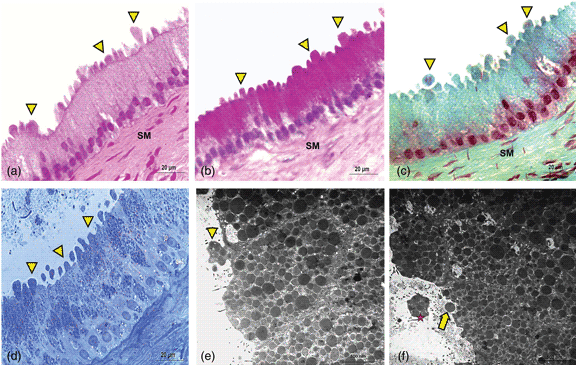
The ductus deferens is a fundamental part of the male genital tract and the continuation of the epididymal duct. As a male secondary sex organ, the ductus deferens plays a crucial role in the nourishment, storage, and maturation of spermatozoa. Some studies have provided information about the ductus deferens structure in reptiles; however, the full description of the ductus deferens remains to be clarified. The current study aimed to describe the Nile monitor lizard (Varanus niloticus) ductus deferens from histological, histochemical, and ultrastructural perspectives. The results revealed that the ductus deferens is formed histologically from two main cell types: principal and basal. The principal cells were tall and filled with periodic acid Schiff (+)/alcian blue (−) cytoplasmic granules. The basal cells were found just above the basement membrane. By transmission electron microscopy, the principal cells exhibited typical protein-secreting cell features. Additionally, some intraepithelial cells, such as halo cells, undifferentiated mesenchymal cells, and agranular leukocytes, were identified. This study presents the first detailed description of the Varanus niloticus ductus deferens. Further immunohistochemical studies are required to explore the function(s) of the cellular components.

Type
Micrographia
Copyright
Copyright © The Author(s), 2021. Published by Cambridge University Press on behalf of the Microscopy Society of America

Access options

Get access to the full version of this content by using one of the access options below. (Log in options will check for institutional or personal access. Content may require purchase if you do not have access.)

References

Awad, M & Mohamadain, D (2019). Histochemical and ultrastructural investigations on the renal parenchyma of the Egyptian Nile Monitor Lizard (Varanus niloticus). Acta Zool 18. doi:10.1111/azo.12308.Google Scholar
Bancroft, JD & Layton, C (2013). Bancroft's Theory and Practice of Histological Techniques, 7th ed. New York, NY: Churchill Livingstone.Google Scholar
Bennett, D (2002). Diet of juvenile Varanus niloticus (Sauria: Varanidae) on the Black Volta River in Ghana. J Herpetol 36, 116117.CrossRefGoogle Scholar
Burns, WA (1978). Thick sections: Technique and applications. In Diagnostic Electron Microscopy, Ch. 4, Trump, BF & Jones, RJ (Eds.). New York, NY: John Wiley & Sons.Google Scholar
Crossmon, G (1937). A modification of Mallory's connective tissue stain with a discussion of the principals involved. Anat Rec 69, 3338.CrossRefGoogle Scholar
de Buffrenil, V, Chabanet, C & Castanet, J (1994). Preliminary data on size, growth and avoidance of the Nile monitor lizard (Varanus niloticus) in the region of Lake Chad (in French: Donne´es pre´liminaires sur la taille, la croissance et la longe ´vite´du varan du Nil (Varanus niloticus) dans la re´gion du Lac Tchad). Can J Zool 72, 262273.Google Scholar
Guerrero, SM, Calderon, ML, de Perez, GR & Ramirez Pinilla, MP (2004). Morphology of the male reproductive duct system of Caiman crocodilus (Crocodylia, Alligatoridae). Ann Anat 186(3), 235245.CrossRefGoogle Scholar
Harris, HF (1900). On the rapid conversion of hematoxylin into haematein in staining reactions. J Appl Microsc Lab Methods 3, 777.Google Scholar
King, D & Green, B (1999). Goannas: The Biology of Varanid Lizards (Australian Natural History Series). Sydney: University of New South Wales Press.Google Scholar
Luiselli, L, Akani, GC & Capizzi, D (1999). Is there any interspecific competition between dwarf crocodiles (Osteolaemus tetraspis) and Nile monitors (Varanus niloticus ornatus) in the swamps of Central Africa? A study from south-eastern. Nigeria J Zool 247, 127131.Google Scholar
Mahfud, M, Winarto, A & Nisa, C (2016). Micromorphological structure of primary reproductive organ of male water monitor lizard (Varanus salvator bivittatus). J Ked Hewan 10(1), 7276.Google Scholar
Moustafa, MAM, Ismail, MN, Mohamed, AEA & Ali, AO (2013). Hematologic and biochemical parameters of free-ranging female Nile monitors in Egypt. J Wildl Dis 49(3), 750754.CrossRefGoogle ScholarPubMed
Mowry, RW (1956). Alcian blue technics for the histochemical study of acidic carbohydrates. J Histochem Cytochem 4, 407408.Google Scholar
Thompson, G (1999). Goanna metabolism: Is it different to other lizards, and if so what are the ecological consequences? Mertensiella 11, 7990.Google Scholar
Tran, D, Golick, M, Rabinovitz, H, Rivlin, D, Elgart, G & Nordlow, B (2000). Hematoxylin and safranin O staining of frozen sections. Dermatol Surg 26, 197199. doi:10.1046/j.1524-4725.2000.09220.CrossRefGoogle ScholarPubMed
Viana, DC, Rui, LA, dos Santos, AC, Miglino, MA, Neto, AC, Aranjo, LPF, Oliveira, AA & Sousa, AL (2014). Seasonal morphological variation of the vas deferens of scorpion mud turtle (Kinosternon scorpioides). Biota Neotrop 14(3), e20130064.CrossRefGoogle Scholar
Wyneken, J & Mader, D (2002). The reproductive system of reptiles—Anatomy, physiology and clinical perspectives. In 2002 Proceedings. Association of Reptilian and Amphibian Veterinarians, p. 187.Google Scholar