


1. Introduction
Microorganisms are ubiquitous in both natural environments and technological applications. They are highly adaptable and inhabit diverse ecosystems such as swamps, oceans and rivers (Schallenberg & Kalff Reference Schallenberg and Kalff1993; Nealson Reference Nealson1997; Herndl & Reinthaler Reference Herndl and Reinthaler2013; Roberto, Van Gray & Leff Reference Roberto, Van Gray and Leff2018; Zhang et al. Reference Zhang, Tu, Li, Lu and Li2020). They also play a significant role in numerous engineering applications (Falkowski, Barber & Smetacek Reference Falkowski, Barber and Smetacek1998; Savage Reference Savage2011; Falkowski Reference Falkowski2012), including fermentation processes for vaccine and food production, and wastewater treatment processes (Swartz Reference Swartz2001; van Loosdrecht & Brdjanovic Reference van Loosdrecht and Brdjanovic2014; Lorenzo et al. Reference Lorenzo, Munekata, Dominguez, Pateiro, Saraiva and Franco2018; Honda et al. Reference Honda, Matsuura, Sorn, Asakura, Morinaga, Van Huy, Sabar, Masakke, Hara-Yamamura and Watanabe2023). Controlling the sedimentation process in biofuel production, for instance, can lead to improvements in algae separation from the fluid medium and an accumulation of algal biomass at the bottom of the production pond (Savage Reference Savage2011).
Suspensions of swimming microorganisms (e.g. E. coli) or active particles (e.g. phoretic colloids) are typical examples of so-called active fluids (Ramaswamy Reference Ramaswamy2010; Marchetti et al. Reference Marchetti, Joanny, Ramaswamy, Liverpool, Prost, Rao and Simha2013; Patteson, Gopinath & Arratia Reference Patteson, Gopinath and Arratia2016a; Moran & Posner Reference Moran and Posner2017). In these fluids, the constituents inject energy, generate mechanical stresses, and create flows within the fluid medium. Thus active fluids are systems that are inherently out of equilibrium even in the absence of external forces (Dabelow, Bo & Eichhorn Reference Dabelow, Bo and Eichhorn2019). Bacterial activity can lead to many complex phenomena such as reduced shear viscosity (Rafaï, Jibuti & Peyla Reference Rafaï, Jibuti and Peyla2010; Gachelin et al. Reference Gachelin, Miño, Berthet, Lindner, Rousselet and Clément2013; López et al. Reference López, Gachelin, Douarche, Auradou and Clément2015), hindrance in mixing and transport of passive scalars (Ran et al. Reference Ran, Brosseau, Blackwell, Qin, Winter and Arratia2021), formation of biofilms (Pradeep & Arratia Reference Pradeep and Arratia2022), and diffusion enhancement (Kim & Breuer Reference Kim and Breuer2004; Leptos et al. Reference Leptos, Guasto, Gollub, Pesci and Goldstein2009; Thiffeault & Childress Reference Thiffeault and Childress2010; Kurtuldu et al. Reference Kurtuldu, Guasto, Johnson and Gollub2011; Miño et al. Reference Miño, Mallouk, Darnige, Hoyos, Dauchet, Dunstan, Soto, Wang, Rousselet and Clement2011; Jepson et al. Reference Jepson, Martinez, Schwarz-Linek, Morozov and Poon2013). In the dilute regime, particle (or tracer) diffusivity has been shown to increase linearly with bacteria concentration (Wu & Libchaber Reference Wu and Libchaber2000), but depends on microorganism swimming behaviour (Chen et al. Reference Chen, Lau, Hough, Islam, Goulian, Lubensky and Yodh2007) and particle size (Patteson et al. Reference Patteson, Gopinath, Purohit and Arratia2016b). Recent work has shown that the enhanced diffusion dynamics in the presence of bacteria can lead to large-scale clustering of particles (Bouvard, Moisy & Auradou Reference Bouvard, Moisy and Auradou2023). However, less understood are the effects of bacterial activity on the settling dynamics of tracer particles.
Initial studies have largely focused on steady-state analysis (Palacci et al. Reference Palacci, Cottin-Bizonne, Ybert and Bocquet2010; Maggi et al. Reference Maggi, Lepore, Solari, Rizzo and Di Leonardo2013; Ginot et al. Reference Ginot, Theurkauff, Levis, Ybert, Bocquet, Berthier and Cottin-Bizonne2015). These include sedimentation studies with active Janus particles that show (steady-state) density profiles that decay exponentially with system height (Palacci et al. Reference Palacci, Cottin-Bizonne, Ybert and Bocquet2010; Ginot et al. Reference Ginot, Theurkauff, Levis, Ybert, Bocquet, Berthier and Cottin-Bizonne2015, Reference Ginot, Solon, Kafri, Ybert, Tailleur and Cottin-Bizonne2018). The resulting length scale is larger than expected for thermal equilibrium systems, which is explained through the introduction of an effective temperature (and diffusivity) with values that far exceed those of passive systems (Palacci et al. Reference Palacci, Cottin-Bizonne, Ybert and Bocquet2010). These findings are corroborated by theory and simulations with no (or limited) hydrodynamic interactions (Tailleur & Cates Reference Tailleur and Cates2008; Nash et al. Reference Nash, Adhikari, Tailleur and Cates2010; Wang et al. Reference Wang, Chen, Sheng and Tsao2014; Ginot et al. Reference Ginot, Solon, Kafri, Ybert, Tailleur and Cottin-Bizonne2018; Vachier & Mazza Reference Vachier and Mazza2019). Experiments with swimming E. coli show that bacteria aggregation (due to extra-cellular polymer) can have significantly enhanced sedimentation rates (Maggi et al. Reference Maggi, Lepore, Solari, Rizzo and Di Leonardo2013), while particles settling in suspensions of C. reinhardtii show the familiar exponential concentration profile but with an effective gravitational length (or diffusivity) that is proportional to algae concentration (Jeanneret et al. Reference Jeanneret, Pushkin, Kantsler and Polin2016). These studies show that the concept of effective diffusivity (and temperature) can be useful in characterizing the steady sedimentation profiles of active suspensions.
Only recently have time-dependent sedimentation (that is, concentration) profiles of bacterial suspensions been investigated; see Singh et al. (Reference Singh, Patteson, Torres Maldonado, Purohit and Arratia2021) and Torres Maldonado et al. (Reference Torres Maldonado, Ran, Galloway, Brosseau, Pradeep and Arratia2022). These studies show that in the dilute regime, bacteria activity can significantly hinder the sedimentation process (Singh et al. Reference Singh, Patteson, Torres Maldonado, Purohit and Arratia2021). At vanishingly low bacteria concentration ( $\phi _b$), the (time-dependent) concentration profiles can be accurately described by an advection–diffusion equation; however, as $\phi _b$ is increased, sink–source terms must be introduced in the formulation to account for bacteria population dynamics, including a dispersivity parameter that increases with $\phi _b$ (Singh et al. Reference Singh, Patteson, Torres Maldonado, Purohit and Arratia2021). In this regime, the bacterial suspension undergoes phase separation, and bacteria settling speed is found to be strongly correlated with a time scale associated with oxygen depletion within the system (Torres Maldonado et al. Reference Torres Maldonado, Ran, Galloway, Brosseau, Pradeep and Arratia2022). These studies highlight the role of oxygen in the sedimentation of living suspensions; microorganisms are known to exhibit aerotaxis, a phenomenon that prompts aerobic bacteria to seek an oxygen source (Wager Reference Wager1911; Baracchini & Sherris Reference Baracchini and Sherris1959; Adler et al. Reference Adler, Erickstad, Gutierrez and Groisman2012; Bouvard et al. Reference Bouvard, Douarche, Mergaert, Auradou and Moisy2022). When suspended in a relatively deep layer of fluid, the microorganism can deplete the oxygen concentration in the water while oxygen continuously diffuses in from the air–water interface above. In response, microorganisms swim towards the oxygen concentration gradient and generate fluid motion, a phenomenon referred to as bioconvection driven by aerotaxis (Hillesdon, Pedley & Kessler Reference Hillesdon, Pedley and Kessler1995; Jánosi, Kessler & Horváth Reference Jánosi, Kessler and Horváth1998). During bioconvection, oxygen-enriched water descends from the interface, giving rise to a complex convection-dependent gradient that guides the swimming bacteria. Analysis predicts that the emergence of bioconvection occurs beyond a critical Rayleigh number (Hillesdon & Pedley Reference Hillesdon and Pedley1996), a dimensionless number that quantifies the ratio of the buoyancy forces to viscous forces (Hill, Pedley & Kessler Reference Hill, Pedley and Kessler1989), although bioconvection can also occur from phototaxis and thus be controlled by a light source (Arrieta et al. Reference Arrieta, Polin, Saleta-Piersanti and Tuval2019). While extensive research has focused on elucidating the mechanisms behind bioconvection formation (Pedley & Kessler Reference Pedley and Kessler1992; Hill & Pedley Reference Hill and Pedley2005; Bees Reference Bees2020), limited experimental work has been conducted to corroborate these theoretical predictions.
In this contribution, we experimentally investigate the sedimentation dynamics of colloidal particles and the formation of bioconvection patterns in dilute bacterial suspensions using particle tracking methods. We find that bacterial activity has a significant impact on particle diffusivity, leading to the identification of two dynamical regimes. Initially, as bacterial concentration is increased, we observed a corresponding linear increase in diffusivity. However, as bacterial concentration is further increased (but still dilute), particle diffusivity becomes independent of bacterial concentration. This transition coincides with the inception of bioconvection patterns above a critical Rayleigh number ( $\varGamma _{cr}$) (Hillesdon & Pedley Reference Hillesdon and Pedley1996).
2. Materials and methods
We experimentally investigate the sedimentation dynamics of spherical colloids in the presence of bacterial activity. Active fluids are mixtures of bacterial suspensions and passive particles suspended in deionized water to reach the desired volume fractions of bacteria and particles, denoted as  $\phi _b$ and $\phi _p$. Bacterial suspensions are prepared using wild-type Escherichia coli (K12 MG1655), which are cultured to saturation ( $10^9\ {\rm cells}\ {\rm ml}^{-1}$) in LB broth (Sigma-Aldrich). Passive particles are polystyrene spheres (density $\rho _p =1.05\ {\rm g}\ {\rm cm}^{-3}$, Thermo Scientific) with diameter $d = 3.2\ \mathrm {\mu }$m, which are gently cleaned by centrifugation (Centrifuge 5430, Eppendorf). All active suspensions are contained within a custom-made quasi-two-dimensional sedimentation cell constructed from optically transparent polymethyl methacrylate (PMMA) material, offering optimal clarity (see figure 1a). The container measures $100$ mm in height, $28$ mm in width, and $400\ \mathrm {\mu }$m in thickness, and is assembled using precision laser cutting and chemical bonding techniques (Sun et al. Reference Sun, Peeni, Yang, Becerril and Woolley2007).

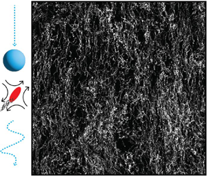
Figure 1. Experimental set-up and sample particle trajectories. (a) A schematic of the set-up and bacteria/particle suspensions (active fluid). Sedimentation experiments are conducted in an optically clear rectangular container. The particles are  $3.2\ \mathrm {\mu }$m polystyrene spheres, and the bacteria are $2\ \mathrm {\mu }$m rod-shaped E. coli; both are subjected to gravity. Particles located at the centre and at a height ${\approx }20$ mm from the bottom of the container are observed through a microscope. These spherical colloidal particles are tracked through a particle tracking velocimetry method. Particle trajectories ( $\phi _p =0.04\,\%$) tracked for total time 10 minutes are shown in (b) without bacteria and in (c) with bacteria ( $\phi _{b} =0.45\,\%$). When particles and E. coli are combined, the passive particle trajectories in the lateral direction undergo significant modifications compared to their trajectories in the absence of bacteria.
Active fluids are homogenized manually using a pipette before being introduced into the custom-made PMMA container. A volume  ${\approx }1.1$ ml of the prepared fluid is then transferred into the container, as depicted schematically in figure 1(a). Subsequently, the container is immersed in a water tank to maintain the fluid at room temperature $24\,^\circ$C. Images are captured at frame rate $30$ fps with a high-resolution camera (IO Industries, Flare 4M180) and a microscope (Infinity, K2) equipped with a $10\times$ objective. The light source used is a single-colour cold visible LED (M490L4, Thor Labs). The depth of field of the microscope is $3.5\ \mathrm {\mu }$m. All the measurements are taken at height 20 mm from the base of the sedimentation cell. Additionally, we focus at the half-distance, at ${\sim }200\ \mathrm {\mu }$m from either side of the walls, to avoid the cell wall effects on the hydrodynamic hindrance of fluctuations from the bacterial activity on our passive colloidal particles. Later in the paper we show that under these experimental conditions, we are able to match the Stokes–Einstein diffusion coefficient and the corresponding settling velocity of a single particle to that of passive colloids in the absence of bacterial activity. This confirms that our experimental conditions are devoid of wall effects.
The initial volume fraction  $\phi _b$ of motile bacteria in the container varies from $0\,\%$ ( $0\ {\rm cells}\ {\rm ml}^{-1}$) to $0.75\,\%$ ( $5.4\times 10^9\ {\rm cells}\ {\rm ml}^{-1}$), while the volume fraction of spherical colloids is maintained consistently at $\phi _p=0.04\,\%$. It is worth noting that both $\phi _b$ and $\phi _p$ concentrations are within the dilute regime ( $\phi <1\,\%$), and no significant macroscopic collective behaviour is observed in these bacteria–particle suspensions under the microscope (Patteson et al. Reference Patteson, Gopinath, Purohit and Arratia2016b; Singh et al. Reference Singh, Patteson, Torres Maldonado, Purohit and Arratia2021). The selected range of $\phi _b$ in this study remains below the threshold at which collective motion is typically observed ( ${\approx }10^{10}\ {\rm cells}\ {\rm ml}^{-1}$) (Kasyap, Koch & Wu Reference Kasyap, Koch and Wu2014). All captured images for the suspensions are acquired within a 30 minute time frame. Importantly, within this designated time frame, no noteworthy reduction in bacteria motility is observed, a finding that has been corroborated by recent investigations (Ran et al. Reference Ran, Brosseau, Blackwell, Qin, Winter and Arratia2021; Singh et al. Reference Singh, Patteson, Torres Maldonado, Purohit and Arratia2021).
3. Results and discussion
The main goal is to understand the interplay between bacterial activity and the settling dynamics of passive spherical particles. To this end, we use particle tracking techniques to visualize the colloidal particle's Lagrangian trajectories (Crocker & Grier Reference Crocker and Grier1996). Sample particle trajectories from passive ( $\phi _p=0.04\,\%$, no bacteria) and active ( $\phi _p=0.04\,\%$, $\phi _b=0.45\,\%$) fluids are shown in figures 1(b) and 1(c), respectively. The passive fluid case shows particle trajectories predominately in the downward direction ( $-y$) with apparent noise along the $x$-axis. For the active fluid, on the other hand, particle paths show amplified displacement along the $x$-axis, indicating that bacterial activity induces modifications to the colloidal particle downward trajectories. This trajectory enhancement (in the $x$-axis) manifests itself consistently across all experiments, providing an explanation for the previously observed hindrance in the speed of the particle front (Singh et al. Reference Singh, Patteson, Torres Maldonado, Purohit and Arratia2021; Torres Maldonado et al. Reference Torres Maldonado, Ran, Galloway, Brosseau, Pradeep and Arratia2022). These qualitative findings show that bacteria modify the settling trajectories of colloidal particles through hydrodynamic interactions, even in the dilute regime ( $\phi _b<1\,\%$).
3.1. Characterizing length scales of particle correlations
We quantify the sedimentation dynamics of our fluid by computing its time-dependent velocity fields using an in-house particle imaging velocimetry method (Li et al. Reference Li, Ran, Wang, Wang, Chen, Niu, Arratia and Yang2021; Brosseau et al. Reference Brosseau, Ran, Graham, Jerolmack and Arratia2022; Ran et al. Reference Ran, Gagnon, Morozov and Arratia2022). The goal is to understand how bacterial activity ( $\phi _b$) alters the spatial correlation functions of particle fluctuations (Segrè, Herbolzheimer & Chaikin Reference Segrè, Herbolzheimer and Chaikin1997; Tee et al. Reference Tee, Mucha, Cipelletti, Manley, Brenner, Segre and Weitz2002). Here, the spatial correlation function of the velocity fluctuations, $C_u(r_x)$, is defined as
 \begin{equation} C_u(r_x)=\langle{u}(x)\,{u}({x}+{r_x})\rangle/\langle u^2\rangle, \end{equation}
where  $x$ represents the direction perpendicular to gravity, $\langle \cdot \rangle$ denotes an ensemble average across all spherical particles, ${u}({x})$ denotes the local velocity of a particle at position ${x}'$, and $r_x$ denotes distance in the direction parallel to the sedimentation. Notably, ${u}({x})$ incorporates contributions from both particle self-diffusivity and hydrodynamic interactions. In the absence of bacteria ( $\phi _b=0\,\%$), we observe a decay in the spatial correlation length scales at small distances (figure 2a). With an increase in $\phi _b$, we identify a corresponding increase in spatial correlation functions over larger distances. However, as $\phi _b\geq 0.45\,\%$, the spatial correlation functions become independent of $\phi _b$. To analyse this trend quantitatively, we calculate the integral length scale of velocity $L_u$ as shown in figure 2(b). This integral length scale is defined as $L_u=\int _0^\infty C_u(r_x)\,{\rm d} r_x$. Results shows that $L_u$ increases linearly as $\phi _b$ is increased (figure 2b), but this relationship weakens and $L_u$ becomes independent of $\phi _b$ for $\phi _b\geq 0.45\,\%$. These findings show a positive correlation between $L_u$ and $\phi _b$ up to $\phi _b\geq 0.45\,\%$. This outcome aligns with previous research, highlighting the presence of two distinct regimes within the sedimentation process (Torres Maldonado et al. Reference Torres Maldonado, Ran, Galloway, Brosseau, Pradeep and Arratia2022).

Figure 2. (a) The spatial correlation functions of particle velocities ( $u$) in the lateral direction across the $x$-axis. (b) The integral length scale of the lateral velocities at different bacteria volume fractions $\phi _b$. The open circular symbols from left to right represent the results at $\phi _b=0.25\,\%$ (black), $\phi _b=0.45\,\%$ (orange) and $\phi _b=0.75\,\%$ (light green). The presence of bacteria leads to an increase in correlation functions, and subsequently to an increase in length scales.
3.2. Mean square displacements and diffusivity
Next, we use experimentally measured particle tracks to compute the mean square displacements of the passive particles as a function of  $\phi _b$. The mean square displacement is defined as $MSD (\Delta t) = \langle |\boldsymbol {r}(t_R + \Delta t) - \boldsymbol {r}(t_R)|^2\rangle$, where $t_R$ is denoted as the reference time. First, we extract the distance vector $\boldsymbol {r}$ in the direction perpendicular to gravity, $r_x$, yielding the mean square displacement along the $x$-axis ( $MSD_x$). As shown in figure 3(a), $MSD_x$ exhibits diffusive behaviour (at sufficiently long time intervals) that increases with $\phi _b$. We can estimate an effective diffusivity $D_{eff}$ by fitting $MSD_x= 2D_{eff}\,\Delta t$ to the experimental data at long times, and find that, similar to $L_u$, $D_{eff}$ grows linearly with $\phi _b$ followed by an asymptote for $\phi _b\geq 0.45\,\%$ (figure 3c). Similarly, we compute the mean square displacements of the tracked particles relative to the $y$-axis ( $MSD_{y^\prime }$) as a function of $\phi _b$. Notably, we subtract the mean sedimentation rates along the $y$-axis since our focus is on understanding how bacteria activity affects particle fluctuations, i.e. $y^{\prime }=y-\langle y \rangle$. Figure 3(b) shows that $MSD_{y^{\prime }}$ exhibits diffusive behaviour that increases with increasing $\phi _b$. We apply a similar fitting approach, relating $MSD_{y^{\prime }}$ to $D_{eff}$ as shown in figure 3(c) (filled symbols). Our results reveal that for the control case ( $\phi _b=0\,\%$), $D_{eff}= 0.150\pm 0.001\ \mathrm {\mu }{\rm m}^2\ {\rm s}^{-1}$ for $MSD_x$, and $D_{eff}=0.145\pm 0.001\ \mathrm {\mu }{\rm m}^2\ {\rm s}^{-1}$ for $MSD_{y^{\prime }}$. These measurements align with the theoretically predicted value from the Stokes–Einstein relation, $D_0=k_B T/(3 {\rm \pi}\mu d)=0.150\ \mathrm {\mu }{\rm m}^2\ {\rm s}^{-1}$ (Einstein Reference Einstein1905), where $k_B$ represents the Boltzmann constant, $\mu$ is the fluid viscosity, and $T$ denotes the temperature. These results indicate that hydrodynamic interactions among passive particles in our study are relatively weak, which is not surprising given the dilute particle volume fraction ( $\phi _p= 0.04\,\%$).

Figure 3. (a) Mean square displacement in the  $x$-axis ( $MSD_x$) of spherical particles at different bacteria volume fractions $\phi _b$. (b) Mean square displacement of particle fluctuations in the $y$-axis ( $MSD_{y^\prime }$) at different $\phi _b$. (c) Effective particle diffusivities $D_{eff}$ from $MSD_x$ (open symbols) and $MSD_y$ (closed symbols) as functions of $\phi _b$. The presence of swimming E. coli increases particle fluctuations in both the $x$ and $y$ directions, resulting in higher $D_{eff}$. (d) Péclet number ( $Pe$) as a function of $\phi _b$. The results demonstrate that the presence of bacteria enhances diffusion transport in the settling process, eventually reaching a plateau regime where advection and diffusion transport are in close balance ( $Pe \approx 1$).
Overall, the mean square displacement measurements of particles in the presence of motile bacteria exhibit an increasing trend as  $\phi _b$ rises, as shown in figures 3(a) and 3(b). This enhancement on particle displacement leads to a corresponding increase in $D_{eff}$, illustrated in figure 3(c). By comparison, a similar experiment was conducted using non-motile bacteria ( $\phi _{d,b}=0.35\,\%$), as shown in figures 3(a) and 3(b). Here, we observe a negligible increase in mean square displacement compared to the absence of bacteria ( $\phi _b=0\,\%$), resulting in $D_{eff}= 0.159\pm 0.001\ \mathrm {\mu }{\rm m}^2\ {\rm s}^{-1}$ along the $x$-axis and $D_{eff}= 0.163\pm 0.002\ \mathrm {\mu }{\rm m}^2\ {\rm s}^{-1}$ along the $y$-axis for $\phi _{d,b}=0.35\,\%$ (figure 3c). These results indicate that the presence of non-motile bacteria exerts negligible impact on particle diffusivity and hydrodynamic interactions. This small but finite effect is likely due to residual motility after bacteria is exposed to ultraviolet (UV) light; that is, there is a small bacterial population that is still motile after the UV treatment. In other words, the observed enhancement in particle diffusivity across the experiments with bacteria is primarily attributed to bacterial activity within the suspension; as mentioned before, $D_{eff}$ exhibits a nearly linear relationship with bacteria concentration up to $\phi _b\leq 0.45\,\%$. Surprisingly, however, $D_{eff}$ (and $L_u$) become independent of $\phi _b$ for $\phi _b\geq 0.45\,\%$; see figures 2(b) and 3(c). The macroscopic signature of this behaviour can be captured by computing the Péclet number, defined as $Pe =V_p d/(2 \langle D_{eff}\rangle )$. Here, $V_p$ is the average settling speed of particles, which has been measured previously for the same fluids and conditions (Torres Maldonado et al. Reference Torres Maldonado, Ran, Galloway, Brosseau, Pradeep and Arratia2022). We note that the sedimentation speed for our control case, $V_p$, is $0.33 \pm 0.0006\ \mathrm {\mu }{\rm m}\ {\rm s}^{-1}$. In comparison, the associated velocity, known as the Stokes settling speed, is given as $V_0=2a^2\,\Delta \rho \,g/9\eta$, where $a$ is the particle radius, $\Delta \rho$ is the density difference between the particle and the solvent, $g$ is the acceleration due to gravity, and $\eta$ is the solvent viscosity, which for 3.2 $\mathrm {\mu }$m particle diameter is $0.33\ \mathrm {\mu }{\rm m}\ {\rm s}^{-1}$ (Russel et al. Reference Russel, Russel, Saville and Schowalter1991). A small $Pe$ signifies the dominance of diffusion in particle transport, while a large $Pe$ indicates the prevalence of advection. Figure 3(d), shows that $Pe$ decreases as $\phi _b$ increases, but it reaches a plateau at $Pe \approx 1$, indicating a point at which advection and diffusion transport are in close balance.
This asymptotic behaviour has also been observed in macroscopic quantities such as the hindering settling function  $H(\phi )$ for the same type of active fluids and in similar range of $\phi _b$ (Torres Maldonado et al. Reference Torres Maldonado, Ran, Galloway, Brosseau, Pradeep and Arratia2022). These results show a different trend compared to prior studies of (passive) particles in active suspensions, where gravity and oxygen gradients do not play a role, and bioconvection is not observed. They demonstrate that $D_{eff}$ increases linearly with $\phi _b$ up to $\phi _b\approx 1\,\%$ (Wu & Libchaber Reference Wu and Libchaber2000; Leptos et al. Reference Leptos, Guasto, Gollub, Pesci and Goldstein2009; Patteson et al. Reference Patteson, Gopinath, Purohit and Arratia2016b). A possible explanation is that motile bacteria likely undergo a transition in their swimming motion, shifting from a random pattern to ‘organized’ trajectories, within the range $\phi _b\geq 0.45\,\%$ (still dilute). One possibility is the development of bioconvection patterns (Hill et al. Reference Hill, Pedley and Kessler1989; Hillesdon & Pedley Reference Hillesdon and Pedley1996). We will explore this possibility below.
3.3. Unveiling the phenomenon of bioconvection
Our study involves a suspension containing E. coli, which is known for its oxygen-seeking behaviour, termed aerotaxis. Following previous work (Hillesdon & Pedley Reference Hillesdon and Pedley1996), we define a dimensionless parameter  $\varGamma$ that is analogous to the Rayleigh number in thermal convection problems:
 \begin{equation} \varGamma=\frac{(\rho_b-\rho_w) \phi_b g L_u^3}{\rho_w \nu D_{eff}}, \end{equation}
where  $\rho _b=1.105\ {\rm g}\ {\rm cm}^{-3}$ (Martínez-Salas, Martín & Vicente Reference Martínez-Salas, Martín and Vicente1981) and $\rho _w = 1.0 {\rm g}\ {\rm cm}^{-3}$ represent the densities of bacteria and water, respectively, $g$ is the acceleration due to gravity, and $\nu$ denotes the kinematic viscosity of water. The only difference between our formulation and prior work is the length scale that we employ (Hillesdon & Pedley Reference Hillesdon and Pedley1996). Our present approach incorporates a length scale associated with the particle dynamics (i.e. $L_u$), which is directly measured from the spatial correlation functions. Figure 4(a) shows that $\varGamma$ increases linearly with $\phi _b$, as expected.

Figure 4. (a) Experimental Rayleigh number ( $\varGamma$), and theoretical critical Rayleigh number ( $\varGamma _{cr}$) as functions of bacteria volume fraction ( $\phi _b$). Results show a crossover of $\varGamma$ across $\phi _b\approx 0.45\,\%$. When $\varGamma < \varGamma _{cr}$, swimming bacteria are not significantly affected by the oxygen at the top of the container. However, when $\varGamma > \varGamma _{cr}$, oxygen plays a significant role in the swimming behaviour of E. coli, leading to bioconvection. This explains the emergence of the plateau regime observed in settling experiments when $\phi _b\geq 0.45\,\%$. The inset at the top shows a schematic of the initial condition (IC) of the dye experiments, where red indicates the suspension with dye, and light blue represents the same suspension without dye. The inset on the left shows an illustrative snapshot of a dye experiment where $\phi _b<0.45\,\%$, and on the right where $\phi _b \geq 0.45\,\%$, displaying bioconvection patterns. (b) Péclet number ( $Pe$) as a function of experimental Rayleigh number ( $\varGamma$), which shows that convective transport of colloidal particles is reduced with increased bacterial-driven convection. The inset shows the validity of (3.5) by using the inequality $\gamma \beta \leq \xi \tan ^{-1}\xi$.
Next, following a linear stability analysis, one can define a critical Rayleigh-like number ( $\varGamma _{cr}$) that describes the relative importance of bacterial diffusion to bioconvection (aerotactic behaviour plus density difference) behaviour (Hillesdon & Pedley Reference Hillesdon and Pedley1996). Initially, it is necessary to establish two dimensionless constants stemming from the equations governing cell and oxygen conservation. The first of these constants, denoted as $\beta$, characterizes the relationship between the rate of oxygen consumption and the rate of oxygen diffusion. It is defined as
 \begin{equation} \beta=\frac{K_0 n_0 L_u^2} {D_{c} C_0},\end{equation}
where  $K_0$ is the oxygen consumption rate, $n_0$ is the initial cell concentration, $D_{c}$ represents oxygen diffusivity, and $C_0$ denotes the initial oxygen concentration. Here, we assume $K_0\approx 2\times 10^{-18}\ {\rm mol}\ {\rm min}^{-1}\ {\rm cell}^{-1}$ (Schwarz-Linek et al. Reference Schwarz-Linek, Arlt, Jepson, Dawson, Vissers, Miroli, Pilizota, Martinez and Poon2016), $D_{c}\approx 200\ \mathrm {\mu }{\rm m}^2\ {\rm s}^{-1}$ (Han & Bartels Reference Han and Bartels1996) and $C_0\approx 0.0058~$ml O $_2/$ml H $_2$O (Carpenter Reference Carpenter1966).
The second dimensionless constant,  $\gamma$, quantifies the strength of oxytactic swimming relative to random diffusive swimming. It is expressed as
 \begin{equation} \gamma=\frac{cV_{sb}} {D_b},\end{equation}
where  $c$ is the chemotaxis constant, $V_{sb}$ represents bacteria swimming speed, and $D_{b}$ is the bacteria diffusivity. Here, we assume $c=V_{sb}\tau _r$, where $\tau _r=0.95$ s stands for the mean run time of E. coli (Patteson et al. Reference Patteson, Gopinath, Goulian and Arratia2015). Additionally, we take $D_{b}\approx 10\ \mathrm {\mu }{\rm m}^2\ {\rm s}^{-1}$ and $V_{sb}\approx 10\ \mathrm {\mu }{\rm m}\ {\rm s}^{-1}$ (Ran et al. Reference Ran, Brosseau, Blackwell, Qin, Winter and Arratia2021).
Our experiment can be approached as a shallow chamber, since it satisfies the inequality  $\gamma \beta \leq \xi \tan ^{-1} \xi$, wherein $\xi$ is defined as $\xi ^2= e^\gamma -1$ (inset of figure 4b). Therefore, $\varGamma _{cr}$, following Hillesdon & Pedley (Reference Hillesdon and Pedley1996), can be expressed as
 \begin{equation} {\varGamma_{cr}}=\frac{576}{\gamma \beta }.\end{equation}
We proceed to compute the experimental values of  $\varGamma$ and $\varGamma _{cr}$ as functions of $\phi _b$, as shown in figure 4(a). The data illustrate an increase in $\varGamma$ with rising $\phi _b$, while $\varGamma _{cr}$ decreases with higher $\phi _b$. This aligns with the expectation that higher $\phi _b$ leads to accelerated oxygen consumption by E. coli within the fluid. Remarkably, an intersection between $\varGamma$ and $\varGamma _{cr}$ occurs at $\phi _b\approx 0.45\,\%$, signifying the onset of bioconvection when $\phi _b\geq 0.45\,\%$. This observation could elucidate the plateau observed in $D_{eff}$ and $L_u$, and in the macroscopic hindering settling function ( $H(\phi )$) seen in previous experiments (Torres Maldonado et al. Reference Torres Maldonado, Ran, Galloway, Brosseau, Pradeep and Arratia2022). Concurrently, with the increase in $\varGamma$, the Péclet number ( $Pe$) reaches a plateau at unity for $\phi _b\geq 0.45\,\%$, as shown in figure 4(b). This strongly suggests that the reduction in convective transport of colloidal particles ( $Pe$) results from the increased convective motion driven by swimming bacteria.
To further illustrate the existence of bioconvection in settling active fluids, we perform dye mixing experiments for a range of  $\phi _b$. These experiments involved introducing $100\ \mathrm {\mu }$l of dye ( $2.5\times 10^{-3}M$ fluorescein aqueous solution) into bacterial solutions at the upper portion of the rectangular chamber within the experimental set-up. Image capture occurred every 2 minutes over a 14 hour period using a Nikon D7100 camera equipped with a 105 mm Sigma lens. Illumination was achieved using black light (USHIO, F8T5/BLB), peaking at $368$ nm in the UV range. Importantly, $90\,\%$ of the UV energy falls within the long-wave UVA-I range (340–400 nm), and it minimally affects suspension activity (Vermeulen et al. Reference Vermeulen, Keeler, Nandakumar and Leung2008; Ran et al. Reference Ran, Brosseau, Blackwell, Qin, Winter and Arratia2021). As expected, experiments without bacteria ( $\phi _b=0\,\%$) demonstrated settling of the dye dominated by diffusion and moving directly downwards, evident in figure 5(a). Similarly, $\phi _b=0.35\,\%$ experiments, with $\varGamma < \varGamma _{cr}$, displayed no significant convection patterns (figure 5b). However, experiments at $\phi _b=0.75\,\%$, where $\varGamma > \varGamma _{cr}$, exhibited pronounced convection patterns (figure 5c). In the early stages, the dye exhibited anticlockwise motion due to convection, transitioning to complex bioconvection patterns in the upward direction at later times.

Figure 5. Representative snapshots of the time evolution of the settling suspensions with dye at the top of the settling container: (a) no bacteria ( $\phi _b=0\,\%$); (b) active bacteria at $\phi _b=0.35\,\%$; (c) active bacteria at $\phi _b=0.75\,\%$. Dye in passive suspensions shows that it settles straight downwards as a function of time. When bacteria are added, as shown in (b) ( $\phi _b<0.45\,\%$), the results show that dye settling is slightly hindered due to the random motion of bacteria. However, no significant bioconvection patterns were observed. When $\phi _b\geq 0.45\,\%$, as shown in (c), dye gets convected in an anticlockwise motion. Dye returns to the field of view from below at long times, showing that bioconvection is observed in this case.
We quantify the dynamics observed in these images by computing the image correlation with respect to the initial image ( $t=0$ min). Figure 6 shows the image correlation for the no-bacteria ( $\phi _b=0\,\%$), $\phi _b=0.35\,\%$ and $\phi _b=0.75\,\%$ cases as functions of time to quantify the dye spreading process in the sedimentation cell. For the no-bacteria case, this process can be described by the diffusion equation plus the effects of gravity (i.e. droplet buoyancy) since there is a small density difference between the dye and the fluid. The solution to this equation, $\partial C/\partial t=D\,\boldsymbol {\nabla } \boldsymbol {\cdot } C + g\,\Delta \rho$, is obtained numerically and shown in figure 6 as an inset; here, $C$ is dye concentration, $D$ is dye diffusivity, $g$ is gravity, and $\Delta \rho$ is the density difference between dye and fluid. The numerical simulation seems to capture the time-dependent decorrelation trend for the passive case relatively well. For $\phi _b=0.35\,\%$, decorrelation rates are initially similar to the no-bacteria case but start to deviate and slow down at $\Delta t \approx 30$ minutes. That is, bacterial activity hinders the settling behaviour of dye particles as the suspension transitions from diffusive-dominated behaviour to one dominated by bacterial-activity-induced convection. Macroscopically, this behaviour manifests itself by decreasing the hindered settling function $H(\phi )$ (where $H(\phi )=V_p(\phi )/V_0$, $V_p(\phi )$ is the mean sedimentation speed of the particles in the presence of bacteria, and $V_0$ is the Stokes settling speed), as reported in our earlier work (Torres Maldonado et al. Reference Torres Maldonado, Ran, Galloway, Brosseau, Pradeep and Arratia2022). As $\phi _b$ is increased further ( $\phi _b=0.75\,\%$), however, the correlation data show two distinct parts. At short times, the correlation shows a significant delay due to bioconvection-induced motion of the dye particles perpendicular to the gravity. At longer time scales, image correlations mirror the samples, with diffusion-like behaviour emerging due to the dye particles following the downward draft in bioconvection rolls. These results show delicate interplay between bacterial activity, oxygen diffusion (i.e. aerotactic behaviour) and density differences. Our results clarify the existence of two regimes in settling dynamics parameters observed in our present study ( $L_u$, $D_p$ and $Pe$) and previous work ( $H(\phi )$) (Torres Maldonado et al. Reference Torres Maldonado, Ran, Galloway, Brosseau, Pradeep and Arratia2022), indicating that active suspensions are purely diffusive at $\phi _b<0.45\,\%$ and are characterized with complex bioconvection patterns for $\phi _b\geq 0.45\,\%$.

Figure 6. Temporal evolution of image correlation in dye experiments as a function of time, for no bacteria ( $\phi _b=0\,\%$), $\phi _b=0.35\,\%$ and $\phi _b=0.75\,\%$. For concentrations $\phi _b<0.45\,\%$, the correlation is reminiscent of pure diffusive behaviour throughout the observation period. However, for $\phi _b>0.45\,\%$, the image correlation showed a significant delay due to bacteria, which convects the dye perpendicular to the direction of gravity and subsequently exhibits diffusive behaviour. The top inset shows the numerical simulation of a point source with an initial condition similar to the image in the top left of figure 5, and the bottom inset shows the corresponding decorrelation with time; see text for details.
Our experimental findings demonstrate that the presence of bacteria leads to an increase in both particle correlation length scales (figure 2) and their effective diffusivity values  $D_{eff}$ (figure 3). As mentioned before, these measurements exhibit a linear increase up to approximately $\phi _b\approx 0.45\,\%$. Beyond this threshold ( $\phi _b\geq 0.45\,\%$), both $L_u$ and $D_{eff}$ remain constant, establishing their independence from $\phi _b$. These experimental results align with our prior findings, wherein the hindered settling function $H(\phi )$ of the particle front showed a linear decrease with bacterial concentration for $\phi _b<0.45\,\%$. The hindered settling function $H(\phi )$ of the particle front ultimately reaches a plateau at $\phi _b\geq 0.45\,\%$ (Torres Maldonado et al. Reference Torres Maldonado, Ran, Galloway, Brosseau, Pradeep and Arratia2022). We assessed the impacts of advection and diffusion by quantifying the Péclet ( $Pe$) number. Within the linear regime ( $\phi _b<0.45\,\%$), we noticed a decrease in advection effects due to particle hindrance, accompanied by an increase in diffusion effects owing to increased particle fluctuations. In the vicinity of the plateau regime, both settling (advection) and random motion (diffusion) exhibit near-equivalence ( $Pe\approx 1$). The emergence of this plateau regime can be explained by (3.2), quantifying $\varGamma$, and (3.5), determining $\varGamma _{cr}$. Interestingly, our results manifest a crossover at $\phi _b\approx 0.45\,\%$ between the experimental Rayleigh number and the estimated critical Rayleigh number (figure 4). This suggests that under $\phi _b<0.45\,\%$, bacteria engage in random swimming, with minimal oxygen-related effects in the upper portion of the container. However, at $\phi _b\geq 0.45\,\%$, oxygen plays a prominent role, giving rise to intricate bioconvection patterns, as depicted in figure 5. In both these regimes, the input of energy via bacterial swimming motions significantly influences the sedimentation process, thereby enhancing particle diffusivities.
4. Conclusion
We experimentally investigated the sedimentation dynamics of colloidal particles within dilute suspensions of swimming bacteria. Our overall observation reveals that bacterial activity enhances the effective diffusivities ( $D_{eff}$) and the correlation length $L_u$ of colloidal particles. Moreover, our experimental analysis of spherical particle behaviour exhibits two distinct regimes. The first regime showcases a linear increase in both $D_{eff}$ and $L_u$ in correlation with bacterial concentration ( $\phi _b$) up to $\phi _b<0.45\,\%$. In contrast, once $\phi _b\geq 0.45\,\%$, both $D_{eff}$ and $L_u$ remain unchanged despite varying the bacterial concentration. To uncover the foundation of these distinct regimes, we calculated the experimental Rayleigh number ( $\varGamma$) and the critical Rayleigh number ( $\varGamma _{cr}$), respectively, using experimentally measured quantities. Solving for these non-dimensional quantities reveals a transition point in bacterial concentration that divides the two observed regimes within our experiments. This transition suggests that the system becomes unstable beyond $\phi _b\geq 0.45\,\%$, leading to complex bioconvection patterns. This inference gains further validation through a separate set of experiments using dye as a tracer within the fluid. For $\phi _b<0.45\,\%$, the variance demonstrates diffusive behaviour. Interestingly, when $\phi _b\geq 0.45\,\%$, the variance initially exhibits diffusive behaviour for short times, followed by a transition to convection-like behaviour.
In summary, our findings show that the reduction in convective transport of colloidal particles is attributed to the increased bacterial-driven convective motion driven by oxygen gradients. This suggests that the experimental boundary conditions could be modified by using oxygen-permeable materials to control the bioconvection phenomenon. Future work involving diverse colloidal sizes and swimmer actuation modes (such as pusher versus puller) holds the potential to elucidate whether the insights obtained from our particle–bacteria interaction study can be generalized to others.
Acknowledgements
The authors thank D. Durian and S. Spagnolie for fruitful discussions.
Funding
This work was partially supported by the National Science Foundation (NSF) DMR-1709763.
Declaration of interests
The authors report no conflict of interest.


