FOREWORD
This review represents work undertaken at the old Medical Research Establishment (MRE) at Porton Down near Salisbury, England. The MRE closed in the late 1970s, and so this work is somewhat historical in nature. The biodefence work undertaken at MRE was transferred to the Ministry of Defence site next door, and the MRE site was handed over to the Public Health Laboratory Service (now the Health Protection Agency). This site now includes the Defence Science and Technology Laboratory (Dstl) and the Centre for Emergency Preparedness and Response (CEPR), respectively. The UK Government has repeatedly stated its commitment to releasing data to the open literature, once it constitutes no risk to the safety of the UK. The eventual release of this research is an example of that commitment, and is also a testament to the tenacity of A. M. Hood in getting this work into the public domain, despite many years in retirement. In reading this review, I hope the audience will gain an insight into how scientists at MRE, faced with unusual challenges such as how to study aerosolized bacteria, developed innovative techniques with technology available at the time. Another interesting aspect is how little we have advanced in our understanding of the factors that influence survival of bacteria in aerosols, a route of transmission common to many pathogens, and particularly the enigmatic open-air factor. Hopefully another generation of researchers will be stimulated to apply modern post-genomic techniques to some of these issues as a result of reading what was done historically.
INTRODUCTION
Unidentified open-air factors (OAFs) were first reported by May et al. [Reference May, Druett and Packman1] who used microthreads (spider escape line) upon which they attached particles of Escherichia coli to expose to open air. They found that on some occasions the strain of E. coli lost viability in contrast to their 100% survival in enclosed air at similar temperature and humidity. They were unable to repeat this observation with open air drawn into the laboratory and thus could not identify the cause of the open-air adverse effect on microbial viability.
All previous studies of OAFs have been confined to its effect on the viability of microorganisms as measured by their capacity to produce colonies or plaques on appropriate media [Reference Hood2–Reference De Mik and de Groot9]. Modifications to the Porton Test Sphere system previously described [Reference Hood2] allowed it to be ventilated at a sufficiently high rate to preserve the OAFs so that studies could be safely made of realistic aerosols of pathogenic microorganisms held in conditions which were equivalent to the open air. This facility could be used to determine their survival and to establish the relationship between viability and virulence via the respiratory route measured by the exposure of animals to such aerosols. Studies of aerosols in conventional laboratory apparatus, in the absence of OAFs, have shown that viability and virulence are not necessarily directly related to each other. Ageing aerosols of viable Francisella tularensis in particular can lose their virulence via the respiratory route in animals whilst retaining viability as measured in vitro [Reference Schlamm10–Reference Hood13]. F. tularensis was chosen for this study because of this characteristic and also because of its potential use as a highly infectious incapacitating bacterial warfare agent. An infective dose of 15–50 cells for humans by the respiratory route has been reported. At the time of the study, some of the factors known to be responsible for the virulence of this organism were not considered essential for growth in vitro [Reference Hood14]. The aims of this study were therefore to determine the effect of OAFs on the viability and respiratory virulence of F. tularensis in realistic aerosols.
MATERIALS AND METHODS
Bacteria
F. tularensis strain Schu S4 was used throughout the study [Reference Eigelsbach, Braun and Herring15]. It was grown as previously described in liquid glucose-cysteine blood agar (GCBA) medium [Reference Hood13, Reference Hambleton16]. E. coli strain MRE 162 was prepared as described for batch culture by Elsworth et al. [Reference Elsworth17]. Bacillus globigii var. niger was grown and spores harvested in water as previously reported [Reference Harper, Hood and Morton18].
Aerosols
Aerosols were produced by a Collison atomizer [Reference May19] from cultures of F. tularensis mixed with B. globigii in similar concentrations of about 2×10¹⁰ c.f.u./ml, and consisted preminately of particles 1–2 μm diameter. Raffinose (5%, w/v) and 2,2-dipyridyl (0·1%, w/v) were added to the spray fluids to enhance viability of F. tularensis in aerosols held under adverse conditions of relative humidity [Reference Larson, Hers and Winkler20]. A three-jet Collison atomizer was used for aerosols contained in the Henderson apparatus [Reference Henderson21] and revolving drum [Reference Goldberg22] and an 18-jet atomizer for certain aerosols held in the Porton Test Sphere system. Aerosols of a wide spectrum of particle sizes were produced in the sphere using a May spray [Reference May23]. Aerosols were held at temperatures ranging from 9°C to l3°C with relative humidities of 72–85%.
Microthread technique
The techniques used for the exposure of E. coli to open air on microthreads were similar to those originally described by May and co-workers [Reference May, Druett and Packman1, Reference Druett and May24]. E. coli was used as a standard reference organism to determine the degree of inactivation being caused by OAFs in the open air during the periods in which tests were in progress in the study of the survival of F. tularensis in aerosols in the ‘open’ air in the ventilated sphere.
Measurement of viability
In this report the term viability refers to the ability of an organism to replicate in vitro as shown by the production of a colony on an agar plate. Percentage viability was measured in aerosols by including the B. globigii tracer in all spray fluids. The ratio of test organisms to B. globigii in the spray fluids was equated to 100% viability and the ratio of test organisms to B. globigii in the aerosol or microthread samples was expressed in terms of this ratio. Colonies of F. tularensis were recovered on blood-glucose-cysteine agar [Reference Hood13] and E. coli and B. globigii on tryptone agar [Reference Anderson25].
Measurement of virulence
Virulence was measured by estimating the LD50 of F. tularensis. Dunkin–Hartley guinea pigs, weighing 350–400 g, were used for all tests of virulence via the respiratory route. The retained dose of cells was calculated as previously described [Reference Hood11]. The LD50 values from control aerosols aged ~1 s were measured using a Henderson apparatus modified to include control of relative humidity [Reference Druett26], housed in a temperature-controlled room. The LD50 values from test aerosols in ‘open’ air were obtained by exposure of 4–5 groups of 8–10 animals for 0·5- to 10-min periods to aerosols aged for various times. After exposure the animals were held for 3 weeks during which time deaths were recorded. Autopsies were performed on about 10% of these to observe gross pathology and make spleen smears for culture of F. tularensis.
Sampling methods
Aerosol samples were collected from the Henderson apparatus in raised Porton impingers [Reference May and Harper27]. All aerosol samples from the sphere were collected in three-stage liquid impingers [Reference May23] to separate the aerosol particles into sizes of <3 μm, 3–6 μm and >6 μm diameters. F. tularensis was collected in liquid medium [Reference Hood11] and E. coli in a phosphate buffer containing 1 m sucrose.
Assay of samples
Measured volumes, 0·25 and 0·5 ml, of bacterial suspensions, diluted when necessary were plated on appropriate culture media. Penicillin was added to 10 IU/ml to prevent growth of B. globigii and allow unrestricted growth of F. tularensis. Mixed growths of B. globigii and E. coli on nutrient agar were easily differentiated by colonial appearance. The latter were incubated at 37°C for 18–24 h and F. tularensis cultures for 2–3 days at the same temperature.
Air trajectory
The wind used for drawing air trajectories was the mean wind between the surface and the gradient wind. The latter was derived from three-hourly synoptic charts and the surface wind was derived from the gradient wind, using accepted relationships which allow for variations in stability, in conjunction with plotted surface winds from the charts. With an experiment timed for example for 09:30 GMT, the 09:00 GMT chart was used to backtrack the parcels of air for 2 h to the position at 07:30 GMT and then the 06:00 hours chart for 3 h to the 04:30 hours position and so on [Reference Norris, Greenstreet and Bishop28].
RESULTS
Viability and particle size
Studies of aerosols of a wide particle size spectrum held in the ventilated (open-air) sphere and in the sealed (no OAFs) sphere were made in similar conditions of temperature and relative humidity. These conditions characterized the response of F. tularensis to humidity with and without the presence of OAFs and allowed the effect of OAFs to be determined. Concurrent exposures of E. coli attached to microthreads held in the open air provided a measure of OAF activity in the air mass being used.
Twelve aerosols of F. tularensis of <3 μm particles held in the ventilated sphere were found to lose viability. This was in contrast to aerosols held in the enclosed sphere under similar conditions of temperature and humidity for the same length of time in which no loss of viability occurred. There was an apparent association between the prevailing wind direction and its effect on the day of the open-air test. Six of the tests in which air from the SW was used were less toxic than air from the NE and NW. After exposure times of 45 min about 80% survival was found in the air from the SW and about 10% in air from the NE and NW (Fig. 1). This association with wind source was also found to apply to all particle sizes together with a relation with particle size. The smaller the particle the more rapid the decay of viability (Figs 2–5). In further tests with <3 μm particles, air mass trajectories were plotted on UK population density maps [panel (b) in Figs 6–9]. Plots of viability showed that 45-min aged aerosols had viability values of around 60, 30, 10 and 80%, respectively, with air masses of westerly, north-easterly, south-easterly and south-westerly directions [panel (a) in Figs 6–9]. The most toxic mass had arrived from an oil refinery about 20 miles from our experimental site.
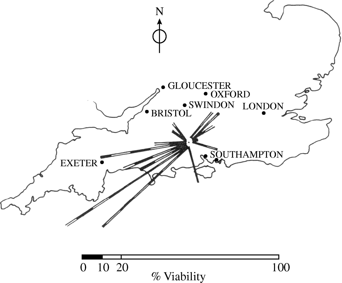
Fig. 1. Viability of F. tularensis in aerosols (<3 μm particles) aged 45 min in relation to air mass history; % viability plotted along wind direction towards Porton.
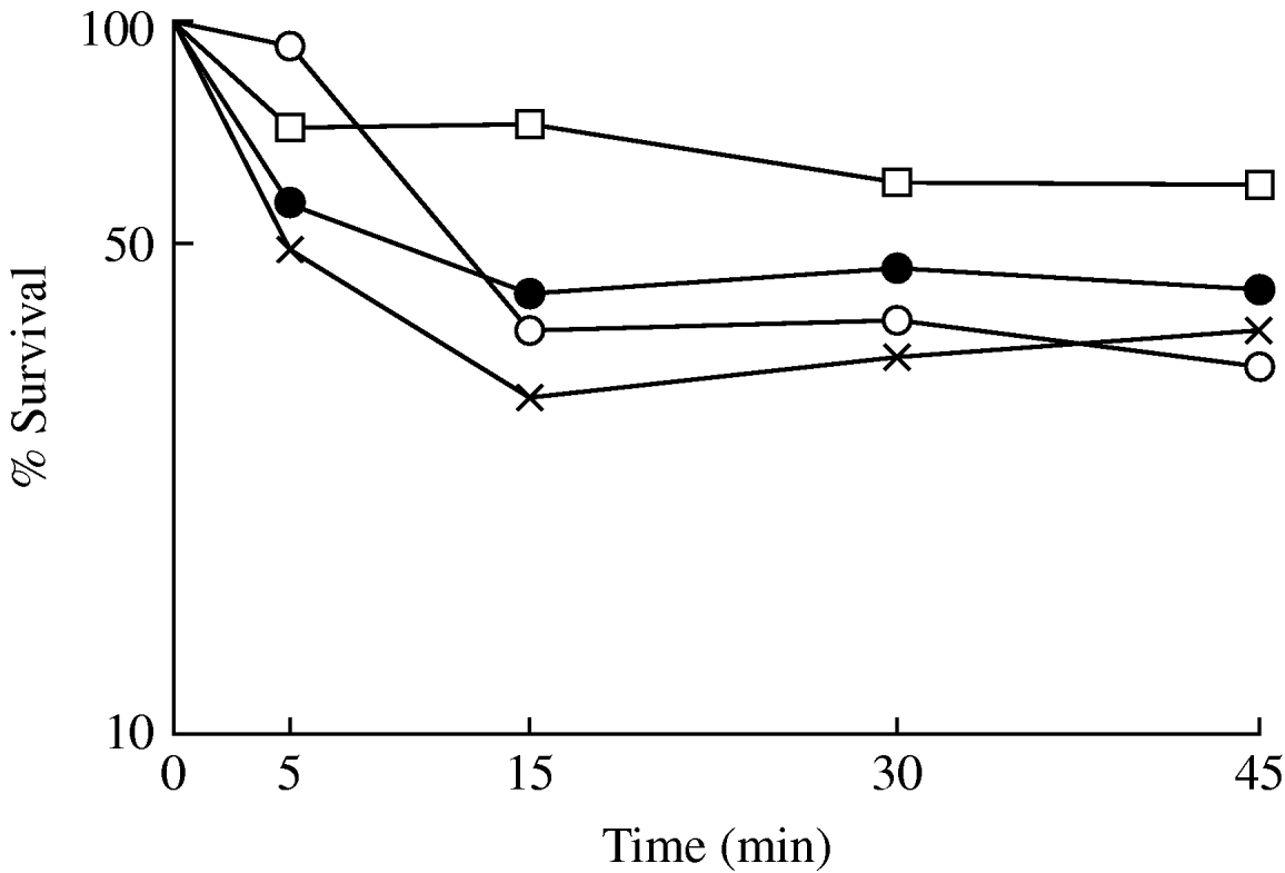
Fig. 2. Survival of F. tularensis in aerosols in open air. Effect of particle size: •, >6 μm; ○, 3–6 μm; ×, <3 μm diameter. □, E. coli (<3 μm particles) exposed to open air on microthreads.
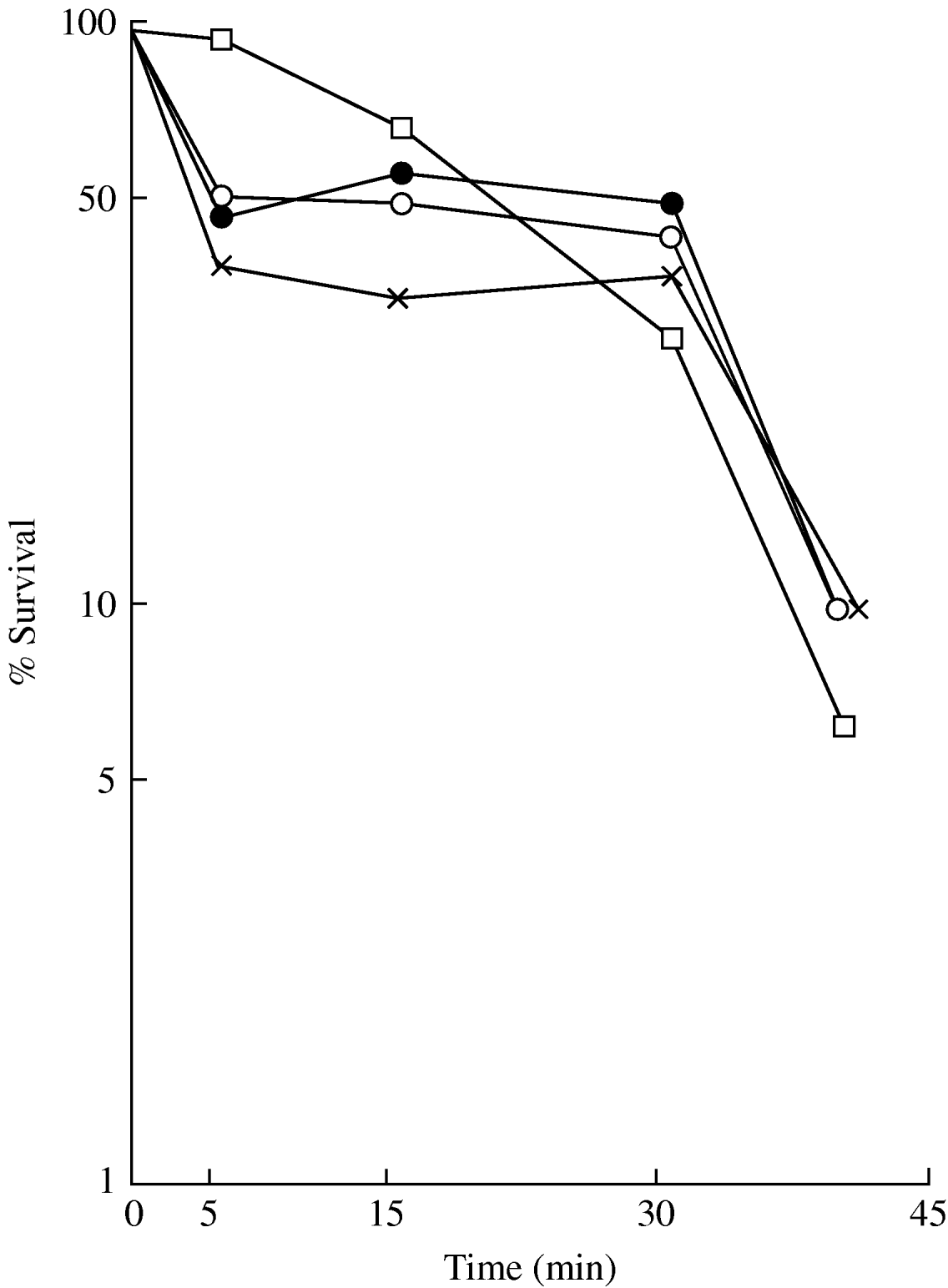
Fig. 3. Survival of F. tularensis in aerosols in open air. Effect of particle size: •, >6 μm; ○, 3–6 μm; ×, <3 μm diameter. □, E. coli (<3 μm particles) exposed to open air on microthreads.
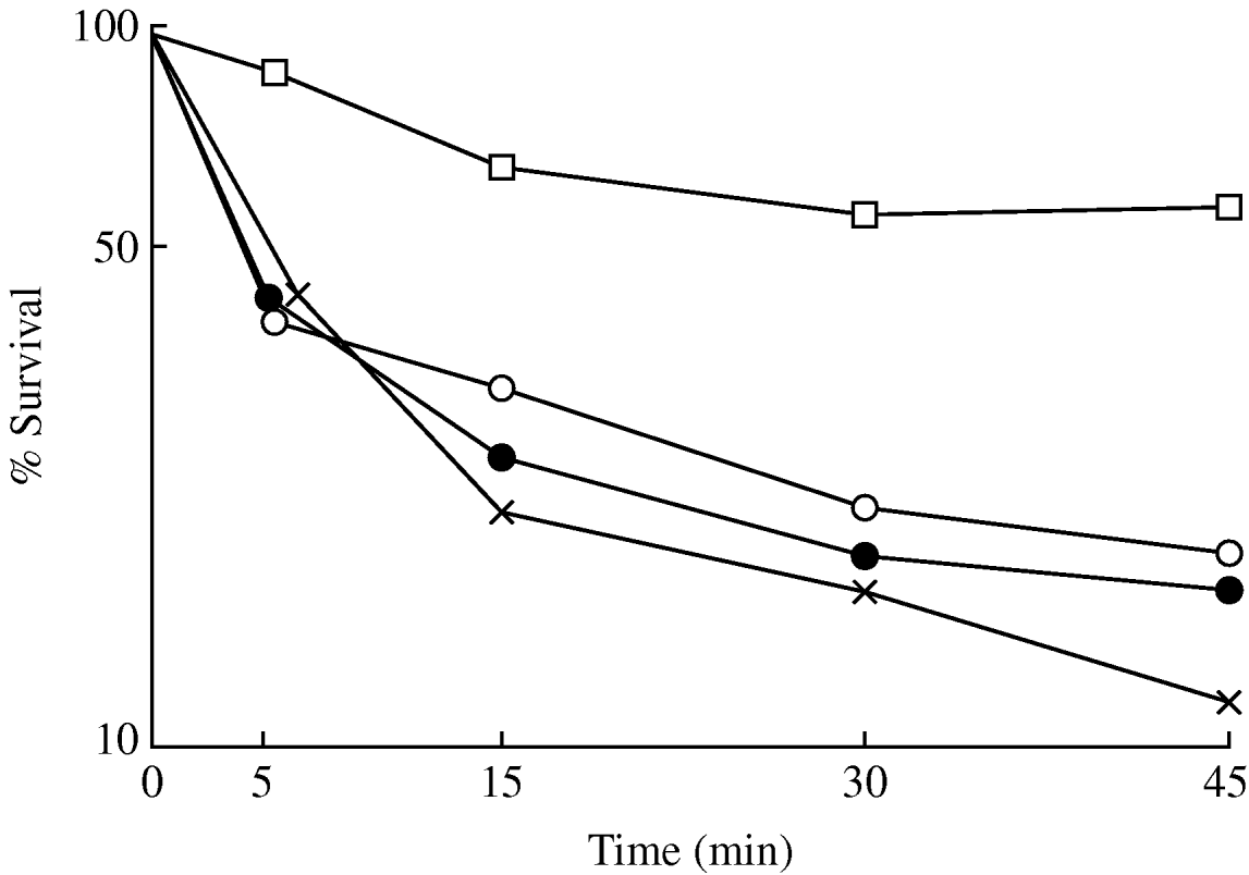
Fig. 4. Survival of F. tularensis in aerosols in open air. Effect of particle size: •, >6 μm; ○, 3–6 μm; ×, <3 μm diameter. □, E. coli (<3 μm particles) exposed to open air on microthreads.
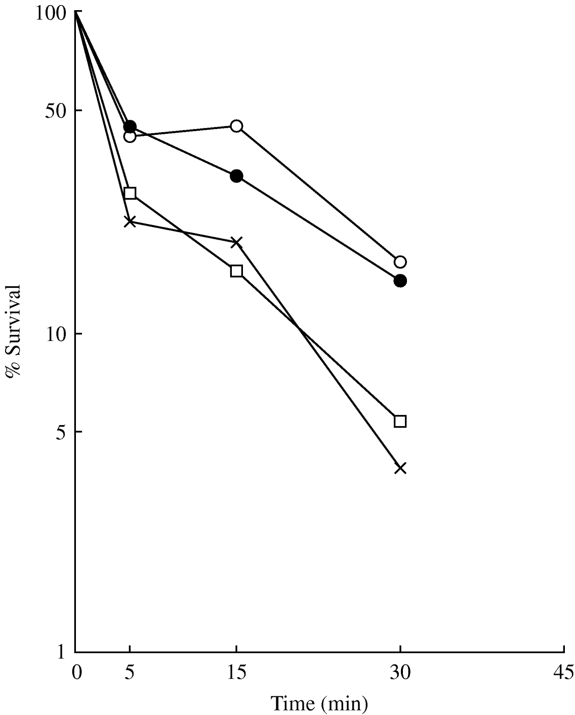
Fig. 5. Survival of F. tularensis in aerosols in open air. Effect of particle size: •, >6 μm; ○, 3–6 μm; ×, <3 μm diameter. □, E. coli (<3 μm particles) exposed to open air on microthreads.

Fig. 6. (a) Survival of F. tularensis (○) in <3 μm particles in aerosols in open air with trajectory as shown in panel (b). ×, E. coli (<3 μm particles) exposed to open air on microthreads. (b) Air trajectory of experiment shown in panel (a). •, 3-h time intervals.

Fig. 7. (a) Survival of F. tularensis (○) in <3 μm particles in aerosols in open air with trajectory as shown in panel (b). ×, E. coli (<3 μm particles) exposed to open air on microthreads. (b) Air trajectory of experiment shown in panel (a). •, 3-h time intervals.

Fig. 8. (a) Survival of F. tularensis (○) in <3 μm particles in aerosols in open air with trajectory as shown in panel (b). ×, E. coli (<3 μm particles) exposed to open air on microthreads. (b) Air trajectory of experiment shown in panel (a). •, 3-h time intervals.

Fig. 9. (a) Survival of F. tularensis (○) in <3 μm particles in aerosols in open air with trajectory as shown in panel (b). ×, E. coli (<3 μm particles) exposed to open air on microthreads. (b) Air trajectory of experiment shown in panel (a). •, 3-h time intervals.
Viability and virulence
For this part of the study aerosols of F. tularensis consisting preminantly of particles of 1–2 μm diameter were generated and held in the ventilated sphere. After a given time period chosen with reference to predicted levels of cell viability guinea pigs were exposed to determine the LD50 of the surviving bacteria. During the animal exposure periods the sphere was sealed so that the aerosols were held in enclosed, OAF-free, air. Thus further loss of viability proceeded at a very slow rate [Reference Hood2]. The physical loss of these very small particles could be ignored. There was sufficient time during these sphere-sealed periods for several groups of animals to be exposed for different time periods to aerosols in a relatively unchanging state. During exposure of the animals air samples were taken to determine the concentration of viable bacteria/l. In calculating the respiratory dose retained by the guinea pig a breathing rate of 129 ml/min [Reference Guyton29] coupled with a retention factor of 0·55 [Reference Harper and Morton6] was used. Thus the calculated dose was taken as one fourteenth of the measured viable bacteria/l×exposure time in minutes.
Tests were confined to periods during which the ambient humidity was relatively high, >70% relative humidity, thus any loss of viability of F. tularensis that occurred could readily be attributed to the adverse effect of OAFs and not to any adverse humidity effect. Aerosols were held in open air in the ventilated sphere for periods ranging from 20 to 60 min. F. tularensis viabilities ranged from 7% to 25%, depending upon the time for which the aerosols were exposed and the OAF activity of the ambient air. This in turn was related to the source of the air as indicated by its trajectory as found above.
Ten control animal exposures to young aerosols, about 1 s old, in conditions of temperature and humidity embracing the range found in the open-air tests were made in enclosed (OAF-free) air in the Henderson apparatus. It was found that the progress and severity of the tularaemia infection resulting from the aerosol exposure, i.e. onset of illness in 3–5 days and death in 5–7 days, was typical of the disease caused by this strain of F. tularensis. There were no apparent differences between animals infected from open-air or indoor air control aerosols. All animals autopsied showed the typical gross pathological changes associated with tularaemia, enlarged spleens, dark red in colour with various amounts of small white necrotic granules and yellowish white necrotic areas on the liver surface. All spleens gave a pure culture of F. tularensis from direct smear inoculations on agar medium. The respiratory LD50 obtained from ten control and six test aerosols was around 1, no significant difference between LD50 values was found (Table 1).
Table 1. The virulence of Francisella tularensis from aerosols in open and indoor air for guinea pigs (respiratory route, LD50)
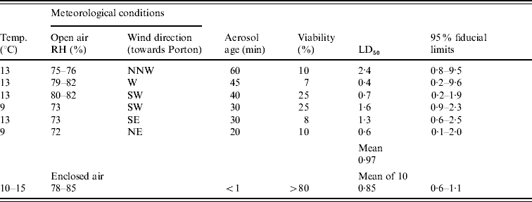
RH, Relative humidity.
DISCUSSION
Now that it has been established, at least with one pathogenic organism that the adverse effect of OAFs on viability correlates with its effect on virulence, further study of the killing mechanism is suggested. Considerable physical damage can be done to this strain of F. tularensis as shown by the effect of chloride ion [Reference Hood11, Reference Hood14] without loss of viability as indicated by its capacity to recover when provided with beneficial conditions such as those presented in vitro (in the GCBA medium) or in vivo (mouse peritoneum). By contrast exposure to OAFs damages the cell beyond repair suggesting chemical destruction of more vital components as occurs with exposure to disinfectants, protein denaturation, enzyme destruction, etc.
The relationship of OAF activity to the history of the air mass found in this area of the UK (Salisbury, Wiltshire) and in The Netherlands and its association with known olefin sources such as oil refineries and dense car populations suggests that it will be a common constituent of much of the open air in Western Europe. Thus the potential of F. tularensis at least as a biowarfare weapon is significantly reduced. General conclusions cannot yet be made of any OAF contribution to hygiene – preventing or at least reducing the incidence of respiratory infections occurring in open air. Studies with the viruses, influenza and Semliki Forest in open air [Reference Benbough and Hood4, Reference Hood, Stagg and Willis30] have shown that these are also sensitive to the adverse effect of OAFs. The indications are that most non-sporing microorganisms would be adversely affected by OAFs. Whatever the destructive mechanism, it should to be appreciated that its action may be sufficiently general for it to have some harmful effect on normal mammalian cells. It is important also to note that OAFs disappear at such an extremely rapid rate from any normally ventilated enclosure [Reference Hood2] that many bacteria will remain viable and infective for at least 24 h depending upon the humidity presented.
OAFs are of such an ephemeral nature that it has not yet been possible to identify it precisely. From previous results in the sphere with various ventilation rates connected with OAFs and its loss from other enclosed vessels it could be calculated that its molecular weight ranges from 50 to 150 [Reference Hood3]. This range would include at least some ozonized olefins. In laboratory tests with 20 different ozonized olefins a wide range of bactericidal activity was found against E. coli suspended on microthreads. This was exhibited in concentrations ranging from 10 to 100 parts per hundred million. Further experiments using one olefin-cyclohexene mixed with ozone and E. coli aerosols showed that breaks were induced in the bacterial DNA [Reference De MIK and de Groot31]. If ‘natural’ OAFs have a similar effect it may well be a carcinogen and might contribute to development of cancer, particularly of the lung. Our current inability to determine the precise chemical nature of OAFs leaves us with the conclusion that since ozonized olefins will exist at times in open air in the UK they may well at least be partly responsible for what we describe as OAFs. We are unlikely to be able to differentiate between these factors in the absence of specific concentration measurements.
ACKNOWLEDGEMENTS
Thanks are due to Mrs H. Willis and A. J. Stagg for technical assistance.
DECLARATION OF INTEREST
None.


