


Stent implantation has been applied in multiple CHDs to allow adequate blood flow towards the distal vessel. Balloon-expandable and self-expanding stents are used. Whilst the latter keep their shape even after external compression, the former can be crushed by external forces. This has been described for large stents used in coarctation of the aorta, the right ventricular outflow tract, and in Mustard tunnels, where the external forces were applied during resuscitation. Reference Haas, Happel and Jategaonkar1 This has also been reported in a patient who received chest compressions after TAVI implantation, in whom the stented valve became stenotic and regurgitant leading to the patient’s death. Reference Kim, Choi, Song and Park2 Deformations of coronary stents due to mechanical chest compressions have also been described, leading to ischaemia of the stented vessel with consecutive death. Reference Vogtmann, Volmar, Kronsbein and Bonzel3,Reference Windecker, Maier, Eberli, Meier and Hess4
We describe a baby with a stented Blalock–Thomas–Taussig shunt which was compressed during mechanical resuscitation, which therefore was not successful.
Case report
Male baby weighing 3.3 kg, antenatal diagnosis of hypoplastic left heart with mitral and aortic atresia, hypoplastic aorta ascendens, and a restrictive foramen ovale. A balloon atrioseptostomy was carried out on day 1 of life, followed by bilateral pulmonary artery banding on day 3, to reduce pulmonary artery flow and thus enhance systemic perfusion.
A Norwood operation was carried out 10 days later at a weight of 3.5 kg, consisting of the aortopulmonary anastomosis, arch augmentation with homograft patch, reconstruction of the pulmonary artery with autologous pericard, and a 4 mm modified Blalock–Thomas–Taussig shunt. On the second post-operative day, more inotropic support was necessary, but oxygen saturations decreased. Echocardiography was suggestive of shunt obstruction, hence the baby was taken to the catheter laboratory, where angiography showed a stenosis of the distal shunt. A balloon-expandable coronary Onyx stent 4.5×12 mm (Medtronic, Minneapolis, United States of America) was placed in the shunt, resolving the stenosis. (Fig 1 a and b) Two days later, the sternum was closed uneventfully, and inotropic support could be weaned. Anticoagulation was continued with heparin with an anti-Xa level of >0.5 U/ml and APTT of >150 seconds. He was extubated 3 days after closure of the sternum. One day later, he acutely deteriorated with low oxygen saturations and extremely low expiratory CO2. He was intubated, received adrenaline boluses, and mechanical resuscitation was commenced. This was unsuccessful. Extracorporeal membrane oxygenation cannulas could not be successfully inserted, and the baby deceased an hour after deterioration.

Figure 1. Angiography, lateral projection. (a) Stenosis at the distal end of the modified Blalock–Thomas–Taussig shunt (arrow). (b) Stented shunt.
A post-mortem was carried out, which confirmed the diagnosis and showed a crushed stent within the modified Blalock–Thomas–Taussig shunt, due to which the shunt was flat from the outside (Fig 2). There were no thrombi seen.
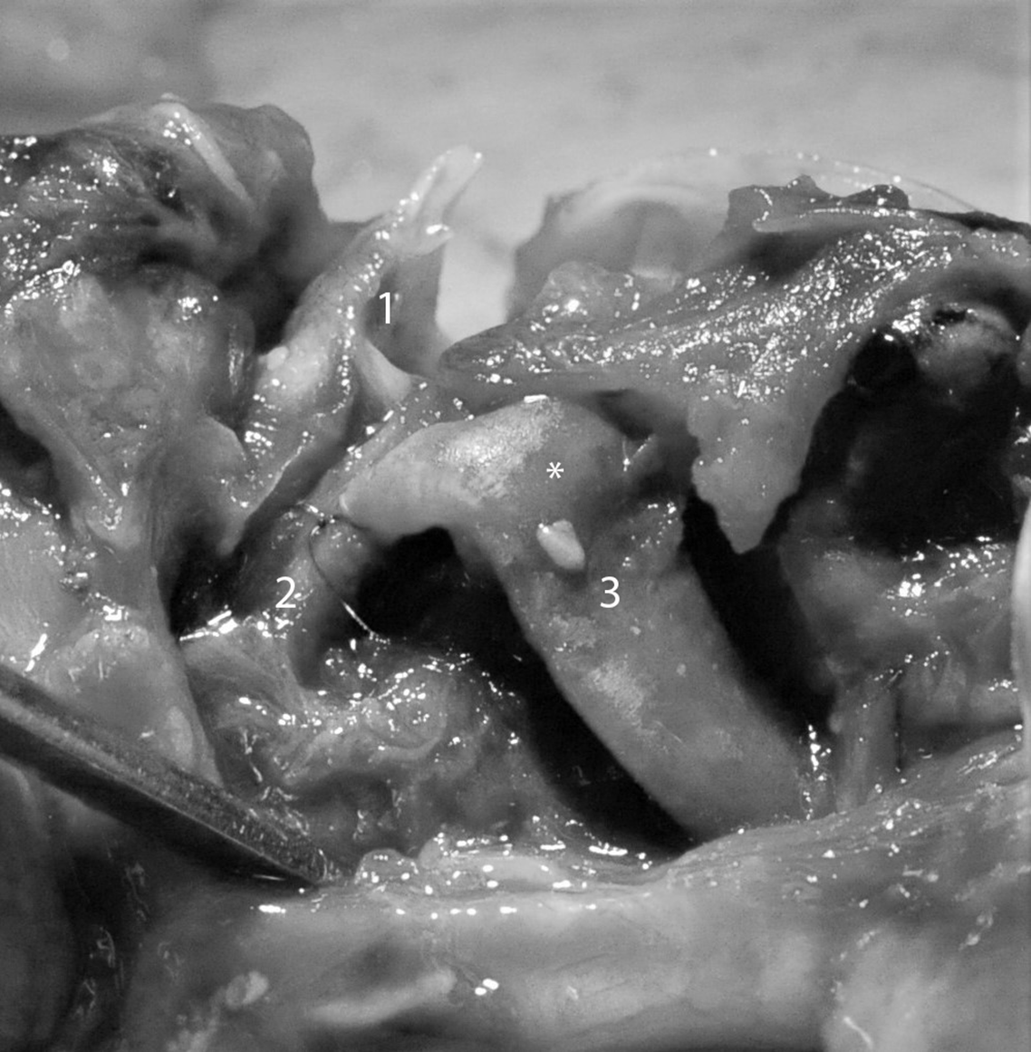
Figure 2. Post-mortem image of a compressed stented Blalock–Thomas–Taussig shunt. Note the flat shape of the shunt over the whole length, best noted at the bend marked with an asterisk (*).
1 = aortic arch, longitudinally opened; 2 = truncus brachiocephalicus, longitudinally opened; 3 = Blalock–Thomas–Taussig shunt.
Discussion
Stents are frequently implanted in patients with CHD. Most of these are balloon-expandable stents, as they have good radial forces. Such stents can be redilated with somatic growth. Reference Duke, Rosenthal and Qureshi5 Other than with self-expanding stents, external compression may lead to the loss of shape of balloon-expandable stents, as seen in our case. Mechanical cardiac resuscitation applies considerable forces to the chest, and to the cardiac structures. Whilst this can (and should) lead to ventricular compression with opening of the semilunar valves, thus contributing to cardiac output, the forces applied to stents are less favourable. As described in adults, Reference Vogtmann, Volmar, Kronsbein and Bonzel3,Reference Windecker, Maier, Eberli, Meier and Hess4 coronary stents can easily be crushed, and even strong large stents can be significantly deformed. Reference Haas, Happel and Jategaonkar1 Fluoroscopical evaluation after thorax compressions has been recommended for patients with previous percutaneous pulmonary valve implantation. Reference Lehner, Kantzis and Haas6 Other than in these large stents, compression of a coronary stent can lead to complete obstruction. Reference Vogtmann, Volmar, Kronsbein and Bonzel3,Reference Windecker, Maier, Eberli, Meier and Hess4 Even if a small amount of blood would have gone through the crushed stent, this would not have been sufficient to fully support the pulmonary circulation in our patient. Unfortunately, we cannot provide cross-sectional images of the crushed stent to prove this. Given the size of the shunt, adequate blood flow towards the pulmonary artery seems highly unlikely.
The deterioration prior to resuscitation may have been caused by thrombosis of the shunt, aspiration, or acute ventricular failure, i.e., due to acute coronary problems. Neither of these could be proven on autopsy, but a thrombus within the shunt may have moved during mechanical resuscitation. As the baby did not recover after the event, it is likely that the crushed shunt contributed to the fatal outcome.
Images of the deformed stents are either fluoroscopic images , or photos of the post-mortem explanted coronary stents. Reference Vogtmann, Volmar, Kronsbein and Bonzel3,Reference Windecker, Maier, Eberli, Meier and Hess4 We present an image of the stent in situ, obtained during the post-mortem, which impressively illustrates that such deformation of a stent leads to disintegration of the Blalock–Thomas–Taussig shunt. Such an image has not been published previously to our recent knowledge.
In patients with balloon-expandable stents in situ, we now add a warning to the patient file that mechanical resuscitation can be dangerous.
Conclusion
Knowledge about stent behaviour during thorax compression is essential, as mechanical forces applied can obstruct the stented vessel considerably.
Funding
No funding was received for this report.
Conflicts of interest
None.


 Open access