


Combined tricuspid atresia and absent pulmonary valve, together with an intact ventricular septum and right ventricular dysplasia, is extremely rare. Surgical management, especially intervention for the non-functional right ventricle and pulmonary artery, has been discussed. In previous reports, right ventriclar exclusion and pulmonary artery division or banding were performed concomitantly with the Fontan operation Reference Szwast, Tian and McCann1,Reference Takahara, Manabe, Mori, Kuroda and Kitagawa2 . Conversely, we avoid intervention because this could cause right ventriclar hypertension and dilation, resulting in left ventricular outflow tract obstruction. We present three cases underwent the Fontan operation with good outcome.
Case report
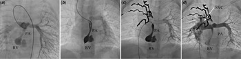
We present three cases of combined tricuspid atresia and absent pulmonary valve detected on fetal echocardiography. All cases underwent right-modified Blalock–Taussig shunt, bidirectional Glenn procedure, and Fontan operation. The right ventricular end-diastolic volume index was measured through cardiac catheterisation. Table 1 summarises the details of the cases. In Case 1, a right ventricular aneurysm was diagnosed during the first echocardiography. The series of right ventriculograms in Case 2 are shown in Figure 1. In all cases, no coronary-to-right ventriclar fistulas were detected. Regarding the Fontan operation, an 18-mm polytetrafluoroethylene graft was used. Aspirin was administered as an anticoagulant. The follow-up periods for the three cases were 10, 8, and 3 years after the Fontan operation. In Case 1 and 2, the latest cardiac catheterizations showed no right ventricular dilation (Table 1) . Until now, no left ventricular outflow tract obstruction was noted nor were thromboembolic events.

Figure 1. Right ventriculogram of Case 2 during cardiac catheterisation before bidirectional Glenn procedure (a), before Fontan operation (b), and 1 year after the Fontan operation (c). Superior vena cava angiogram after the Fontan operation (d). PA, pulmonary artery; RV, right ventricle; SVC, superior vena cava.
Table 1. Details of the cases

PA = pulmonary artery; RVEDVI = right ventricle end-diastolic volume index.
Discussion
Cases of tricuspid atresia and absent pulmonary valve with an intact ventricular septum and right ventricular dysplasia are extremely rare. Reference Szwast, Tian and McCann1 Only a few cases undergo Fontan operation because of the rarity of the disease and the poor prognosis. Reference Szwast, Tian and McCann1
Characteristically, the right ventriclar myocardium, similar to that in Uhl’s disease, gradually increases in size gradually and affects the left ventricle. Reference Takahara, Manabe, Mori, Kuroda and Kitagawa2 Another morphological feature is asymmetrical ventricular septal hypertrophy, which could also lead to left ventricular outflow tract obstruction. Furthermore, pulsatile flow from the right ventricle to the pulmonary artery causes unnatural backward flow in systemic veins, which is a limitation for Fontan circulation. Reference Klimes, Abdul-Khaliq and Ovroutski3 Thus, surgical interventions for right ventricle and pulmonary artery remain controversial.
In our practice, they are not manipulated because right ventriclar dilation may cause right ventricular hypertension. The inflow to the right ventricle, such as from the Thebesian veins, could contribute to the increased pressure. The presence of significant coronary-to-right ventriclar fistulas would have further increased the inflow. We believed that right ventricular hypertension would have exacerbated right ventricular hypertrophy, and pressure needs to be reduced by opening the tricuspid valve or retaining pulmonary artery forward flow. This is a completely different approach to those mentioned in previous reports. To date, the volume of the right ventricle has not increased to such a point so as to cause left ventricular outflow tract obstruction. Regarding the flow in the systemic veins after the Fontan operation, cardiac catheterisation detected no significant pulse flow in the superior or inferior vena cava pressure line. This indicates that the pulmonary artery forward flow does not affect Fontan circulation.
It is difficult to apply this strategy to all cases. From our experience, we believe that this approach is suitable for cases in which right ventricular end-diastolic volume index is less than 30 mL/m2. We will continue to monitor for right ventriclar dilation, left ventriclar function, and the overall condition of these patients.
Acknowledgements
The authors thank Editage (www.editage.jp) for English language editing.
Financial support
This research received no specific grant from any funding agency, commercial or not-for-profit sectors.
Conflicts of interest
None.
Ethical standards
This article does not contain any studies with human participants.
Informed consent
All participants provided informed consent.


 Open access