29 results
Prediction of psychosis in prodrome: development and validation of a simple, personalized risk calculator
-
- Journal:
- Psychological Medicine / Volume 49 / Issue 12 / September 2019
- Published online by Cambridge University Press:
- 14 September 2018, pp. 1990-1998
-
- Article
- Export citation
Multimodal Imaging in Psychiatry: The Electroencephalogram as a Complement to Other Modalities
-
- Journal:
- CNS Spectrums / Volume 4 / Issue 8 / August 1999
- Published online by Cambridge University Press:
- 07 November 2014, pp. 44-57
-
- Article
- Export citation
Organization
-
- Book:
- Rapid Eye Movement Sleep
- Published online:
- 07 September 2011
- Print publication:
- 14 July 2011, pp xviii-xviii
-
- Chapter
- Export citation
Acknowledgments
-
-
- Book:
- Rapid Eye Movement Sleep
- Published online:
- 07 September 2011
- Print publication:
- 14 July 2011, pp xvii-xvii
-
- Chapter
- Export citation
Index
-
- Book:
- Rapid Eye Movement Sleep
- Published online:
- 07 September 2011
- Print publication:
- 14 July 2011, pp 460-478
-
- Chapter
- Export citation
Section I - Historical context
-
- Book:
- Rapid Eye Movement Sleep
- Published online:
- 07 September 2011
- Print publication:
- 14 July 2011, pp -
-
- Chapter
- Export citation
Preface
-
- Book:
- Rapid Eye Movement Sleep
- Published online:
- 07 September 2011
- Print publication:
- 14 July 2011, pp xv-xvi
-
- Chapter
- Export citation

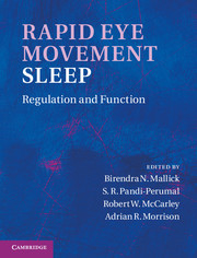
Rapid Eye Movement Sleep
- Regulation and Function
-
- Published online:
- 07 September 2011
- Print publication:
- 14 July 2011
Section V - Functional significance
-
- Book:
- Rapid Eye Movement Sleep
- Published online:
- 07 September 2011
- Print publication:
- 14 July 2011, pp -
-
- Chapter
- Export citation
Section VI - Disturbance in the REM sleep-generating mechanism
-
- Book:
- Rapid Eye Movement Sleep
- Published online:
- 07 September 2011
- Print publication:
- 14 July 2011, pp -
-
- Chapter
- Export citation
Contents
-
- Book:
- Rapid Eye Movement Sleep
- Published online:
- 07 September 2011
- Print publication:
- 14 July 2011, pp vii-ix
-
- Chapter
- Export citation
Section IV - Neuroanatomy and neurochemistry
-
- Book:
- Rapid Eye Movement Sleep
- Published online:
- 07 September 2011
- Print publication:
- 14 July 2011, pp -
-
- Chapter
- Export citation
Section III - Neuronal regulation
-
- Book:
- Rapid Eye Movement Sleep
- Published online:
- 07 September 2011
- Print publication:
- 14 July 2011, pp -
-
- Chapter
- Export citation
Contributors
-
- Book:
- Rapid Eye Movement Sleep
- Published online:
- 07 September 2011
- Print publication:
- 14 July 2011, pp x-xiii
-
- Chapter
- Export citation
Plate section
-
- Book:
- Rapid Eye Movement Sleep
- Published online:
- 07 September 2011
- Print publication:
- 14 July 2011, pp -
-
- Chapter
- Export citation
Section II - General biology
-
- Book:
- Rapid Eye Movement Sleep
- Published online:
- 07 September 2011
- Print publication:
- 14 July 2011, pp -
-
- Chapter
- Export citation
29 - Neuronal models of REM-sleep control: evolving concepts
- from Section IV - Neuroanatomy and neurochemistry
-
-
- Book:
- Rapid Eye Movement Sleep
- Published online:
- 07 September 2011
- Print publication:
- 14 July 2011, pp 285-300
-
- Chapter
- Export citation
Frontmatter
-
- Book:
- Rapid Eye Movement Sleep
- Published online:
- 07 September 2011
- Print publication:
- 14 July 2011, pp i-vi
-
- Chapter
- Export citation
Contributors
-
-
- Book:
- Foundations of Psychiatric Sleep Medicine
- Published online:
- 01 June 2011
- Print publication:
- 23 December 2010, pp vii-x
-
- Chapter
- Export citation
Chapter 2 - Neuroanatomy and neurobiology of sleep and wakefulness
- from Section II - Normal Sleep
-
-
- Book:
- Foundations of Psychiatric Sleep Medicine
- Published online:
- 01 June 2011
- Print publication:
- 23 December 2010, pp 13-35
-
- Chapter
- Export citation


