


Case
The patient is an 8-year-old girl with KBG syndrome (a rare genetic syndrome characterised by characteristic facial features, short stature, developmental delay, and behavioural issues). Her medical history is also notable for benign giant cell granulomas in the bilateral maxillae, initially diagnosed in 2018, who underwent surgical debulking and was subsequently treated with denosumab for 2 years. After initial response to treatment, she developed recurrence in January 2022. She was started on imatinib at a dose of 340 mg/m2 once daily, 9 days prior to hospital presentation.
A few days after starting imatinib, she began complaining of palpitations. She presented to outpatient care, where her electrocardiogram revealed sinus tachycardia. A Holter monitor was placed, and workup for sinus tachycardia was initiated including complete blood count and thyroid panel, which were normal. Her tachycardia persisted, and she subsequently developed tachypnoea and increased work of breathing and inability to lay flat, for which she presented to the Emergency Department.
Upon presentation, she was tachycardic to 155 beats per minute, tachypnoeic to 36 breaths per minute, oxygen saturation 98%, and blood pressure 110/71, without pulsus paradoxus identified. She was sitting in a stretcher with the head of the bed raised, in moderate respiratory distress. Cardiac exam revealed distant heart sounds without murmur, rub, or gallop. There was tachycardia with regular rhythm. She exhibited increased work of breathing with belly breathing and accessory muscle usage. Her extremities were mildly cool but well perfused with 1+ distal pulses. She was alert and interactive, answering questions appropriately, though speaking with shortness of breath.
A chest radiograph was performed which revealed cardiomegaly and small bilateral pleural effusions. Echocardiogram was then performed which revealed a very large circumferential pericardial effusion with echocardiographic findings suggestive of cardiac tamponade, with prominent collapse of the right atrial free wall and collapse of the right ventricular free wall (Fig 1).

Figure 1. Echocardiogram: Subcostal long (left) and apical (right) views revealing very large circumferential pericardial effusion with collapse of the right atrial and right ventricular free wall.
She was brought emergently to the cardiac catheterisation lab for pericardiocentesis, with drainage of 460 millilitres of milky-appearing fluid and placement of a pericardial drain (Fig 2). Analysis of the pericardial fluid was consistent with chyle (250 nucleated cells/μL with 79% lymphocytes, total protein 4.7 g/dL, cholesterol 66 mg/dL, triglycerides 946 mg/dL, lactate dehydrogenase 289 U/L).
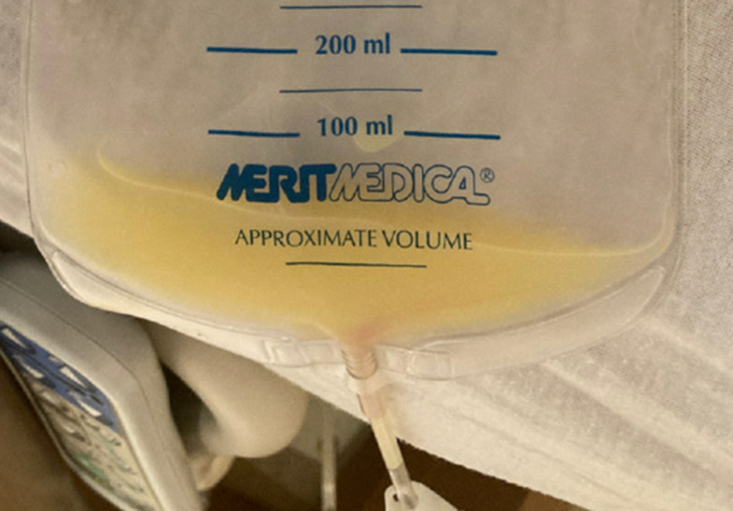
Figure 2. Image of pericardial fluid draining from pericardial drain in the days following pericardiocentesis.
Her imatinib was discontinued. Over the following days, she continued to have chylous output from the pericardial drain and recurrence of a small-to-moderate pericardial effusion on echocardiogram. She returned to the catheterisation lab 1 week later for drain manipulation and replacement, with another 250 ml of chyle removed. The chylous drainage persisted, and she was transferred to another institution for possible lymphatic intervention. Prior to further lymphatic imaging being obtained, the drainage stopped and echocardiogram showed resolution of the effusion. She was ultimately discharged 18 days after her initial presentation. She has since been followed as an outpatient without significant reaccumulation of pericardial fluid.
Discussion
This case details the development of a large chylous pericardial effusion causing cardiac tamponade that is temporally associated with the initiation of imatinib, which resolved in the weeks following discontinuation of imatinib therapy without recurrence.
Imatinib is an oral ABL-class tyrosine kinase inhibitor which is used for a number of oncologic indications, most notably chronic myelogenous leukaemia. Imatinib is known to cause fluid retention and oedema, and can occur acutely soon after initiation of therapy, with rapid improvement following discontinuation of therapy. Reference Kim, Shinagare and Krajewski1 Pleural effusions have been described as rare side effects of treatment with imatinib, though are much more commonly seen with other more potent medications in the same class, such as the second-generation ABL-class tyrosine kinase inhibitor dasatinib. Reference Banka and Udwadia2 Pericardial effusions have also been reported, most commonly in the adult population, but with rare reports in children. Reference Kelly, Swords and Mahalingam3–Reference Terry, Avery and Morton6 Pericardial effusions secondary to tyrosine kinase inhibitor treatment can lead to cardiac tamponade, either from acute onset, large size, or a combination of both. Reference Barton, Jones and Lamberth7–Reference Wattal, Rao and Chandra8 The mechanism of pleural and pericardial effusions secondary to tyrosine kinase therapy is not entirely clear, but appears to be the result of serosal inflammation. Reference Kelly, Swords and Mahalingam3
Chylous effusions have been described much more rarely, with case reports seemingly limited to therapy with dasatinib, which has 325-fold greater activity against BCR-ABL as compared to imatinib. Reference Wattal, Rao and Chandra8 Chylous pericardial effusions are particularly important to manage, as they carry a high risk of the development of scarring and fibrosis, potentially resulting in constrictive pericarditis. Reference Yacoub, Quintanilla Rodriguez and Mahajan9 Treatment consists primarily of drainage of chyle from the pericardial space, often followed by continuous drainage via a pericardial drain until resolution, as well as a low-fat diet to minimise chyle production. Reference Rochefort10 In our case, cessation of imatinib therapy led to fairly prompt and complete resolution of chylous effusion in a period of 2 weeks, without recurrence noted at multiple follow up visits.
Conclusion
Pleural and pericardial effusions are known side effects of therapy with tyrosine kinase inhibitors. Chylous effusions are much less described and are mostly limited to more potent second-generation medications. This case describes a massive chylous pericardial effusion leading to cardiac tamponade requiring emergent pericardiocentesis in a child recently started on imatinib, which resolved promptly following cessation of imatinib therapy. This case adds to the limited body of literature regarding chylous effusions secondary to tyrosine kinase inhibitors and is the first known case of chylous pericardial effusion causing tamponade secondary to imatinib in a child. Providers, including paediatric providers, should be aware of this association and monitor closely with a high vigilance for effusions if symptoms develop.
Acknowledgements
The authors would like to acknowledge those who contributed to the patient's care, including the PICU, cardiac cath, and sonography teams.
Financial support
This research received no specific grant from any funding agency, commercial, or not-for-profit sectors.
Conflicts of interest
None.


 Open access