Introduction
Recent progress in the development of InGaN-based lasing structures was highlighted by Nichia’s announcement of “engineering sample shipments” of their blue laser diode (LD) [1]. However, due to the lack of ideal substrates for the growth of thin film nitrides, a large number of dislocations and cracks are naturally formed in the epitaxial layer to alleviate the lattice mismatch and the strain of postgrowth cooling. This does not always negatively affect lasing characteristics but sometimes introduces interesting lasing properties due to self-formed high-finesse microcavities [Reference Bidnyk2] that could be utilized for the development of near- and deep-UV LDs.
In this work we describe the results of optical pumping experiments on GaN/AlGaN separate confinement heterostructures in the presence of a large number of naturally formed microcavities. We achieved ultra-violet lasing in these structures over a wide temperature range. The spacing, directionality, and far-field patterns of the lasing modes were correlated to the geometry of microcavities. The temperature sensitivity of the lasing wavelength was measured and compared to that of bulk-like GaN films.
Experimental Details
The GaN/AlGaN separate confinement heterostructure (SCH) samples used in this work were grown by metalorganic chemical vapor deposition on 6H-SiC (0001) substrates with ∼3-μm-thick GaN epilayers deposited prior to the growth of the SCH region. The SCH sample under discussion has a 150-Å-thick GaN active layer, surrounded by 1000-Å-thick Al0.05Ga0.95N cladding layers and 2500-Å-thick Al0.10Ga0.90N waveguide layers symmetrically located on each side. For the purpose of comparison, we also studied a 4.2-μm-thick GaN epilayer grown on (0001) 6H-SiC. The samples were mounted on a copper heat sink attached to a wide temperature range cryostat. Conventional photoluminescence (PL) spectra were measured in the back-scattering geometry using a frequency-doubled Ar+ laser (244 nm) as the excitation source. In order to study the lasing phenomena, a tunable dye laser pumped by a frequency-doubled, injection-seeded Nd:YAG laser was used as the primary optical pumping source. The excitation beam was focused to a line on the sample using a cylindrical lens. The emission from the edge of the sample was coupled into a 1-m spectrometer with a side-mounted optical multi-channel analyzer and photomultiplier tube. Special precautions were taken to avoid fluctuations in sample position due to the thermal expansion of the mounting system. This allowed us to spatially “pin” the sample and obtain lasing modes from a single microcavity over the entire temperature range studied.
Discussion
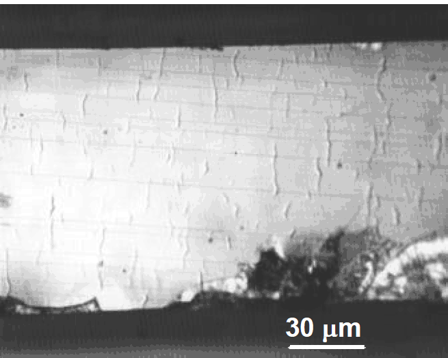
In order to evaluate the effects of cracks on the optical properties, we cleaved our samples into submillimeter-wide bars (note that when GaN is grown on SiC, it can be easily cleaved along the (11

Figure 1. A picture of the sample surface of a GaN/AlGaN separate confinement heterostructure after cleaving. Before cleaving, the sample exhibited no noticeable defects on the surface. After the cleaving process, however, cracks can be seen running parallel to the length of the bar.
When the sample was excited above the lasing threshold, high-finesse cavity modes were observed. Typical emission spectra at pump densities above and below the lasing threshold are depicted in Figure 2. A series of equally spaced and strongly polarized (TE:TM ≥ 300: 1) lasing modes with full width at half maximum of ∼3 Å appears on the low energy side of the GaN-active-region peak. We note, however, that when the spontaneous emission was collected from the sample edge instead of the surface, the lasing modes appeared on the higher energy side of the spontaneous emission peak [Reference Schmidt, Yang, Shan, Song, Salvador, Kim, Aktas, Botchkarev and Morkoç4]. This phenomenon is due to strong re-absorption that introduces a shift of several nanometers to the spontaneous emission peak. Lasing in our samples occurs in the wavelength range of 360 to 364 nm at room temperature, which is much deeper in the UV than for InGaN/GaN-based structures. We were able to correlate the spacing between the modes in Figure 2 to the distance between the cracks depicted in Figure 1. Assuming that the unity round-trip condition is satisfied and there is no loss due to absorption in the GaN layer, the threshold gain can be estimated from [Reference Bagnall and O’Donnell5]:


Figure 2. Lasing and spontaneous emission in the GaN/AlGaN separate confinement heterostructure at room temperature. Lasing is believed to be of microcavity origin.
where R is the mirror reflectivity (20% for a GaN-air interface) and L is the distance between the cracks. Alternatively, L could be extracted through an analysis of the mode spacing Δν through the relation Δν = c/(2nL). We obtained a gain value of ∼500 cm−1 (corresponding to a threshold pump density of 105 kW/cm2), which is on the same order of magnitude as the near-threshold gain values measured in GaN by the variable stripe technique (see Refs. [Reference Wiesmann, Brener, Pfeiffer, Khan and Sun6,Reference Dingle, Shaklee, Leheny and Zetterstrom7] for example). We note, however, that due to the limited quality of GaN and AlGaN layers, the photon flux inside a microcavity encounters additional losses associated with absorption and scattering of light. In view of this, the value for gain calculated through Eq. 1 represents the lower limit for the actual gain at threshold.
It was found that by pumping different areas along the length of the bar, the emission exhibited varying degrees of cavity finesse. The finesse is directly linked to the end mirror reflectivities and internal losses. Thus the different degree of finesse is presumably due to the presence of cavities of varying quality formed by parallel cracks running through the active layer. Areas exhibiting high finesse consistently had a narrower far-field pattern than those exhibiting low finesse possibly indicating larger scattering loss.
We further note that the high finesse emission exits the sample at an angle of φ ∼ 18 °rather than parallel to the sample surface. In order to understand this phenomenon, the samples were examined in cross section using a scanning electron microscope. A large number of cracks were noticed lying at small angles (α ∼ 7 °) to the c axis. Taking into account the refractive index of GaN, we were able to correlate the geometry of the cracks to the emission angle through Snell’s law:
A dramatic decrease in the lasing threshold of our SCH structure in comparison to a bulk-like GaN epilayer was observed over the entire temperature range studied. For one of the SCH samples, the lasing threshold was measured to be as low as 65 kW/cm2 at room temperature. We made a direct comparison of threshold carrier densities in GaN/AlGaN SCHs and bulk-like GaN epilayers and concluded that lasing in SCHs occurs at carrier densities < 5×1017 cm−3, which is considerably lower than that required for the formation of an electron-hole plasma. By further examining the relative energy position between the spontaneous emission and lasing modes we concluded that the exciton-exciton scattering mechanism remains dominant in SCH lasing structures even at room temperature. The details of carrier density calculations and photon energy analysis are given elsewhere [Reference Bidnyk, Lam, Little, Kwon, Song, Bulman, Kong and Schmidt8].
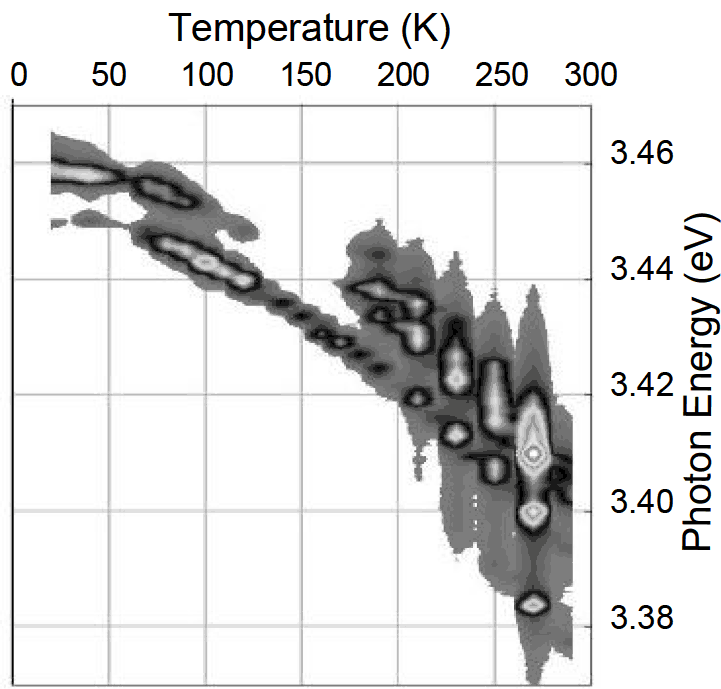
The temperature dependence of the energy position of the lasing modes was also studied. To avoid spatial displacement of the sample due to a non-zero temperature expansion coefficient of the cryostat system, we chose to mount the cryostat horizontally so that the rod expansions occurred in the direction parallel to the direction of the laser beam. This allowed us to obtain lasing from the same microcavity over the entire temperature range. We also used the optical multi-channel analyzer to consistently acquire spectra at 1.3 × Ith for each temperature. Figure 3 shows a plot of mode energy position versus temperature. The gray pallet corresponds to different emission intensities. We note that the energy position of the gain maximum in the GaN/AlGaN SCH changes by about 50 meV when the temperature is varied from 10 to 300 K, which is twice smaller than that expected from a bulk-like GaN epilayer [Reference Bidnyk, Schmidt, Little and Song9]. Such a reduced temperature sensitivity of the laser emission suggests that the GaN/AlGaN SCH could be well suited for UV applications that require increased temperature stability of the emission wavelength.

Figure 3. Lasing modes as a function of temperature for a single microcavity. This image was obtained by consistently taking lasing spectra with an optical multi-channel analyzer at 1.3 × Ith(T) while the temperature was gradually varied from 20 K to 300 K.
Conclusions
We achieved ultra-violet lasing in optically pumped GaN/AlGaN separate confinement heterostructures over a wide temperature range. The spacing, directionality, and far-field patterns of the lasing modes were shown to be the result of microcavities that were naturally formed in the lasing structures due to strain relaxation. The temperature sensitivity of the lasing wavelength was found to be twice lower than that of bulk-like GaN films, indicating that the separate confinement heterostructures could be used in applications that require increased temperature stability of the emission wavelength.
Acknowledgements
The authors would like to thank ONR, DARPA, AFOSR, and ARO for financial support of this research project.