


Pediatric arteriovenous malformations (AVM) typically present with a higher incidence of intracerebral hemorrhage (ICH)/intraventricular hemorrhage (IVH) than their adult counterparts, and carry higher mortality rates with hemorrhage.Reference Niazi, Klimo, Anderson and Raffel 1 - Reference Foy, Wetjen and Pollock 3 Debate exists around the most appropriate treatment option for AVMs in both the pediatric and adult populations. Available treatment options include: microsurgical resection, endovascular embolization, stereotactic radiosurgery (SRS), and combinations of these approaches.Reference Niazi, Klimo, Anderson and Raffel 1 - Reference Rubin, Santillan, Greenfield, Souweidane and Riina 4 Microsurgical resection, considered the gold standard, is typically reserved for small superficially located lesions without deep venous drainage, with obliteration and morbidity/mortality rates in children reported around 90% and 3 to 5%, respectively.Reference Rubin, Santillan, Greenfield, Souweidane and Riina 4 The use of endovascular embolization of AVMs in pediatric patients has been associated with poor obliteration rates; typically below 20%, with mobidity/mortality rates upwards of 11.8%.Reference Niazi, Klimo, Anderson and Raffel 1 , Reference Frizzel and Fisher 5 With the advent of SRS, the treatment of all cerebral AVMs has changed drastically in the last 20 years, offering a safer option for deep seated and eloquently located lesions.
Small numbers of retrospective case series have appeared in the literature over the last ten years reporting AVM response to Gamma Knife (GK) in the pediatric population.Reference Dinca, De Lacy and Yianni 6 - Reference Pollock, Kondziolka and Flickinger 12 Obliteration rates for pediatric AVMs with GK SRS range from 65% Reference Pan, Kuo and Guo 7 to 82.7%Reference Dinca, De Lacy and Yianni 6 at three years follow-up. Reported complication rates range from 1.5%Reference Borcek, Emmez and Akkan 9 to 15.5%.Reference Borcek, Emmez and Akkan 9 The majority of these complications are reported as post-GK hemorrhage, that are well within the expected natural history of these lesions.
Within this study we retrospectively reviewed our institutional series of pediatric AVMs treated with GK in order to determine our obliteration and complication rates.
Methods
Study Design
Our study population of interest was all pediatric patients treated with GK for AVMs at our institution between November 2003 (initiation of GK in Winnipeg) up to and including September 2014. We performed a retrospective review of our institutional GK database in order to identify those patients treated for AVMs. We then selected those patients 18 years of age and under treated for AVMs with GK. This age range was deemed to represent the “pediatric” population.
Data Acquisition
Two independent reviewers reviewed the patient records and GK treatment plans for all the patients meeting the inclusion criteria, as defined above. Nineteen patients were identified. The following data was recorded: patient demographics, presenting features, AVM characteristics, Spetzler-Martin grade,Reference Spetzler and Martin 13 Pollock radiosurgery grade,Reference Pollock and Flickinger 14 , Reference Pollock and Flickinger 15 treatment response, timing of response, and complications. All data was stored within a secure electronic database.
Statistical Analysis
Detailed statistical analysis was not performed given the small number of patients identified. Simple descriptive statistics were presented, including mean and median values.
Ethics
Local research health ethics approval was obtained for this study.
Results
Patient Demographics and AVM Characteristics
We identified 19 pediatric patients treated with GK for AVMs between November 2003 and September 2014. The average age at presentation/treatment with GK was 14.2 years (range: 7-18 years; median: 15.0 years), with 10 male (52.6%) and 9 female patients (47.4%). Treatments prior to GK at our institution included: attempted surgical resection followed by embolization in one patient (5.35%), attempted embolization alone in patients (10.5%) and GK at a different center in two patients (10.5%). The average time from attempted embolization to GK was 12.3 months (range: 2 to 24 months). The remaining 14 patients (73.7%) had not received any other treatment for their AVMs prior to GK at our institution. The mean follow up post-GK was 62 months (range: 13 to 118 months; median: 51 months). Follow-up clinic visits were conducted at six to eight weeks post treatment. Imaging was conducted at one, two, and three years post treatment with magnetic resonance imaging (MRI) or computed tomographic angiography (CTA). Initially, when either MRI or CTA demonstrated the absence of an AVM nidus, subsequent digital subtraction angiography (DSA) was conducted in order to determine AVM patency or obliteration. However, more recently we have relied on MRI and/or CTA alone to demonstrate obliteration, in the absence of the use of DSA. Avoiding the use of DSA was based on the literature demonstrating the efficacy of MRI and its ability to predict AVM patency.Reference Pollock, Kondziolka and Flickinger 12 A summary of patient demographics can be seen in Table 1.
Table 1 Demographic Data for Patient Population and Three Year Follow-Up Group

SM=Spetzler-Martin Grade, AVM=Arteriovenous Malformation, Gy=Gray, n=number. Serious complications were defined as complications that had clinical manifestations and excluded minor conditions associated with frame placement, example pin site bleeding, edema, etc.
Four patients (21.1%) were lost to follow-up, with all being referred from out of province sources. All of these patients returned to their respective province of origin post treatment. Attempts were made to contact each patient post-GK for follow up via telephone, but were unsuccessful. The demographic data for those patients lost to follow up can be seen in Table 2.
Table 2 Characteristics of Four Patients Lost to Follow Up

M=Male, ICH=intracerebral hemorrhage, H/A=head ache, Lt=left, SM=Spetzler-Martin, AVM=arteriovenous malformation.
Clinical presentation for the reviewed patients included: ICH or IVH in nine (47.4%), headache in five (26.3%), seizure in two (10.5%), loss of consciousness in two (10.5%), and motor tic in one (5.3%). The locations of the AVMs were as follows: frontal lobe in seven (36.8%), basal ganglia/thalamus in three (15.7%), brainstem in three (15.7%), temporal lobe in two (10.5%), occipital lobe in two (10.5%), parietal lobe in one (5.3%), and cerebellum in one (5.3%). Ten patients (52.6%) had left sided AVMs, eight (42.1%) had right sided AVMs, and one (5.3%) had a midline AVM.
The mean diameter and volumes for the AVMs treated with GK were 2.68 cm (range: 0.85-3.50 cm; median: 2.40 cm) and 3.10 cm3 (range: 0.32-14.14 cm3; median: 0.96 cm3), respectively. The mean Spetzler-MartinReference Spetzler and Martin 13 and PollockReference Pollock and Flickinger 14 , Reference Pollock and Flickinger 15 grades of the AVMs treated were 2.4 (range: 1-4; median: 2.0) and 0.99 (range: 0.28-3.63; median: 0.84), respectively.
Eleven of 15 patients (73.3%) with follow-up data available had a minimum of three years follow-up post GK. We defined three years as a sufficient time period to allow for AVM response post-GK. Within this subpopulation of patients, the mean age was 13.0 years (range: 7-18 years; median: 15.0), with seven females (63.6%). The mean diameter and volume of the AVMs treated were 2.02 cm (range: 1.00-3.50 cm; median: 2.2) and 1.92 cm3 (range: 0.37-5.60 cm3; median: 0.86 cm3), respectively. The locations for these AVMs included: frontal lobe in three (27.3%), brainstem in two (18.2%), thalamic in two (18.2%), temporal lobe in one (9.1%), parietal lobe in one (9.1%), occipital lobe in one (9.1%), and cerebellum in one (9.1%). Finally, the mean Spetzler-Martin and Pollock grades of these AVMs were 2.5 (range: 1-3; median: 3.0) and 1.06 (range: 0.28-3.63; median: 0.90), respectively. Demographic data for the three year follow-up group can be seen in Table 1.
Treatment Characteristics
The mean maximum dose was 39 Gy (range: 32-44 Gy; median: 40 Gy). The mean marginal dose at the 50% isodose line was 19.5 Gy (range: 16-22 Gy; median: 20 Gy). We relied mainly on CTA and/or MRI scanning in treatment planning. All patients, however, did have a DSA as part of their initial diagnostic work up. Treatment planning and dose assignment were conducted by a neurosurgeon, radiation oncologist, and medical physicist.
One patient had two GK treatments for the same AVM at our facility. He was a 16 year male with a SM grade 3 left temporal AVM, presenting with a history of headaches. His initial treatment with GK was in 2004, displaying a good response and decrease AVM size over the course of three years. Given residual AVM on 36 month follow-up DSA, it was elected to repeat GK treatment to the remaining nidus. At 24 months post second GK, his AVM was confirmed to be obliterated by DSA.
AVM Response to GK
All of the patients with follow-up experience a decrease in AVM size post-GK. We defined AVM obliteration as the absence of an AVM on follow-up MRI or DSA. Nine of the 15 patients (60.0%) with follow-up demonstrated obliteration of their AVMs post-GK. Of the remainingsix patients, all have displayed a decrease in AVM size on follow-up MRI post-GK, indicating some response to therapy. Follow-up for them is ongoing, with a current mean follow-up duration of 46.1 months (range: 13-118). The mean time to any radiological response post-GK was 15 months (range: 3-30 months).
Obliteration was confirmed with MRI and DSA in six patients, and MRI alone in three patients. In those patients who displayed AVM obliteration post-GK, the mean time to MRI resolution was 17.0 months (range: 6-27 months; median: 18.0 months). Arteriovenous malformation obliteration as confirmed via DSA occurred at a mean of 24 months (range: 12-30 months; median: 24.0 months) post-GK.
Within the three year follow-up group, 9 of 11 (81.8%) experienced AVM obliteration on MRI or DSA. The remaining two patients have experienced a decrease in nidus size post-GK treatment, with follow-up ongoing. There was a trend between obliteration rates and the Spetzler-Martin and Pollock grades in the three year follow-up group, as seen in Tables 3 and 4.
Table 3 Spetzler-Martin Grades and Obliteration Rates for Three Year Follow-Up Group
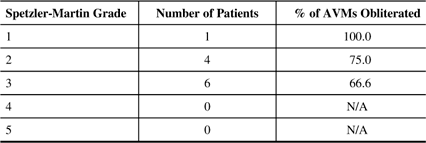
AVM=Arteriovenous Malformation, N/A=Not applicable due to no patients in category.
Table 4 Pollock Grades and Obliteration Rates for Three Year Follow-Up Group
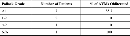
AVM=Arteriovenous Malformation, N/A=no Pollock grade due to insufficient data on the patients AVM volume.
Table 3 displays the Spetzler-Martin grades of 1, 2, and 3 with corresponding obliteration rates of 100%, 75%, and 66% respectively. Similarly, Table 4 displays the number of patients with Pollock radiosurgery grades of less than 1, 1 to 2, and greater than 2 with obliteration rates of 85.7%, 0%, and 0% respectively in the three year follow-up group. One patient had insufficient data to calculate an AVM volume for Pollock grade determination. They did however proceed onto AVM obliteration post-GK.
Of the three patients identified with previous embolization attempts prior to GK, one was lost to follow-up, and the remaining two displayed obliteration of their AVMs post-GK. The marginal dose at the 50% isodose line for these two cases were 18 and 20 Gy respectively.
Complications
Complications were reported in 7 of the 15 patients (46.7%) with follow-up. We defined major complications as those that caused the patient clinical deficit enough to require either emergency room visit or non-scheduled imaging as an outpatient. Only two patients displayed major complications leading to post-GK edema. One developed transient hand weakness, the other only headache. Both resolved with a short course of dexamethasone without permanent complications.
The remaining complications described were minor and included: pin site edema/pain in four patients and transient headache/dizziness in one. These minor complications resolved after 48 hours post-GK. No hemorrhages occurred in any of the patients during the post-GK follow-up period.
Discussion
Radiosurgery for AVMs in the pediatric population is emerging as a first line treatment option for these lesions. Concerns with operative morbidity/mortality for microsurgical resection and endovascular embolization, especially in the developing brain, has fostered the interest in SRS as it is a non-invasive, and highly accurate radiation based treatment of AVMs. To date only a few studies focusing on the use of GK SRS for pediatric cerebral AVMs exist, with obliteration rates reported upwards of 82.7%.Reference Dinca, De Lacy and Yianni 6 Complication rates with GK in pediatric AVMs have been variable in the literature, with the upper limit approaching 15%.Reference Borcek, Emmez and Akkan 9 The interpretation of these complication rates must occur with caution however, given the AVM hemorrhage rates are reported as complications post-GK in the majority of studies. These hemorrhage rates post-GK are within the values for the natural history of AVMs. Recent literature has indicated that the hemorrhage rate for AVMs post-GK is unchanged compared to natural history.Reference Pollock, Flickinger and Lunsford 16
Our institutional experience with AVMs was previously reported in 2011,Reference Zeiler, McDonald, Kaufmann, Fewer, Butler, Schroeder and West 17 with obliteration rates at that time of 87.8% in a population consisting mainly of adult patients. The goal of this current study was to focus our results for GK of pediatric cerebral AVMs from November 2003 up to and including September 2014.
Through our review, we were able to identify 19 pediatric patients treated with GK for AVMs within the defined time period. Four patients were lost to follow-up. The mean age of patients treated was 14.2 years (median 15.0 years), with the mean/median AVM diameter and volumes being 2.68 cm/2.40 cm and 3.10 cm3/0.96 cm3 respectively. The mean/median Spetzler-Martin and Pollock grades of the treated AVMs were 2.4/2.0 and 0.99/0.84 respectively. Of the 15 patients with follow-up (mean follow up of 62 months; median 51 months), 9 (60.0%) were obliterated, with 9 of 11 (81.8%) of those patients with a minimum of three years follow-up displaying obliteration. Major complications occurred in two patients. No AVM hemorrhage occurred during the follow-up period.
We were able to identify some important points from our results. First, our obliteration rate for those patients with three years of follow-up is approaching the upper limit of that reported in the literature to date. This likely stems from appropriate case selection, and maintaining unfractionated treatment plans for AVM diameters and volumes under 3cm and 10 cm3 respectively. This is highlighted by our median AVM volume of 0.96 cm3. Furthermore, our success rate also reflects maintenance of marginal doses near 20 Gy (our mean was 19.5 Gy; median 20 Gy), as lower dosing has been specifically demonstrated in the pediatric population to lead to reduced obliteration rates.Reference Potts, Sheth and Louie 18 Given recent literature demonstrating ongoing obliteration in those patients observed beyond four and five years post-GK,Reference Kano, Kondziolka and Flickinger 19 it could be argued that, with longer follow-up, more of our patients would experience AVM obliteration. Second, our mean follow-up of 62 months for all patients is amongst the longest reported in the pediatric AVM GK series to date. The sustained obliteration in our patients during this long follow-up supports long term sustained obliteration of these malformations with GK. Third, only three patients treated at our institution had prior embolization, which has been linked to statistically significant reduced GK response and worse obliteration rates.Reference Kano, Konziolka and Flickinger 20 - Reference Back, Vollmer and Zeck 22 Thus, our obliteration rate mostly reflects that of lesions presenting without prior treatment. For those two patients with previous embolization whom experience obliteration of their AVMs, there was no appreciable difference in their treatment parameters compared to the non-embolized patients. Fourth, low Spetzler-MartinReference Spetzler and Martin 13 and Pollock gradesReference Pollock and Flickinger 14 , Reference Pollock and Flickinger 15 seemed to improve the chance of obliteration, as seen in our previous study.Reference Zeiler, McDonald, Kaufmann, Fewer, Butler, Schroeder and West 17 Fifth, none of our pediatric patients treated with GK for their AVMs had hemorrhages during their follow-up period. This further emphasizes the literature evidence that GK treatment does not alter the hemorrhage rate during the period post-GK prior to AVM response.Reference Pollock, Flickinger and Lunsford 16 Finally, our major and permanent complication rates were low and within the range reported in the literature. This represents vigilance in maintaining tight conformity and minimizing the total 12 Gy volume, as this has been very well demonstrated to reduce the chance of complications in at risk individuals.Reference Flickinger, Kondziolka and Lunsford 23 Similarly, we avoided dosing above 25 Gy at the 50% isodose line, as this has been shown to increase post-GK complications without providing an increase in in-field obliteration rates.Reference Flickinger, Kondziolka and Maitz 24 , Reference Flickinger, Pollock and Kondziolka 25
Despite these promising results, there are some significant limitations to our review. First, this is a retrospective single-center study with a small number of patients. Thus the results here cannot be generalized to all pediatric AVMs treated with GK and may not reflect those seen in larger series. Second, four patients were lost to follow-up. This represented 21.1% of the population treated. Despite our best efforts, contact with the patients was unsuccessful. It is possible that all of these patients failed treatment and may have had complications we are currently unaware of. Third, we defined obliteration of the AVM based on absence of the lesion on either MRI or DSA; this is not common convention in all radiosurgery centers. Obliteration via MRI was confirmed by both the interpreting neuroradiologist and treating neurosurgeon, looking for the presence of flow voids on T2 imaging and presence of contrast uptake consistent AVM presence on pre-operative studies. Furthermore, we have recently been utilizing perfusion based imaging to aid in determining perfusion asymmetries concerning for an otherwise occult persistent AVM, when such T2 and contrast based changes are absent. If there is any concern of persistent AVM based on MRI at our institution we then discuss with the patient (and family) the risk and benefits of formal angiographic confirmation. Thus, it can be argued, that those patients without DSA confirmation of obliteration may still have a small nidus present if interrogated by formal angiography. Currently, DSA is the gold standard in confirming AVM obliteration post-treatment. Recently, literature suggests MRI is sufficientReference Zadeh, Andrade-Souza and Tsao 11 and current series are starting to report obliteration rates in patients based on MRI only.Reference Kano, Konziolka and Flickinger 8 , Reference Pollock, Flickinger and Lunsford 16 Therefore, despite some controversy, we still believe in MRI confirmation of AVM obliteration post-GK. Fourth, although we have not seen long term significant radiation induced effects, given the young age of this population and potential years of life left to live, it is possible that they may suffer side effects related to GK in the future. Such potential long-term complications include: post-GK edema, radiation necrosis, and impairment of normal neuro-development. Similarly, the potential for radiation induced neoplasms, though rare,Reference Rowe, Grainger, Walton, Silcocks, Radatz and Kemeny 26 has been described during long term follow-up.Reference Shamisa, Bance and Nag 27 The exact incidence of post-radiosurgery neoplasm formation is currently unknown, with recent literature confirming its very rare nature.Reference Rowe, Grainger, Walton, Silcocks, Radatz and Kemeny 26 Finally, only two patients presented with seizures. Thus, formal comments on seizure control post-GK for pediatric AVMs cannot be made at this time.
Conclusions
GK radiosurgery for pediatric AVMs affords a high obliteration rate at three years, with low permanent complication rates during early follow-up.
Disclosures
The authors do not have anything to disclose.


