


Alberta has been recognized as a hotbed for the study of dinosaurs since the late 1800s. Early collectors sent boxcars full of dinosaur specimens back to cities in eastern North America, especially New York, Ottawa, and Toronto, which became featured exhibits in their museums. In 1981, the provincial government decided to establish a museum in which Alberta's fossil resources could be seen in Alberta, and the Royal Tyrrell Museum of Palaeontology was born. The mandate of the Tyrrell Museum was to preserve, protect, and present the fossil record of the province. It was recognized that this couldn't be done without a research component, so as well as being a facility that exhibits spectacular dinosaurs, it is a research institution that is trying to understand what the fossils say about the world of the dinosaurs. It was soon recognized that the fossil record in Alberta is exceptional as a series of fossil-bearing beds that document a succession of five successive vertebrate assemblages from the last 15 million years of the time of the dinosaurs, with each of these assemblages containing abundant fossils of all kinds (Figure 1). With this database to work from, research at the Tyrrell Museum soon began to focus on reconstructing the communities and how the communities changed over time, leading to the end of the Cretaceous. For this, it was necessary to understand all members of the community. While dinosaurs are often preserved as complete skeletons, most other members of the community are not. This is especially true for smaller animals. Lizards, salamanders, frogs, and mammals are just some of the animals present that are represented only by isolated elements, such as jaws, vertebrae, and limb bones. Fortunately, many of the kinds of animals that are preserved have living relatives, so it is possible to identify the fossils by comparing with the skeletons of these relatives. So, for example, we have a good understanding of the kinds of amphibians, lizards, and turtles that were present in Alberta 65–80 million years ago. However, understanding the fossil record of teleost fish, the group of fish that is dominant in today's aquatic communities, has been much more challenging. Until recently, members of this group were represented only by isolated elements. It was clear that many different kinds are present, but how many and how they are related to modern fish is unknown.

Figure 1: Stratigraphic distribution of fossil assemblages in the Late Cretaceous of Alberta, Canada. Localities that yielded material used in this study are indicated with a fish symbol.
Teleost fish from the end of the time of dinosaurs are of particular interest because living groups of teleost fishes are thought to have diversified during this time. In marine environments the diversification of crown teleosts appears to be directly related to the extinction event. The most prominent casualties of marine teleost fishes at the K-Pg (Cretaceous – Paleogene) boundary are ecological analogs of modern, large-bodied predatory teleosts such as scombroids (tunas and mackerels), xiphioids (billfishes), sphyraenids (barracudas), and carangoids (jacks and dolphinfishes), all of which make their first appearance in the early Paleogene [Reference Patterson1], suggesting that they might have radiated to fill the functional roles vacated by extinction victims [Reference Cavin, Buffetaut and Koeberl2]. The radiation of teleost fishes to fill newly available habitats appears to have been a result of a rapid burst of evolution that occurred shortly after the K-Pg mass extinction [Reference Ribeiro3]. As well, the extinction event marked a profound change in the structure of marine fish communities around the globe since, based on the relative abundance of shark denticles and ray-finned fish teeth in deep-sea sediments, there is a dramatic increase in the importance of ray-finned fish in the Paleocene communities [Reference Sibert and Norris4].
The impact of the K-Pg extinction event on teleost fish of freshwater environments is much less well understood. Inventories of fish fossils from non-marine beds have shown that different kinds of jaws and other tooth-bearing elements are present, pointing to the presence of a high diversity of teleost fish. Many of the lower jaws are 4 mm long or less so would have come from fish of very small size, likely with a total length of 5 to 10 cm. Some can be placed in modern groups based on comparison with living fish, but most remain problematic. Recently, the potential to resolve some of these problems emerged with the discovery of a locality in which small fossil fish are preserved as articulated skeletons (Figure 2). Because they are complete, or nearly so, their relationships with modern groups of fish can be established. Also, because isolated elements of teleost fish have been found in the same beds, it should be possible to identify the isolated elements by comparing them with the articulated skeletons. However, such comparisons often cannot be made because the relevant elements in the articulated skeletons are embedded in a rock matrix or covered by other parts of the skeleton so they cannot be seen.

Figure 2: Map showing the localities that yielded the fossil specimens scanned in this study. 1) Pisces Point locality, which yielded the articulated skeleton of the small esocid shown in Figure 3. 2) Dinosaur Park locality, which yielded the isolated dentary shown in Figures 6 and 7. 3) Onefour locality, which yielded the isolated dentary shown in Figure 5. Stratigraphic position of these localities is shown in Figure 1.
There is an increasing desire to study specimens using non-destructive techniques, preserving them for the future. This is particularly important for fossils when removal from the rock matrix will likely cause irreparable damage. Because of these challenges, an analysis of small fossil fish preserved in a rock matrix using synchrotron radiation computed tomography (SR-CT) was undertaken. SR-CT brings new capabilities because of the monochromatic and high-intensity X-ray beam provided by a synchrotron. The high intensity means that fossils can be analyzed rapidly. The intense monochromatic beam also allows for very high-resolution analyses, capable of resolving structures that previously could not be identified using laboratory CT systems [Reference Tafforeau5].
The goal was to digitally dissect skeletons of the fossil fish so individual elements can be imaged in three dimensions and compared with the three-dimensionally preserved isolated elements. As part of this study, some of the isolated jaws of small size (length approximately 3 mm) were scanned to try to identify internal anatomical features that could aid in their identification.
Specimens
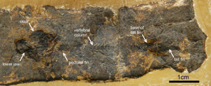
A fossilized fish about 6 cm in length was selected for CT scanning (Figure 3). Based on the external anatomy, the fish was thought to be a member of the Esocidae, the group of living fish that includes the northern pike. The skull and much of the vertebral column was present, although the lower jaws were nearly completely embedded in rock matrix. This specimen was of particular interest because it differs from living members of the family Esocidae in having a short skull and short vertebral column. Thus, if correctly identified, it is new and likely a primitive member of the group.
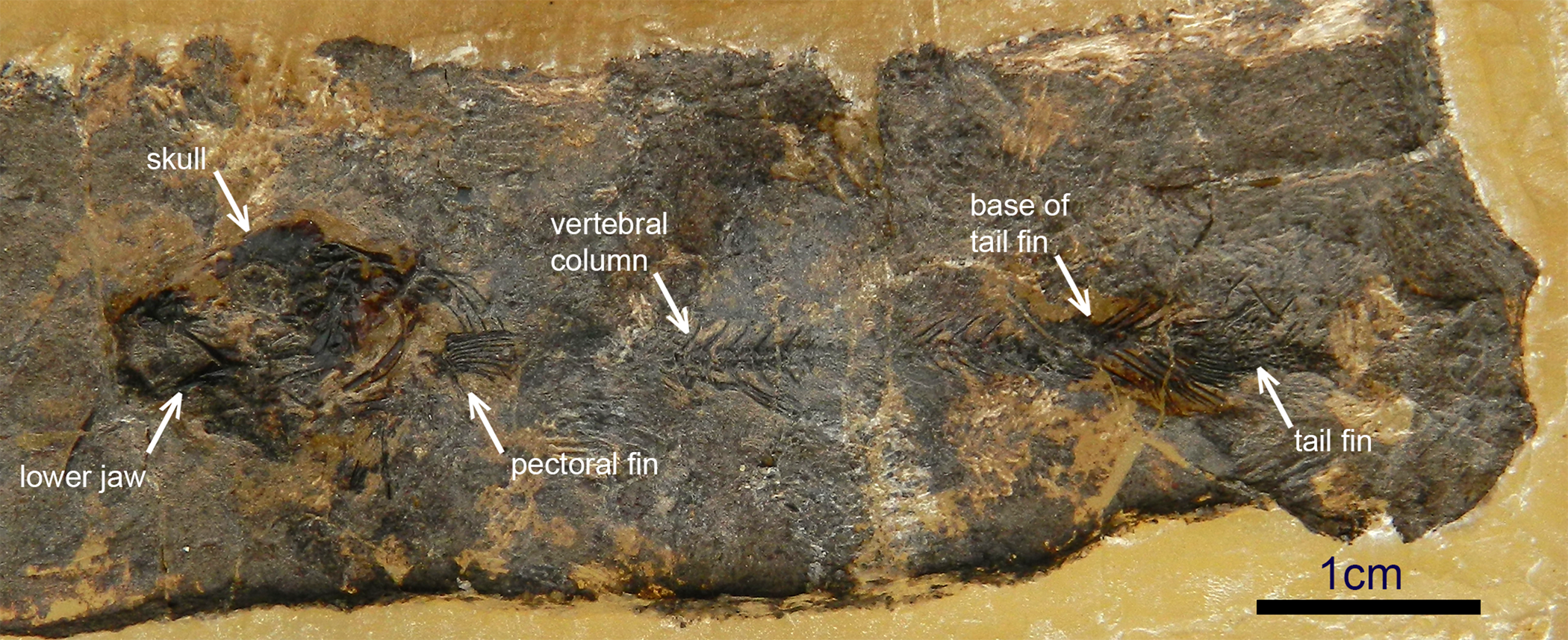
Figure 3: Fossilized fish about 6 cm in length embedded in rock matrix, specimen TMP 2013.45.1433. Visual inspection suggests it is an early member of the pike family (Esocidae).
In addition, three lower jaws from additional fossils were also selected for scanning. One of these had previously been identified as a member of the Esocidae on the basis of features related to the teeth and had been placed in the genus Estesesox. It is elongate, indicating that it was similar to the living northern pike (genus Esox) in having an elongate skull. The other two are from unknown species of fish. They were selected because both showed lower jaw features similar to the dentary of Estesesox, but are different in that both are short. Additional features needed to be found to resolve whether they also represent an early member of the Esocidae, and if so, are they from a fish closely related to the one represented by the articulated skeleton.
Synchrotron micro-CT
Micro-CT scanning of the specimen was conducted at the Argonne National Laboratory Advanced Photon Source (APS) XSD-IMG 2-BM beamline. The 2-BM beamline is fully dedicated to microtomography and can perform large field-of-view imaging with applications in the life sciences, geosciences, and paleontology. The incident energy used was 25 keV with a 4.2 mm (horizontal) × 1.2 mm (vertical) field of view. Over the 180° rotation, 1500 projections were acquired with an exposure time of 140 milliseconds per projection. Data were collected and reconstructed using custom software at the beamline [Reference Gürsoy6]. The specimens were visualized and segmented using the program Amira.
Results
The scans of the articulated skeleton showed strong contrast between the skeleton and the rock matrix, allowing digital reconstructions of the skeleton with clear definition of the skeleton. Examples of individual elements are shown in Figures 4A and B. Figure 4A shows the base of the tail, a part of the skeleton that is important for establishing relationships to other members of the family. Both sides are visible, which is rare in an articulated specimen of such a small size. Figure 4B shows the lower jaw. This element was almost fully encased in rock matrix, so its shape was unknown. As anticipated from the proportions of the skull, it is short. Despite this difference compared to living members of the group, esocid relationships are confirmed by the upper jaw, which lacks teeth, a feature of the Esocidae.

Figure 4: 3D reconstructions of isolated elements from the articulated skeleton in Figure 3. A) Base of the tail (left side in upper image, right side in lower image). Individual elements in the base of the tail were impossible to differentiate by eye. B) Dentary shown in internal and external views. Synchrotron micro-CT allows for both sides of the lower jaw to be seen. Only a small piece of the jaw was visible in the rock matrix. The features indicate that this specimen is a relative of the pike family.
Scans of three isolated lower jaws (Figures 5–7) revealed previously unknown features that aid in identification. One of these, Estesesox (Figure 5), was known to be a member of the Esocidae and thus was used as a reference for comparison with the other two jaws (Figures 6 and 7) to determine if they are also members of this family. Three sets of canals within the jaw could be recognized: a set of canals in the middle of the jaw for the Meckelian cartilage, which is the embryonic precursor of the jaw (shown in blue in Figures 5–7); a set of canals in the lower portion of the jaw, which contains sensory organs (shown in yellow in Figures 5–7); and a set of canals in the upper portion of the jaw (shown in red in Figures 5–7). The reconstructions of canals within the jaw show that one of the unidentified jaws (Figure 6) was a member of the Esocidae. As in Estesesox, relatively few canals are present in the upper portion of the jaw (red canals in Figures 5–7), and these do not lead to the bases of the teeth. In the second unknown jaw (Figure 7), a complex series of canals is present dorsally that leads to the bases of the teeth, and a long canal is present ventrally, a pattern very different from that of Estesesox. However, we do not yet know what kind of fish this jaw is from. For this, additional jaws must be scanned to see how these features vary in modern groups of teleost fish.

Figure 5: Photo and 3D reconstruction of canals in the dentary of the pike Estesesox, specimen TMP 1995.157.42. Upper row: dentary shown in lateral and medial views. Second row: dentary with foramena on the external and internal surface of the jaw visible. Third row: dentary semi-transparent, so the position of the canals relative to the external surface of the dentary can be seen. Bottom row: dentary transparent, so the canals are visible. The canals in the upper portion of the dentary are in red. The Meckelian canal (canal in the center of the dentary) is in blue. The canals in the lower portion of the dentary (sensory canals) are in yellow.

Figure 6: Photo and 3D reconstruction of a jaw hypothesized to be from a member of the Esocidae, specimen TMP 1995.181.117. Esocid relationships are confirmed because it is similar to Estesesox in not having canals leading to the teeth and in having a reduced sensory canal.

Figure 7: Photo and 3D reconstruction of a jaw initially hypothesized to be from a member of the Esocidae, TMP 1995.181.47. The scans revealed the jaw to be different than the Estesesox specimen shown in Figure 5 in the development of a series of canals leading to the teeth and the presence of a large sensory canal.
Conclusion
The 3D visualizations of fish fossils of small size made possible by SR-CT reveal new aspects of their morphology that help to resolve questions of diversity and relationships of small fish from freshwater environments. By digitally dissecting the skeletons, individual elements can be imaged while preserving the specimen. By scanning both articulated specimens and isolated elements, features can be identified that aid in their identification and understanding of the diversity of teleost fish that swam with the dinosaurs. With this information, the history of living groups of freshwater fish can be traced and questions such as the following answered: Did the extinctions at the end of the Cretaceous lead to the development of modern groups of freshwater teleost fishes, or were they already present in the Cretaceous? Did teleost fish faunas change rapidly over time during the late Cretaceous? How widely distributed were different kinds of teleost fishes? All are questions that we have not been able to answer before now.
Acknowledgements
We thank Dr. P. Shevchenko, Dr. F. De Carlo, and Dr. V. Nikitin for their support with data collection and analysis. This research used resources of the Advanced Photon Source, an Office of Science User Facility operated for the U.S. Department of Energy (DOE) Office of Science by Argonne National Laboratory, and was supported by the U.S. DOE under Contract No. DE-AC02-06CH11357, and the Canadian Light Source and its funding partners. Funding for this project was provided by grants from the National Sciences and Engineering Research Council of Canada (NSERC) to N.R. Banerjee.


