


Monochorionic twin pregnancies have a risk of adverse outcomes and specific complications because of the vascular anastomoses resulting in twin-to-twin transfusion syndrome (TTTS), twin anemia polycythemia sequence (TAPS) and selective intrauterine growth restriction (sIUGR). Accurate determination of chorionicity is vital in the clinical management of twin pregnancies. Although ultrasound determination of chorionicity in the first trimester has a high accuracy, it also has some pitfalls. This report presents the case of a monochorionic diamniotic (MCDA) twin pregnancy with two gestational sacs (GSs) and a lambda sign caused by a folded chorionic membrane in the first trimester. The folded chorionic membrane extended toward both sides and eventually flattened during the pregnancy. Furthermore, the report demonstrates the dramatic changes in the shape of the intertwin membrane using ultrasound.
Case Report
A 32-year-old woman, gravida 2 para 0, was referred to our institution at 7 weeks of gestation. Transvaginal ultrasonography identified two GSs, suggesting a dichorionic twin pregnancy, and two embryos with fetal heartbeats (Figure 1).
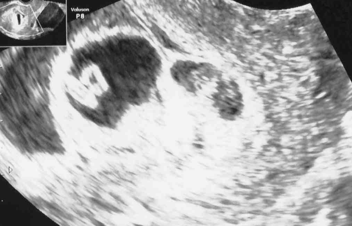
Fig. 1. Two gestational sacs and two embryos with heartbeats were observed at 7 weeks’ gestation.
At 9 weeks’ gestation, an ultrasound image showed a lambda sign composed of thick triangular tissues without consecutive membranes from both sides, resulting in the diagnosis of an MCDA twin pregnancy with chorionic membrane folding (CMF; Figure 2A). Figure 2B shows a graphic representation of the ultrasound image.

Fig. 2. (A) Lambda signs were detected on both sides of the base of the intertwin membrane at 9 weeks’ gestation. An amniotic membrane swelled toward one side between the chorionic membrane folding (arrows). (B) A graphic representation of the ultrasound image.
At 11 weeks’ gestation, an ultrasound image showed a lambda sign at one portion of the septum (Figure 3A) and a T sign at another portion (Figure 3B). This change suggested that the folded chorionic membrane had partially flattened.
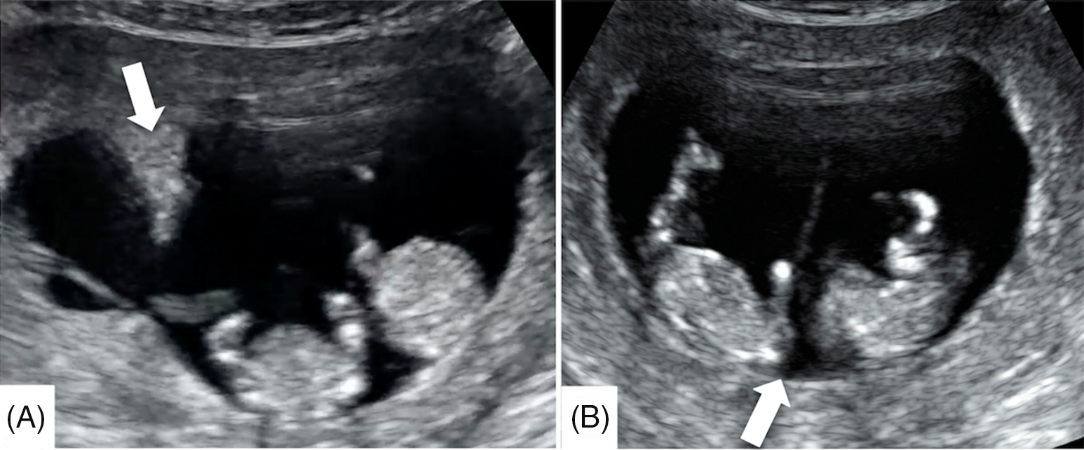
Fig. 3. (A) One part of the septum with chorionic membrane folding shows a lambda sign (arrow) at 11 weeks’ gestations. (B) Another part of the septum without chorionic membrane folding shows a T sign (arrow).
We managed this case as an MCDA twin pregnancy, and no complications such as TTTS, TAPS and selective IUGR developed.
At 35 weeks’ gestation, an emergency cesarean section was performed because of labor onset and breech presentation. Two healthy male neonates weighing 1646 g and 1974 g were delivered.
Macroscopic placental examination revealed that the placenta was composed of a single placental mass and two thin amniotic membranes without the interposition of the chorionic membrane, which formed an intertwin membrane at the middle of the placenta (Figure 4A). The placental vascular anastomoses could not be identified due to the thickening of the chorionic plate caused by the Breus’ mole that appeared in the late second trimester (Figure 4B).
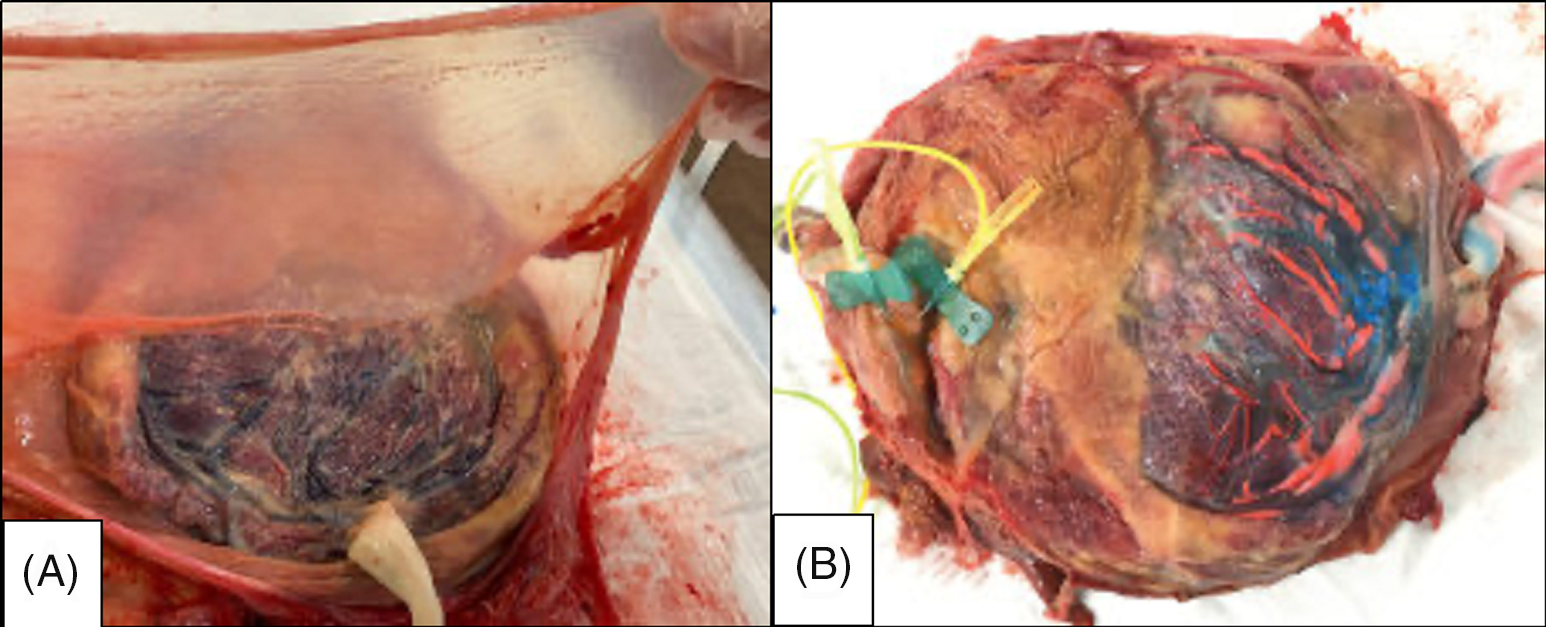
Fig. 4. (A) Postpartum macroscopic evaluation revealed a thin intertwin membrane containing two amniotic membranes. (B) The placental vascular anastomoses could not be identified due to the thickening of the chorionic plate caused by the Bleus mole.
Histological examination confirmed that the intertwin membrane was composed of two amniotic membranes without a folded chorionic membrane, confirming the diagnosis of an MCDA twin pregnancy.
Discussion
To the best of our knowledge, this is the first case report that demonstrates changes in ultrasound findings during a monochorionic pregnancy with CMF. This report describes two important ultrasound chorionicity findings: monochorionic pregnancies with CMF can show two GSs and a lambda sign and the CMF can flatten or change during the pregnancy.
Before 10 weeks’ gestation, the relationship between the number of GSs and the number of embryonic heartbeats provides a strong indication of chorionicity (Morin & Lim, Reference Morin and Lim2017). The presence of two GSs is strongly believed to indicate a dichorionic twin pregnancy, but the presented monochorionic twin pregnancy case also showed two GSs. This is consistent with a previous case report of a monochorionic twin pregnancy with two GSs in the first trimester and is complicated by TTTS (Yamashita et al., Reference Yamashita, Ishii, Hidaka, Hayashi, Takeuchi and Mitsuda2015). The presence of CMF from the base of the intertwin membrane suggests that the chorionic membranes were very close to each other, mimicking two GSs early in the first trimester.
The lambda sign, also called as the twin peak sign, is a triangular tissue projection extending from the base of the intertwin membrane consisting of chorionic tissue between two amniotic membranes (Finberg, Reference Finberg1992). A recent meta-analysis showed that the presence of the lambda sign was very sensitive and specific in predicting chorionicity during ultrasonography in the first trimester (Maruotti et al., Reference Maruotti, Saccone, Moriando and Martinelli2016). Generally, the presence of the lambda sign indicates a dichorionic twin pregnancy. In this case, ultrasonography performed at 9 weeks’ gestation detected the lambda sign on both sides of the base of the intertwin membrane and amniotic membrane swelling between the two chorionic membranes. This finding supports the concept that CMF can show the lambda sign and that the form of CMF can change during pregnancy. The difference in the lambda sign between dichorionic monoamniotic (DCDA) twin pregnancies and MCDA twin pregnancies is that the triangular tissue projection extending from the base of the intertwin membrane is followed by a relatively thick membrane to the other side in DCDA twin pregnancies, whereas the CMF in MCDA twin pregnancies does not follow chorionic membrane from the lambda sign and the thick chorionic membrane appears to be interrupted.
In the absence of chorionic tissue, the bottom of the intertwin membrane is composed of two amniotic membranes and it is called a ‘T sign’. A similar case has been reported of an MCDA twin pregnancy that showed lambda and T signs on the first-trimester ultrasonography (Galjaard et al., Reference Galjaard, Moerman, Corveleyn, Devlieger and Lewi2014). Histological examination of the intertwin membrane is characterized by coexistent monochorionic (amnion-amnion) and dichorionic (amnion-chorion-chorion-amnion) components, and such cases are classified as partial monochorionic/dichorionic twins (Chmait et al., Reference Chmait, Floyd and Benirschke2011; Galjaard et al., Reference Galjaard, Moerman, Corveleyn, Devlieger and Lewi2014; Lu et al., Reference Lu, Cheng, Law and Leung2018). The lambda sign is found at the part with CMF, and the T sign is found at the other part without CMF. The mechanism causing CMF is unknown, but this complication is probably rare. In this case, both the lambda sign and T sign coexisted at 11 weeks’ gestation, suggesting that CMF can be flattened over time, and ultrasonography can show both the lambda and T signs depending on the timing of examination and location of scanning.
This case demonstrates that ultrasonography during monochorionic twin pregnancies with CMF can show various expressions. The CMF can change dramatically throughout the pregnancy and eventually become flat. These findings suggest that ultrasonography should be performed more frequently, and scanning should be more thorough to obtain accurate chorionicity.
Conflict of interest
None.
Ethical standards
Written informed consent was obtained from the patient for publication of this case report.


