


Clostridium difficile is an anaerobic spore-forming and toxin-producing bacteria that was initially discovered in 1935. It was not until 1978 that it was identified as a cause for antibiotic-associated diarrhea.Reference Bartlett 1 Currently, it is considered to be the most common cause of hospital- and antibiotic-associated diarrhea, with a more severe form causing pseudomembranous colitis.Reference Wilkins and Lyerly 2 Recently described strains of C. difficile have been associated with increased morbidity and mortality—even in previously healthy individuals. The strain alternatively known as North American pulsed-field gel electrophoresis type 1, restriction endonuclease analysis group BI, and polymerase chain reaction (PCR) ribotype 027 (NAP1/BI/027) is linked to several C. difficile infection (CDI) epidemics in North America, including the Quebec CDI outbreak that peaked between 2001 and 2003.Reference O'Donoghue and Kyne 3 Asymptomatic colonization of the intestinal tract with C. difficile is common, but disease usually occurs only following a disruption in the gastrointestinal microbiota—for example, following antibiotic use, which then allows for C. difficile to proliferate and produce toxins A and B. Toxin A is an enterotoxin responsible for tissue damage and toxin B is a potent cytotoxin.Reference Guilbault, Labbe, Poirier, Busque, Beliveau and Laverdiere 4 – Reference Taur and Pamer 6
Early diagnosis and treatment of CDI are important to limit morbidity, healthcare costs, and nosocomial transmission; patients with CDI will require isolation measures, ancillary housekeeping services, and additional antimicrobial therapy.Reference Dubberke and Olsen 7 , Reference Dubberke, Reske, Olsen, McDonald and Fraser 8 The gold standard tests for the diagnosis of CDI include toxigenic culture and cell culture cytotoxicity assay. Toxigenic culture detects the presence of toxigenic C. difficile that has the capacity to produce toxin, whereas culture cytotoxicity assay detects the presence of produced toxin in the stools. However, these techniques are labor-intensive, require expertise, and have a turn-around time that requires more than 48 hours. For that reason, enzyme immunoassays (EIA) were developed to allow for the easy and rapid detection of C. difficile toxin.Reference Delmee, Van Broeck, Simon, Janssens and Avesani 9 , Reference van den Berg, Bruijnesteijn van Coppenraet, Gerritsen, Endtz, van der Vorm and Kuijper 10 Recently, nucleic acid amplification techniques—such as PCR and loop-mediated isothermal amplification—that are based on the detection of the toxin B gene (tcdB) have been developed to improve sensitivity over EIA, while maintaining a short turn-around time for the diagnosis of CDI.Reference van den Berg, Bruijnesteijn van Coppenraet, Gerritsen, Endtz, van der Vorm and Kuijper 10 , Reference van den Berg, Bakker and Kuijper 11 Several studies showed that PCR had an equivalent sensitivity and specificity (up to 100%) compared with toxigenic cultures.Reference Peterson, Manson and Paule 12 – Reference Samra, Luzon and Bishara 14 However, PCR may not be useful when trying to differentiate carrier status from true CDI.Reference Dupont 15 Moreover, given that clinical manifestations of CDI are milder in children compared with the adult population,Reference Morinville and McDonald 16 and given the current absence of testing strategies that accurately and optimally diagnose CDI,Reference Cohen, Gerding and Johnson 17 we aimed to determine the proportion of pediatric patients diagnosed as having CDI by PCR who would also be diagnosed by EIA, and to compare the clinical characteristics of PCR+/enzyme-linked immunosorbent assay (ELISA)+ vs PCR+/ELISA− patients. We also determined the impact of switching from an EIA-based to a PCR-based testing strategy on the proportion of positive samples, as a secondary analysis.
METHODS
Study Design and Setting
We performed a retrospective observational cohort study at the Montreal Children’s Hospital, a tertiary care facility in Quebec, Canada, with hematopoietic stem cell and solid organ transplant programs. Using the microbiology laboratory information system, a retrospective cohort of patients with diarrhea or change in stool consistency deemed clinically significant by the treating team and with positive C. difficile PCR assay between June 2010 and July 2014 was created. Our laboratory protocol rejects stool samples sent for C. difficile in infants younger than 6 months.Reference Gerding, Johnson, Peterson, Mulligan and Silva 18 Stool samples were thus from patients aged 6 months to 18 years from inpatient and outpatient settings. Duplicate stool testing for C. difficile—defined as 1 or more tests performed for the same patient within a 14-day window after the initial positive test—were excluded.
Diagnostic Assays
PCR
All soft or liquid stool samples sent to the microbiology laboratory were tested within 24 hours of reception for toxin B gene using the BD GeneOhm C diff assay, according to the manufacturer's instructions (BD Diagnostics) and the McGill University Health Center laboratory protocol. PCR-based method for the detection of C. difficile was implemented in our hospital as a routine test in June 2010. The BD GeneOhm is a real-time PCR that amplifies the toxin B (tcdB) gene from C. difficile with fluorogenic target-specific hybridization probes for the identification of amplified target DNA. An internal control was employed and interpreted using the SmartCycler instrument (Cepheid). Results are reported as positive, negative, or indeterminate—in which case a repeated sample is requested. After the procedure, all stool samples were routinely stored in a −20°C non–frost-free freezer.
ELISA
C. difficile TOX A/B II (TechLab) was performed a posteriori on all available samples in a single batch to only thaw samples once. Toxin-detecting antibodies consisted of a mixture of toxin A monoclonal mouse antibody and toxin B polyclonal goat antibody. To perform the test, stool samples were thawed and diluted. The supernatants from stool suspensions were collected and placed in 96-well plates according to the manufacturer’s instructions. An optical density of 0.8 or greater was considered positive.
Clinical characteristics
Using a piloted case report form, a medical chart review was performed to extract patients’ demographic characteristics, clinical characteristics, and laboratory data. Data collected included recent antibiotic use, clinical manifestations of CDI (abdominal pain, frequency of diarrhea, leukocytosis, fever, elevated C-reactive protein, elevated erythrocyte sedimentation rate, lactic acidosis, elevated stool leukocytes, and presence of blood in the stool), development of complications (intensive care unit admission, shock, or colectomy), management of CDI, and presence of comorbidities or CDI risk factors (Table 1). Targeted CDI risk factors included the presence of inflammatory bowel disease (IBD), malignant tumor, gastric acid suppression medication (either proton-pump inhibitor or histamine-2 inhibitor), immunosuppressive agents, feeding device, cystic fibrosis, Hirschprung disease, immunodeficiency, and bone marrow transplant. Also, the total of C. difficile episodes for each patient was obtained up to the time of medical chart review.
TABLE 1 Demographic and Clinical Characteristics of 136 Patients With Clostridium difficile Detected by PCR, Montreal Children Hospital, Canada, 2010–2014

NOTE. Data are no. (%) of patients unless otherwise indicated. aOR, adjusted odds ratio; CDI, Clostridium difficile infection; CRP, C-reactive protein; ELISA, enzyme-linked immunosorbent assay; ESR, erythrocyte sedimentation rate; HPF, high-power field; IBD, inflammatory bowel disease; IQR, interquartile range; PCR, polymerase chain reaction; PO, per os (by mouth); WBC, white blood cells.
a Denominator as in heading, unless otherwise indicated.
b Adjusted for age category (older or younger than 5 years), gender, and CDI risk factors (healthy, malignant tumor, immunosuppressive agents, gastric acid suppressive agents, feeding device, bone marrow transplant, and IBD).
c 7 congenital heart disease, 7 immediate postsurgical period (appendectomy, posterior spinal fusion, ureteric implantation), 4 neurologic disease, 3 post–renal transplant, 3 hepatopancreaticobiliary disorders, 3 genetic disorders (trisomy 21, cri-du-chat), 2 chronic ear and sinus infection, 2 chronic hepatosplenic candidiasis, 18 miscellaneous.
Statistical analysis
We used summary statistics to describe clinical characteristics of patients who were positive for C. difficile by both PCR and ELISA with those who were positive only by PCR and used χ2 tests to compare proportions. Continuous variables were compared using the t test. Multivariable analysis was performed on crude odds ratios to adjust for age category (<5 years or ≥5 years), gender, and CDI risk factors (healthy, malignant tumor, immunosuppressive agents, gastric acid suppressive agents, feeding device, bone marrow transplant, and IBD). Statistical analyses were performed using SAS, version 9.3 (SAS Institute), and R, version 3.1.1. A 2-tailed P≤.05 was considered statistically significant.
Ethical considerations
This study was approved by the Montreal Children’s Hospital Research Ethics Board.
RESULTS
During the study period, 310 consecutive stool samples tested positive for tcdB gene by PCR. Of those, 154 samples were retrieved from the microbiology laboratory freezer. A larger proportion of samples obtained before 2012 was missing, whereas most of the samples collected in 2014 were retrieved (Table 2). When comparing missing specimens with those that were found, the same proportion of samples in both categories came from ambulatory or inpatient settings (Table 3). In addition, the clinical background was comparable in both groups. Among the samples retrieved, 17 were excluded because they were duplicates and 1 was excluded because the patient was never evaluated in our institution (sold service) (Figure 1). Figure 2 illustrates the proportion of stool specimens that tested positive for C. difficile by month during the 13 months prior to PCR implementation and the 50 months following PCR implementation. The switch from EIA to PCR was associated with an increase in the proportion of positive samples, from 5.8% to 11.3% (P=.003).

FIGURE 1 Flowchart of specimens tested and results. C. difficile, Clostridium difficile; EIA, enzyme immunoassay; PCR, polymerase chain reaction.

FIGURE 2 Proportion of stool samples that were positive for Clostridium difficile at the Montreal Children’s Hospital, by month (April 1, 2009, to August 1, 2014). The arrow indicates when polymerase chain reaction testing for C. difficile was implemented.
TABLE 2 Proportion of Specimens Found to Total Samples in the Microbiology Laboratory Database
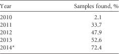
* Until July 2014.
TABLE 3 Characteristics of Patients With Lost vs Retrieved Specimens

NOTE. IBD, inflammatory bowel disease.
a Congenital heart disease, immediate postsurgical period, neurologic disease, post–renal transplant, hepatopancreaticobiliary disorders, genetic disorders and syndromes, chronic sinopulmonary infections, chronic fungal infections, chronic kidney diseases, and renal transplant.
Of the 136 PCR-positive samples that were tested by EIA, fewer than half (54 [40%]) were positive for toxins A or B. The population’s mean age was 8.5 years and approximately half of the sample were boys (Table 1). There was no difference in the mean age of EIA-positive and EIA-negative cases. Although not statistically significant, there was a trend toward having younger patients (<2 years of age) in the EIA-negative group compared with EIA-positive patients: 25.6% vs 18.5% (P=.33). There was also a trend toward fewer lifetime C. difficile recurrence in the EIA-negative group: 23.1% had more than 3 documented CDIs compared with 33.3% in the EIA-positive group (P=.19).
There was no significant difference in terms of clinical manifestations of CDI and underlying comorbidities between the EIA-positive and EIA-negative cases, looking at the crude and the adjusted odds ratio (OR). EIA-positive patients were more likely to have been recently exposed to antibiotics: 67.9% of EIA-positive compared with 50% of EIA-negative (crude OR, 2.1 [95% CI, 1.03–4.28]). However, this did not remain statistically significant when the OR was adjusted for CDI risk factors (adjusted OR, 2.13 [95% CI, 0.91–5.10]), likely due to lack of power. This difference was not noted for any specific antimicrobial classes. There were fewer patients with IBD in the EIA-positive group compared with the EIA-negative group (13% vs 25.6%; adjusted OR, 0.36 [95% CI, 0.12–0.96]). There were no significant differences in the treatment choice between the two groups. No complications related to CDI were identified.
DISCUSSION
In our study, only 40% of patients with a diagnosis of CDI by PCR had a toxin-producing C. difficile detectable by EIA. PCR detects the presence of the toxin B gene but not necessarily toxin production. This means that many patients diagnosed by PCR may in fact not have a real CDI but rather another cause for their diarrheal episode. Unlike other studies,Reference Patel, Randhawa, Nanavati, Marton, Baddoura and DeBari 19 we were not able to demonstrate a statistically significant difference in clinical manifestations between EIA-positive and EIA-negative patients. The 2 groups were similar in their presenting symptoms and management. Upon initial analysis, the absence of difference in the 2 groups’ clinical manifestations was believed to be partly attributed to a confounding effect of patients with IBD or alternate diagnoses for diarrhea that were not the focus of this study. Patients with IBD can present with symptoms similar to CDI and hence carriers may be misclassified as having CDI. However, no significant difference in the OR was noted when patients with IBD were excluded from the analysis. It is also possible that the small sample size did not allow for differences to be identified.
The Montreal Children’s Hospital healthcare-associated infections surveillance program noticed a slight increase in the incidence of CDI in the year following the implementation of PCR for C. difficile. Our hospital healthcare-associated annual CDI rate was 1.1 cases/10,000 patient-days in 2009–2010 (April 2009 to March 2010) and increased to a pooled mean rate of 3.39 cases/10,000 patient-days from April 2010 to March 2015. Even though the number of positive cases was comparable with those of the previous years, the highest peak was seen after the introduction of PCR in June 2010 and this finding was similar in both the outpatient and inpatient populations.Reference Gonzales, Quach, Perpete and Moore 20 , Reference Quach, Perpete and Lacharite 21
Adult and pediatric centers that implemented PCR-based assays for the detection of C. difficile in replacement of EIA noticed an increase in the proportion of positive C. difficile tests by 2- to 10-fold.Reference Luna, Boyanton and Mehta 22 – Reference Khanna, Baddour and Huskins 24 For instance, Luna et alReference Luna, Boyanton and Mehta 22 reported an increase in the proportion of C. difficile positivity from an average of 8% to 16.2% after switching from EIA to PCR. We similarly show that switching from ELISA to PCR increased the number of identified cases by more than 2-fold. This finding is consistent with other studies that compared PCR with toxin detection in the diagnosis of CDI. It is unclear whether this increase in CDI was due only to the increased sensitivity of the assay and decreased specificity for clinical disease, to a change in circulating strains, or to an indication bias where hospitals in areas with increasing CDI incidence due to outbreaks decided to implement the test.Reference Luna, Boyanton and Mehta 22 , Reference Khanna, Baddour and Huskins 24
Polage et alReference Polage, Gyorke and Kennedy 25 documented that, in adults, PCR has a positive predictive value of 44.7% compared with EIA. The increase in the number of cases cannot be explained solely by the difference in performance characteristics of the 2 tests: the sensitivity (68%–90% for EIA vs 88%–100% for PCR) and specificity (95.3%–99% for EIA vs 92.6%–98.4% for PCR) of the 2 tests are comparable.Reference Peterson, Manson and Paule 12 – Reference Samra, Luzon and Bishara 14 , Reference Snell, Ramos, Longo, John and Hussain 26 , Reference Eastwood, Else, Charlett and Wilcox 27 The sudden increase in the incidence of CDI following the implementation of PCR could be partly explained by the fact that PCR detects toxin genes but cannot determine whether the organism is actively producing toxin. For that reason, some studies concluded that PCR is unreliable in differentiating CDI cases from asymptomatic carriers of a potentially toxigenic organism.Reference Dupont 15 Planche et alReference Planche, Davies and Coen 28 documented that CDI severity and related mortality was higher in patients with positive toxin assay compared with patients who were positive by toxigenic culture but toxin assay negative. More false-positive results may be seen with PCR compared with toxin detection methods.Reference Polage, Gyorke and Kennedy 25 This is supported by the fact that in adult populations, the complication rate in patients with CDI is 40%–50% higher if they were diagnosed with EIA or cytotoxicity assay compared with PCR.Reference Longtin, Trottier and Brochu 23 , Reference Polage, Gyorke and Kennedy 25 In pediatrics, the complication rates are significantly lower compared with adults.Reference Morinville and McDonald 16
Toxigenic C. difficile carriage has been documented in healthy adults with a rate that was reported to be as high as 14% in hospitalized patients.Reference Shim, Johnson, Samore, Bliss and Gerding 29 , Reference Kyne, Warny, Qamar and Kelly 30 The asymptomatic carriage rate in children, especially in those younger than 3 years, was found to be even higher than in adults. Rousseau et alReference Rousseau, Poilane, De Pontual, Maherault, Le Monnier and Collignon 31 demonstrated in a small cohort that almost 40% of infants were carriers of toxigenic C. difficile by the age of 6 months. A study by Matsuki et alReference Matsuki, Ozaki and Shozu 32 showed a carriage rate of 48% for children younger than 5 years, with the highest carriage rate (80%–100%) being in infants. In a case-control study that looked for causes of diarrhea in children, the proportion of C. difficile positives was higher in controls compared with cases.Reference Denno, Shaikh and Stapp 33 Our study’s objective was not to document carriage rate.
The effect of single or multiple freeze-thaw cycles on the performance characteristics of the EIA have not been well studied. A study by LyerlyReference Lyerly 34 concluded that multiple freeze-thaw cycles could lead to toxin degradation. A similar finding was shown by Freeman and WilcoxReference Freeman and Wilcox 35 where the toxin titer dropped from 1×10⁶ toxin units to 1×101 toxin units after 56 days with multiple freeze-thaw cycles. However, this is unlikely to account for the current study result because only 1 freeze-thaw cycle was performed. In a study by Arrow et al,Reference Arrow, Croese, Bowman and Riley 36 the immunogenic effect of C. difficile was preserved, with a superiority of EIA over toxigenic cultures, after a single freeze-thaw cycle. Also, Sharp et alReference Sharp, Ruden, Pohl, Hatcher, Jayne and Ivie 37 showed that a glutamate dehydrogenase and A/B Toxin Combination test remained positive in 26 of 27 samples after a single freeze-thaw cycle.
To our knowledge, this is the first study that aimed to evaluate the clinical implications of implementing PCR for the diagnosis of CDI in children. However, it has its limitations. The major limitation of this study is its retrospective nature. Because it was limited to information written in the medical chart, it is possible that clinical manifestations were not well documented. However, it is likely that severe manifestations would have been recorded. This could explain why it failed to show a significant difference between EIA-positive and EIA-negative patients in terms of clinical characteristics. Another limitation is that it was not possible to perform toxigenic cultures to determine definitely whether PCR-positive but EIA-negative samples were true- or false-positive results. Despite all that, one can presume that PCR is likely overestimating the rate of CDI, at least in the pediatric population. The difference between the 2 groups cannot be explained merely by the difference between the tests’ performance. For that reason, some authors suggested validating PCR testing by developing a quantitative PCR or adding another step in the diagnostic process, which would include either a test that detects functional toxins or a test for a fecal marker of inflammation.Reference Dupont 15 , Reference Vaishnavi, Bhasin, Kochhar and Singh 38 – Reference Dubberke and Burnham 40
ACKNOWLEDGMENTS
Financial support. Alera (supply of ELISA kits).
Potential conflicts of interest. C.Q. reports that she has received funding from GlaxoSmithKline, Pfizer, Sage, and AbbVie (all for research grant or support for unrelated research projects). Y.L. reports that he has acted as a consultant for AMG Medical and Oxypharm and received funding from GenePOC. All other authors report no conflicts of interest relevant to this article.


