

X-ray ptychography differs from more commonly known x-ray techniques that rely on attenuation in x-ray intensity to detect matter. In contrast, ptychography functions on diffraction of coherent x-rays. Diffraction patterns result from interference (i.e., superposition) of x-rays scattered by the structural features of the material under investigation. Accordingly, ptychography can provide information at much smaller dimensions than radiography. In principle, this diffraction imaging technique offers wavelength-limited resolution. However, in practice, spatial resolutions are also limited by inaccuracies while positioning the incident beam. Junjing Deng and his colleagues at Argonne National Laboratory have introduced an instrument called the Velociprobe to relax this limitation and advance ptychography imaging. They reported this design in a recent issue of the Review of Scientific Instruments (doi:10.1063/1.5103173).
Unlike the traditional ptychography, the Velociprobe provides continuous scanning with about two orders of magnitude improved positional accuracy down to a couple of nanometers (traditional ptychography performs “step scan” where the scanning stage is first moved and then scanned, while the Velociprobe scans as it moves). Such advanced spatial and temporal control is facilitated by (1) a granite stage for higher stiffness, thermal stability, and vibrational response; (2) laser interferometry for accurate position detection; and (3) a specialized control algorithm to interpret position data and effect adjustments without time lags. The name—Velociprobe—implies fast (velocity) measurements with x-rays as a probe.
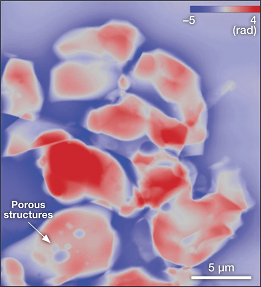
Ptychographic reconstruction of a perovskite sample of about 21 µm × 24 µm dimensions with a fine resolution close to 6.2 nm. Credit: Junjing Deng.
The Velociprobe exhibits high spatial resolution down to the pixel size, as shown in the Figure where a LaFe0.3Co0.7O3 perovskite structure is imaged. As the x-rays used in the Velociprobe penetrate the sample, structural information is retrieved rather than just from the surface. For a given sample placement, the probe beam moves to scan an area (typically a raster-like motion), and then the sample is repositioned to image the adjoining portion. The resulting array of diffraction images are stitched to reconstruct the sample. For example, the perovskite structure shown in the Figure is composed of nine (3 × 3) scanning tiles. The continuous scanning allows entire imaging in 7 minutes (∼46 seconds for each tile).
The fast sampling opens the possibility of in situ and in operando imaging. Alternatively, three-dimensional ptychography also provides an interesting avenue. Deng says that the Velociprobe is also a proof-of-concept for technologies and techniques to be incorporated into an upgrade of the instruments used at the Advanced Photon Source, a synchrotron facility at the Argonne National Laboratory, such as the PtychoProbe. Orders-of-magnitude improvement in the x-ray flux from the soon-to-be upgraded synchrotron is anticipated.
Jeff Gelb from Sigray, Inc., an industrial developer of laboratory x-ray systems, says that coherent diffraction imaging techniques, such as ptychog-raphy, are the future of synchrotron x-ray imaging. Gelb, who was not involved in this study, says the lensless approaches represent a major breakthrough in overcoming the limits imposed by x-ray optics, and that researchers have only begun to scratch the surface of what is possible with coherent radiation.
