- CFU
colony forming units
- DC
dendritic cells
- LAB
lactic acid bacteria
- PBMC
peripheral blood mononuclear cells
With increasing affluence and food abundance, there has been a rising interest in functional food components, offering benefits beyond mere nutrition by beneficially influencing the health of those who ingest them. Among these, lactic acid bacteria (LAB), especially in the form of probiotics, have long been known to enhance the body's ability to ward off infections especially of the gastrointestinal tract. Administration of certain LAB to infants was repeatedly shown to reduce the severity and duration of viral diarrhoea and even to prevent it(Reference Heyman1, Reference Gupta and Garg2). In this regard, LAB are also relevant for the nutrition in developing countries, where diarrhoea and related diseases are common and contribute particularly to child mortality. However, there is also evidence for an improved defence against other infections such as influenza and the common cold even though no prevention was achieved(Reference de Vrese, Winkler and Rautenberg3) LAB have also been suggested as a treatment for atopic diseases, inflammatory bowel diseases and the irritable bowel syndrome and showed beneficial effects in some studies in these conditions(Reference Miraglia del Giudice, Rocco and Capristo4–Reference Minocha7).
The mechanisms behind the immune-modulating effects of LAB are still partly unknown. Peripheral blood mononuclear cells (PBMC) incubated in vitro with LAB showed a higher secretion of cytokines, proteins or peptides acting as mediators and regulators of the immune response(Reference Oppenheim, Feldmann, Oppenheim and Feldmann8), and the type of cytokines produced was strain dependent. A higher cytotoxic activity of natural killer cells, involved in the defence against viruses and tumour cells, was shown as well as an increase in phagocytic activity(Reference Delcenserie, Martel and Lamoureux9).
Direct co-culture of peripheral immune cells with LAB does not reflect the physiological situation especially in healthy subjects. However, PBMC and natural killer cells isolated from the blood of subjects following consumption of LAB also showed increased phagocytic and cytotoxic activity, respectively, after appropriate immunological stimuli(Reference Sheih, Chiang and Wang10, Reference Parra, Martínez de Morentin and Cobo11). This is in accordance with the observed better resistance against various infections.
These immune-modulating properties of LAB have been used as a health claim on foods containing such organisms, particularly probiotic ones. While LAB strains may differ in their suitability for the treatment of some diseases, healthy individuals may benefit just as much from conventional fermented milk as suggested by the long tradition of such foods as health promoting. Indeed, the earliest evidence of the protective effect of LAB came from observations in regular consumers of fermented milk such as yoghurt or kefyr. Furthermore, traditional yoghurt starter cultures are considered probiotic by some experts(Reference Minocha7, Reference Senok, Ismaeel and Botta12).
This led our working group to study the immunological effects of conventional and probiotic LAB strains in more detail. In a first approach, the immunological effects of a probiotic fermented milk product were compared with those of a conventional product in vivo in healthy subjects. The mechanisms behind the observed effects were further investigated in an in vitro study using cell culture.
An overview of both studies is given in the following.
In vivo effects of daily ingestion of fermented milk by young healthy women
Study design, materials and methods
The aim of this study was to compare the immunological effects of a commercially available probiotic fermented milk product with those of a conventional fermented milk. As the main target group for over-the-counter probiotic products is healthy persons, these were chosen as the study population. The study was approved by the Ethical Committee of the City of Vienna (protocol number EK 04-0060204) and the participants provided written informed consent. Results of the study have been published(Reference Meyer, Micksche and Herbacek13, Reference Meyer, Elmadfa and Herbacek14).
Thirty-three non-smoking young women (aged 22–29 years) were randomly divided into two groups: one received the probiotic product and the other the conventional product. Both products contained the classic yoghurt starter cultures Lactobacillus delbrueckii ssp. bulgaricus and Streptococcus thermophilus albeit at different concentrations, with additional Lactobacillus casei DN114001 in the probiotic product. The bacterial contents were determined as follows: in the conventional product, 6·4×107 colony forming units (CFU) L. delbrueckii ssp. bulgaricus per ml and 3·9×107 CFU S. thermophilus per ml; in the probiotic product, 2·0×108L. delbrueckii ssp. bulgaricus per ml, 107 CFU S. thermophilus per ml and 3·7×108 CFU probiotic L. casei DN114001 per ml. Following a one-week equilibrating period during which fermented foods were avoided, participants consumed daily one portion (100 g) of the respective product for two weeks. The amount was doubled to 200 g/d for another two weeks. A wash-out period with no fermented products lasting two weeks concluded the intervention. Blood samples were taken at the end of each period. Whole blood aliquots were incubated with pokeweed mitogen (40 μg/ml), a stimulant of T-lymphocytes, for 4 h at 37°C and 5% CO2. The percentage of helper T cells expressing the activation marker CD69 on their surface was measured by flow cytometry (FACSCalibur dual laser system (Becton Dickinson Biosciences, San Jose, CA, USA)). To assess natural cytotoxicity, PBMC were isolated by gradient centrifugation using Ficoll Paque Plus (Amersham Biosciences AB, Uppsala, Sweden) and co-cultured with myelogenic leukaemic K562 cells as targets in ratios of 25:1, 50:1 and 100:1 (effector to target) for 4 h at 37°C and 5% CO2. 7-Aminoactinomycin D (7-AAD, Cell Technology Inc.) was used to stain the lysed target cells and determine their percentage by flow cytometry.
To induce cytokine production, whole blood was incubated separately with phytohaemagglutinin from Phaseolus vulgaris (T-lymphocyte stimulant) (5 μg/ml) and lipopolysaccharide from Escherichia coli O26:H6 (macrophage and B lymphocyte stimulant; 1 μg/ml) for 24 h at 37°C and 5% CO2. Interferon-γ, IL-6 and IL-10 concentrations were measured using the cytometric bead array and flow cytometry, whereas IL-1β and TNFα concentrations were measured with ELISA (both kits from BD Pharmingen, Becton Dickinson Biosciences, San Diego, CA, USA).
Results
After the consumption of both products, the percentage of CD69+ helper T cells increased, but this was statistically significant only in the conventional group. After intake cessation, levels decreased again. Likewise, natural cytotoxicity was higher following yoghurt consumption with significant differences in both groups. After the wash-out period, activity rose even further, particularly in the probiotic group, suggesting a longer-lasting effect in this group(Reference Meyer, Micksche and Herbacek13). Stimulated production of the proinflammatory cytokines interferon-γ, IL-1β and TNFα increased following yoghurt consumption with effects seen in both groups, albeit to varying degrees. Generally, increases were stronger in the conventional group. In contrast, no changes were observed for IL-6, while secretion of IL-10 was slightly but significantly reduced after probiotic yoghurt intake and increased markedly after the wash-out compared with the baseline levels(Reference Meyer, Elmadfa and Herbacek14). An overview of the results is given in Table 1. No cytokine production was found in unstimulated blood samples and no marked differences in cell numbers occurred, suggesting that LAB improve the body's reaction to pathogens without modifying the basal cell numbers (in blood) and functions.
Table 1. Effects of daily intake of fermented probiotic and conventional yoghurt on markers of immune function in young healthy women

E:T, effector to target ratio; IFN, interferon
Each intervention phase as well as the wash-out period lasted for 2 weeks. Cytokine secretion and expression of CD69 were measured following stimulation with LPS/PHA and pokeweed mitogen, respectively. Data were analysed for statistical differences using repeated measures ANOVA with post hoc contrast testing. Values are rounded.
a Significantly different from baseline.
b Significantly different from 100 g/d.
c Significantly different from 200 g/d.
There were no significant differences between the conventional and the probiotic group.
For the conventional group, n 16, and for the probiotic group, n 17.
In vitro stimulation of human dendritic cells with lactic acid bacteria
To further investigate the mechanisms behind the earlier-described immune stimulation obtained with probiotic and conventional LAB in vivo, their effects on dentritic cells (DC) were studied at the cellular level. The human epithelial colorectal adenocarcinoma cell line Caco-2 differentiates to an enterocytic phenotype and, hence, has been repeatedly used as a model for the physiological conditions in the gastrointestinal tract.
Study design, materials and methods
The probiotic LAB strain used was Lactobacillus rhamnosus GG (ATCC 53103). L. delbrueckii ssp. bulgaricus (ATCC 11842) was chosen as the conventional strain (LGC Promochem, Wesel, Germany). Caco-2 cells were purchased from DSMZ (Braunschweig, Germany). PBMC were isolated from the blood of four volunteers by gradient centrifugation (Ficoll Paque Plus, GE Healthcare, Vienna, Austria) and monocytes were gained by negative isolation through a magnetic bead system (Dynabeads, MyPure Monocyte Kit 2, Invitrogen, Lofer, Austria). Monocytes were then cultured with granulocyte-macrophage colony-stimulating factor and IL-4 (each at 100 ng/ml) for 7 days to differentiate to immature DC. These cells were incubated separately with the two LAB strains at 1:10 (DC:CFU LAB) for 24 h either directly or separated by a Caco-2 cell monolayer. Analysis of the cytokines interferon-γ, TNFα, IL-1β, IL-10 and IL-12 in the supernatants of DC incubated with LAB was done with a Cytometric Bead Array (Human Soluble Protein Flex Set for CBA, Becton Dickinson Biosciences, San Diego, CA, USA). Cell surface markers on DC were analysed by flow cytometry (FACSCalibur dual laser system).
Results
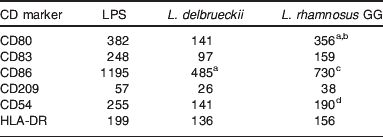
Expression of the co-stimulatory molecules CD80, CD86 and CD54 as well as the maturation marker CD83 was induced by direct incubation with LAB. However, except for CD86, effects were weaker or absent with L. delbrueckii and not statistically significant. Expression of HLA-DR, a molecule of MHC-II involved in antigen presentation, showed a tendency to increase but without reaching statistical significance. On the contrary, CD209 (DC-SIGN), involved in T cell activation and ingestion of antigens by DC, was significantly down-regulated following incubation with both LAB strains with no significant differences between them (see Table 2). The observed modifications are typical for DC maturation. Secretion of IL-1β, IL-10, IL-12 and TNFα increased significantly after direct incubation of DC with L. delbrueckii, while a weaker non-significant effect or, in the case of IL-12, no effect could be seen for L. rhamnosus. In turn, interferon-γ was not produced (data not shown). When DC were incubated with LAB separated by a Caco-2 monolayer, no effects on cell surface markers or cytokine production were observed (P Klein and I Elmadfa, unpublished results).
Table 2. Change (%) in expression of CD markers on dendritic cells following direct incubation with lipopolysaccharide (LPS; positive control, 1 μg/ml) or Lactobacilli (1:10) for 24 h compared with the negative control (De Man Rogosa Sharpe broth (MRS) medium only)

Data were analysed for statistical differences using one-way ANOVA and post hoc Bonferroni's multiple comparison testing.
a P<0·01 compared with the negative control.
b P<0·05 compared with L. delbrueckii.
c P<0·001 compared with the negative control.
d P<0·05 compared with the negative control.
Conclusion
The immune-stimulatory effects of LAB are well recognised. While there is much evidence for the influence of various probiotic strains, conventional cultures are also beneficial to health as suggested by the findings described above. Indeed, while the studied strains differed slightly in their effects, all were able to activate immunological functions. Moreover, for most of these latter, the in vivo approaches showed no significant differences between the probiotic and the conventional product. Importantly, the response to exogenous stimuli was increased without major changes in the cell numbers.
Under healthy conditions, the gut mucosa presents a natural barrier to ingested exogenous micro-organisms and antigens(Reference Heyman15). Nevertheless, as also suggested by the in vivo findings described above, these latter can influence the systemic immune system although direct contact between LAB and the host's cells is normally limited to the gastrointestinal tract. Cytokines play a central role in cell signalling, transmission of stimuli and directing the immune response. Indeed, the cytokine pattern depends not only upon the producing cell, but also on the stimulus. Thus, the different types of helper T cells that are essential in specific immune defence only differ by their cytokine pattern with helper T cells of type 1 stimulating a cellular proinflammatory response, and T helper type 2 cells a humoral antibody-mediated one(Reference Mosmann and Sad16). It was shown that LAB have an influence on which cytokines are secreted depending on the particular strain(Reference Fujiwara, Inoue and Wakabayashi17). A number of studies have revealed a role for DC, which are one of the most important antigen-presenting cell populations. Indeed, gastrointestinal DC are able to cross the epithelium and get into direct contact with bacteria in the intestinal lumen(Reference Corthésy, Gaskins and Mercenier18). Presentation of bacterial antigens by DC activates T lymphocytes. Furthermore, co-stimulatory molecules on the surface of DC as well as the cytokines they secrete influence their differentiation. In turn, LAB were shown to have effects on the cytokine pattern and the expression of surface markers by DC(Reference Hart, Lammers and Brigidi19, Reference Mohamadzadeh, Olson and Kalina20). Besides, intestinal epithelial cells have an influence on the response to ingested micro-organisms as well through the secretion of cytokines and the expression of co-stimulatory surface molecules. Notably, secretion of proinflammatory cytokines was down-regulated(Reference Haller, Bode and Hammes21, Reference Parlesak, Haller and Brinz22).
In our in vitro study, both, probiotic and conventional LAB, showed effects at the cellular level, albeit in different ways. This is in accordance with the described strain-specific activity of these micro-organisms. Interestingly, in both approaches, L. delbrueckii was a more potent inducer of proinflammatory cytokines IL-1β and TNFα. This is possibly reflected in the slightly higher in vivo production in the conventional group although a direct comparison is impossible due to the use of different probiotic strains.
The various different effects of single LAB strains on the immune response appear particularly promising with regard to the treatment of some diseases like inflammatory bowel disease or atopic disturbances, but there is still a need for further research.
Acknowledgements
The authors declare that there are no conflicts of interest. The in vivo study was partly funded by the Institut Danone für Ernährung, Germany, and Danone Austria. Danone Austria and the Niederösterreische Molkerei Genossenschaft (NÖM AG) kindly provided the probiotic and the conventional yoghurt, respectively. P.K. was responsible for the laboratory and data analyses of the in vitro study and A.M. was responsible for the laboratory and data analyses of the in vivo study. I.E. was involved in the design and supervision of both investigations. All the authors contributed to the manuscript and approved it.


