


Introduction
Monogeneans stand out as important threats in fish farms, where infections caused by pathological agents are recurrent (Nowak, Reference Nowak2007). Among monogenean parasites, microcotylids are considered particularly hazardous in marine aquaculture because of their impacts on fish health and growth (Ternengo and Katharios, Reference Ternengo and Katharios2008; Ternengo et al., Reference Ternengo, Agostini, Quilichini, Euzet and Marchand2010; Ogawa, Reference Ogawa2015; Shinn et al., Reference Shinn, Pratoomyot, Bron, Paladini, Brooker and Brooker2015). In the Mediterranean, the microcotylid Sciaenacotyle pancerii (Sonsino, Reference Sonsino1891) was originally reported in shi drum [Umbrina cirrosa (L.)] (see Sonsino, Reference Sonsino1891; Parona, Reference Parona1912; Palombi, Reference Palombi1949) and subsequently found in wild meagre [Argyrosomus regius (Asso, 1801)] (see Ktari, Reference Ktari1970). Recent studies also report massive infections of S. pancerii in farmed meagre (Merella et al., Reference Merella, Cherchi, Garippa, Fioravanti, Gustinelli and Salati2009; Ternengo et al., Reference Ternengo, Agostini, Quilichini, Euzet and Marchand2010), an emerging species in Mediterranean aquaculture (Rigos and Katharios, Reference Rigos and Katharios2010). Therefore, related studies deal with parasite effects in farmed hosts (Merella et al., Reference Merella, Cherchi, Garippa, Fioravanti, Gustinelli and Salati2009; Ternengo et al., Reference Ternengo, Agostini, Quilichini, Euzet and Marchand2010) and usually disregard morphological analysis, despite its relevance for diagnosis and infection management.
In the field, all life-history stages of S. pancerii (i.e. adults, juveniles and post-larvae) occur in mixed infections, but specific identification relies on the morphological analysis of adult specimens (Sonsino, Reference Sonsino1891; Ktari, Reference Ktari1970). Understanding sources of morphological variability, such as the development in monogeneans, may help determine consistent diagnostic features (Thoney, Reference Thoney1986) and can be critical for decision making in aquaculture. Previous morphological analyses on S. pancerii include the descriptions of the oncomiracidium, the adult and a post-larval stage with 12 pairs of clamps (Ktari, Reference Ktari1970), but the development of this species or any other microcotylid with asymmetrical haptor is still unexplored. Due to the relevance of the haptor morphology in parasite attachment (Kearn, Reference Kearn and Kearn2004), further developmental studies are timely and required. Therefore, this study aims at understanding the attachment strategy and improving the morphological description of S. pancerii by describing the main morphological changes occurring during the post-larval development and comparing the development of symmetrical and asymmetrical microcotylids.
Materials and methods
Specimens of the current study were provided and identified by the authors of a previous study (Merella et al., Reference Merella, Cherchi, Garippa, Fioravanti, Gustinelli and Salati2009). Sciaenacotyle pancerii individuals were obtained from 36 heavily infected meagre (A. regius) collected during an epizootic episode (mean intensity above 100 parasites/fish) in a fish farm off the north-east of Sardinia (41°00′N, 8°52′E to 39°59′N, 9°41′E) between September and October 2007. Based on the quality of the specimens and their developmental stage, a total of 114 ethanol-fixed parasites were selected for morphological analysis. Worms were examined on permanent or temporary mounts, according to their developmental degree. Most parasites (N = 94, > 6 pairs of clamps) were stained with iron acetocarmine, dehydrated through a graded alcohol series, cleared in dimethyl phthalate and mounted in Canada balsam. Additionally, some early post-larval stages (N = 20, ⩽ 28 pairs of clamps) were mounted unstained in Kaiser's glycerol-gelatine (Sigma-Aldrich, USA). Morphological analysis of post-larvae specimens of S. pancerii was performed using a Leica DMR light microscope (Wetzlar, Germany; 100–1000×) and data on the main developmental events were recorded. Morphological changes in key structures for attachment (oral suckers, larval haptor and clamps), feeding (pharynx, oesophagus, gut and caeca) and reproduction (genital atrium, vitellaria, uterus, vagina, germarium, testes and intrauterine eggs) were registered. The clamp pair number of each parasite and measurements of the main structures were obtained from drawings using ImageJ 1.48v software (Rasband, 1997–Reference Rasband2016). Following previous studies on microcotylids, the clamp pair number ‘CPN’ was used as an estimator of the age of the specimens. Parasites will be hereafter referred to according to their developmental degree as ‘CPN#’ in symmetrical stages or ‘CPN#short side/#long side’ in asymmetrical stages, being ‘#’ the number of clamps in each haptor side. The correlation between body length (mm) and the CPN during development was analysed. To account for post-maturity variability, bivariate relationships between the CPN and body size, haptor length and the number or size of the morphological structures in mature stages of S. pancerii were also analysed with the statistical significance set at P < 0.05. For statistical purposes, the number of clamps at the longer side of the haptor side is used as CPN. Kendall's correlation coefficient was calculated in R v.3.1.2 software (R Development Core Team, 2014). The size of the morphological structures is expressed as length × width in micrometres unless otherwise stated.
Results
One hundred fourteen parasites were morphologically analysed to describe the main developmental events in the post-larval development of S. pancerii. Most of the parasites were immature stages (from CPN 1 to CPN 91; N = 68), while 40% of the specimens were mature stages (from CPN 94/96 to CPN 132/137; N = 46).
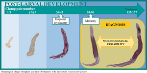
The youngest specimen (CPN 1) is 275 long × 50 wide. The first pair of clamps (42 × 52) develop anterior to the posterior hooks (35 long) and larval hamuli (34 × 21) (Fig. 1A). The CPN 1 specimen shows the postero-lateral hooklets in the posterior-most region of the haptor whereas lateral hooklets are not observed. As parasite grew, new clamps develop towards the anterior end of the haptor, where between 1 and 5 developing pairs of clamps can be observed: 1–2 up to CPN 21; 2–4 up to 50; 2–5 up to CPN 111; 2–4 up to CPN 114/116; 1–2 up to CPN 132/137. The terminal lappet with posterior hooks and hamuli falls between CPN 10 and CPN 17 stages (N = 20; Table 1; Figure 1B). Posterior hooks (35–42 long) and larval hamuli (33–42 × 21–25) barely grow through post-larval development from CPN 1 to CPN 17 stages (N = 26). The posteriormost clamps are the smallest and the first to be developed and do not grow throughout development, overlapping in size from CPN 1 to CPN 132/137 stages (32–48 × 50–62). Germinal clamps develop at the anterior haptor end and increase gradually in size. The largest clamp pair is generally located between the anterior third and half of the haptor. Clamps anterior and posterior to the largest clamp pair are progressively smaller. The size of the anteriormost clamps is always larger than the posteriormost clamps. The size of the largest clamp increases throughout development from 39 × 58 at CPN 11 to 72 × 140 at CPN 109/112 parasites. Both sides of the haptor show a similar number and size of clamps until CPN 39 stages. A slight haptoral asymmetry in the number of clamps, either on the left or the right side, was detected in older parasites, especially in mature stages (Fig. 1I and J). The difference in the number of clamps between the long and the short haptor side ranges between 2 and 13 (CPN 63/61 and CPN 103/116, respectively). The clamps in the longer side of the haptor are also slightly wider (between +5 and +10 μm width, overall), especially in the anterior half of the haptor. The specimen with the maximum number of clamps accounts for 132/137 clamps.

Fig. 1. Morphology of different post-larval stages of Sciaenacotyle pancerii. (A) Earliest clamp-bearing stage (CPN1). (B) CPN 13 with the terminal lappet. (C–D) CPN 36 with a detail of the genital atrium, (E–F) CPN 50 with a detail of the testes. (G–H) CPN 60 with a detail of the uterus and germarium. (I) Mature specimen without eggs. (J) Mature specimen with intrauterine eggs. Scale bars = (A, D) 50 μm, (F, H) 100 μm, (B) 200 μm, (C, E, G) 500 μm, (I) 1000 μm, (J) 2000 μm.
Table 1. Developmental stage and body length of the specimens at the occurrence of the main events in the post-larval development of Sciaenacotyle pancerii. Developmental stages designated by the clamp pair number (CPN)

CPN in asymmetrical stages referred to as the number of clamps at the short/long haptor side.
A pair of oval-shaped buccal suckers (20 × 14), as well as a circular pharynx (13 × 13), are already visible at CPN 1 stages (Fig. 1A). The septum is differentiated in each sucker from CPN 11 stages. Biloculate suckers (77 × 52) are completely defined at CPN 36 stages and reach their maximum size (150 × 109) at CPN 111/119 specimens. The pharynx increases in size and changes from circular in CPN 1 stages to pear shaped in juveniles before maturity (CPN 81; 76 × 71). Maximum size is reached after maturity (CPN 123/128; 124 × 94). The gut is diffuse and extends to the haptor in the early post-larval stages (CPN 1 – CPN 6; N = 4). From CPN 10 stages, the bifurcated gut, as well as the oesophagus and caeca, are clearly distinguishable.
Regarding the reproductive structures, S. pancerii follows a protandrous development sequence (Table 1). The genital atrium and the vagina are the first structures developed and are distinguishable from < 35 CPN. The primordium of the genital atrium, unarmed and circular shaped, was first recognized at CPN 31 stages. By the CPN36 (Fig. 1C and D) the genital atrium, transversally elongated and constricted, was clearly defined (80 × 84) and the genital spines had developed. The genital atrium grows and reaches the definitive size (190 × 387) at CPN 124/134. The primordia of testes are also apparent at CPN36. Testes are completely defined (21 × 38; N = 38) with visible spermatids at CPN 51 (Fig. 1E and F). Testes increase in size and number through development to their maximum (79 × 110; N = 71) between CPN 122/131 and CPN 124/134 stages (N = 6). Female reproductive organs begin to develop between CPN 50 and CPN 59 stages (N = 4; Figure 1G and H) when the uterus is developed and the primordia of the germarium and vitelline reservoir were first observed. In course of development, the germarium grows from 682 long at CPN 84/90 to 1604 long at CPN 111/119 and vitelline glands extend. Completely mature parasites (Fig. 1I and J) were detected from CPN 94/96 when the first intrauterine eggs (N = 1) are observed. A maximum of 111 intrauterine eggs (mean per egg = 180 × 69; N = 23) were found in CPN 109/112 specimens.
The clamp pair number strongly correlates with body length throughout parasite development (Fig. 2). The relationship between the 2 variables is described by a linear function (R 2 = 0.939; Y = 0.0809x – 0.2077). Positive correlations between total body size (length and width) (P < 0.001), the largest clamp size (P < 0.05) and haptor length (P < 0.001) with the CPN were recorded throughout post-maturity development. The size of suckers (P < 0.05), pharynx (P < 0.05), genital atrium (P < 0.01), testes (P < 0.01) and germarium (P < 0.01) and the number of testes (P < 0.01) also increased significantly with the CPN after parasite maturity. No significant correlations were found between the rest of the morphological variables of S. pancerii nor the number of intrauterine eggs in mature stages and the CPN (P > 0.05).

Fig. 2. Body length of Sciaenacotyle pancerii as a function of the number of clamp pairs. The clamp-pair number in asymmetrical stages referred as the number of clamps at the longer haptor side. Dotted lines represent the 95% confidence interval. Dashed lines represent 95% prediction interval. Oncomiracidia length from Ktari (Reference Ktari1970).
Discussion
Developmental changes in the morphology of S. pancerii are poorly known though they can be helpful for understanding parasite behaviour and effects and assessing the infection status. The post-larval development of S. pancerii is herein described for the first time. New records on S. pancerii provide novel developmental information on a genus with special morphological features among microcotylids (i.e. large body length, high clamp pair number and asymmetrical haptor) and thus, enhance the current knowledge on the former family.
The analysis of the post-larval development of S. pancerii provides for the first time the size variability of the larval hooks, hamuli and germarium during development (only available in figures to date; see Ktari, Reference Ktari1970), and updates the dimensions of the parasite body and attachment structures of adult specimens. The size of the main morphological features of adult stages of S. pancerii is generally consistent with the original taxonomic description (Sonsino, Reference Sonsino1891) and the redescription (Ktari, Reference Ktari1970) of the species as well as with recent reports where it was identified (Merella et al., Reference Merella, Cherchi, Garippa, Fioravanti, Gustinelli and Salati2009). Posteriormost clamps are, however, smaller (shorter and narrower) than previously described (60 × 60 μm; Ktari, Reference Ktari1970) and tend to be wider than long, as described for the rest of microcotylids (Mamaev, Reference Mamaev and Lebedev1989). The size variability of most morphological structures of adult S. pancerii was higher than previously reported. To date, metrical data on S. pancerii is exclusively based on specimens infecting U. cirrosa (Sonsino, Reference Sonsino1891; Ktari, Reference Ktari1970) while this is the first morphological analysis of this monogenean on A. regius, thus the higher variability can be related to the host species. Sample size, host size (Thoney, Reference Thoney1988) or environmental factors at the sampling locality (Brazenor and Hutson, Reference Brazenor and Hutson2015) may also contribute to this size variability. The new measurements of adult specimens of S. pancerii allow widening the morphological ranges of some taxonomic characters at the top and low margins and should be updated and considered for species identification (see Table 2). To date, 20 microcotylid species, belonging to the genera Anchoromicrocotyle, Cynoscionicola, Diplostamenides, Microcotyloides, Pauciconfibula and Sciaenacotyle have been described from sciaenid fish (see Gibson et al., Reference Gibson, Bray and Harris2005; WoRMS Editorial Board, 2022). The genus Sciaenacotyle differs from other microcotylids from sciaenid hosts mainly by the shape and armature of the genital atrium (Fujii, Reference Fujii1944; Price, Reference Price1962; Unnithan, Reference Unnithan1971; Bravo-Hollis, Reference Bravo-Hollis1981; Mamaev, Reference Mamaev and Lebedev1989; Chisholm et al., Reference Chisholm, Beverley-Burton and McAlpine1991). The same feature allows for discriminating Sciaenacotyle spp. together with its hosts and geographic distribution (Hayward et al., Reference Hayward, Bott, Itoh, Iwashita, Okihiro and Nowak2007). Moreover, present findings on S. pancerii development reveal that some morphological features of adults, commonly used for species diagnosis within the Microcotylidae (e.g. the number of testes or clamps) (Mamaev, Reference Mamaev and Lebedev1989) can be difficult to ascertain or be variable after maturity. Testes in S. pancerii have a poorly defined contour, are closely overlapped and are partially covered by the vitelline fields thus hindering the counting. The number of testes increases from 38 in young to 71 in old adults, which may also be confusing. Based on the size variations of S. pancerii throughout adult development, we recommend setting the number of clamps on the short side to a minimum of 116 to offer consistent and comparative morphological data.
Table 2. Measurements (in micrometres) of distinctive features of mature stages of Sciaenacotyle pancerii

Data expressed as mean ± s.d. (range).
*Intrauterine eggs.
a Haptor with 100–130 clamps in each side with a minimum difference from 4 to 8 between both sides (Ktari, Reference Ktari1970).
Post-larval development of S. pancerii is characterized by: progressive acquisition of haptoral clamps combined with increases in body size, expansion and bifurcation of the gut, loss of the larval haptor, protandrous development of the genitalia and finally formation of the vitellaria. This developmental sequence is generally consistent with previous studies on microcotylids (Sproston, Reference Sproston1946; Thoney, Reference Thoney1986; Thoney and Munroe, Reference Thoney and Munroe1987; Ogawa, Reference Ogawa1988; Roubal and Diggles, Reference Roubal and Diggles1993; Repullés-Albelda et al., Reference Repullés-Albelda, Raga and Montero2011). Nevertheless, some developmental events, such as terminal lappet loss or parasite maturity, occurred in older stages than in other microcotylids. The terminal lappet is generally lost at 2–13 CPN stages in other microcotylids (Thoney, Reference Thoney1986) whereas it was retained up to 17 CPN stages in S. pancerii. These differences are likely related to the comparatively higher clamp pair number reached by S. pancerii at the end of development (i.e. 130–132/137 clamps per haptor side; Ktari, Reference Ktari1970; present study). In fact, the terminal lappet loss in S. pancerii occurs when about 12% of the maximum clamp pair number has been developed, which fits with previous records on other microcotylids (between 10 and 20% of the maximum clamp pair number; Remley, Reference Remley1942; Thoney, Reference Thoney1986; Thoney and Munroe, Reference Thoney and Munroe1987; Roubal and Diggles, Reference Roubal and Diggles1993; Repullés-Albelda et al., Reference Repullés-Albelda, Raga and Montero2011). By contrast, parasite maturity is achieved at comparatively older stages in S. pancerii than in most microcotylid species, even when the proportion between CPN at maturity and the maximum CPN is considered. While parasite maturity generally occurred between 15 and 45 CPN stages, when 50–70% of the clamps are developed (Repullés-Albelda et al., Reference Repullés-Albelda, Raga and Montero2011), S. pancerii reach maturity at 94/96 CPN stages with at least 70% of the maximum CPN (Ktari, Reference Ktari1970; present study). Despite fitting within the general range of microcotylids, S. pancerii is one of the latest to reach maturity, only coinciding with Polylabroides multispinosus Roubal, 1981 (Roubal and Diggles, Reference Roubal and Diggles1993) and Microcotyle hiatulae Goto, 1894 (Thoney and Munroe, Reference Thoney and Munroe1987) which, however, are characterized by a smaller size and a lower clamp pair number.
Developmental data have been reported for 8 out of the 218 microcotylid species listed in the World Register of Marine Species (WoRMS Editorial Board, 2022), but only one of them, Diplostamenides spinicirrus (MacCallum, 1918), infects a sciaenid host. The current study describes the development of the unique asymmetric microcotylid reported to date. The combination of morphological features in S. pancerii, i.e. haptoral asymmetry, large body size and a high number of clamps is rare among microcotylids (Yamaguti, Reference Yamaguti1963). Indeed, only 3 of the microcotylids with known development compare well for at least one of the exclusive features (i.e. body length, clamp pair number or the maximum length/CPN ratio). Regarding body size, S. pancerii coincides in length with D. spinicirrus, a parasite of the freshwater sciaenid Aplodinotus grunniens (Rafinesque, 1819), but the maximum CPN of the latest (99 pairs of clamps; Remley, Reference Remley1942) is substantially lower. According to the ratio between maximum length and CPN, increments of body length per CPN in S. pancerii (ratio = 80–100 for the long haptor side) can partially overlap those of Bivagina tai (Yamaguti, 1938) (ratio = 61–80, max. length = 4000 μm and max. CPN = 50–65; Ogawa, Reference Ogawa1988) and Microcotyle sebastis Goto, 1894 (ratio = 92, max. length = 3300 μm and max. CPN = 36; Thoney, Reference Thoney1986), although both have a lower length and CPN. The large CPN of S. pancerii stands out as exceptional among microcotylids overall because only 2 other species within this family bear more than 100 pairs of clamps; Cynoscionicola heteracanta (Manter, 1938) and Cynoscionicola longicauda (Goto, 1899), both from sciaenid fish (based on the morphological descriptions from Yamaguti, Reference Yamaguti1963). The high clamp number of these microcotylids from sciaenid fish suggests that host traits affect their attachment strategy. Morphological features of the gills of the sciaenids may lead to similar adaptations such as the increase in the number of clamps which may occur relatively fast, based on the high number of developing clamps (between 2 and 5 clamps) observed throughout post-larval development.
The haptor of monogeneans determines the attachment to their hosts and their behaviour (Kearn, Reference Kearn and Kearn2004). In S. pancerii, the haptor is characterized by a high number of clamps, but this species also exhibits a slight haptoral asymmetry in the arrangement and size of the clamps, which is one of the key differential features of Sciaenacotyle spp. within the Microcotylidae (Mamaev, Reference Mamaev and Lebedev1989). The asymmetry in the clamp arrangement is a common diagnosis feature of other mazocraeid families as the heteraxinids (Yamaguti, Reference Yamaguti1963), which also show differences in the clamp size at both haptor sides. Detailed analysis of the asymmetry during the development of microcotylid and heteraxinid species, however, reveals relevant differences related to the first register of the asymmetry. The asymmetrical haptor of S. pancerii was mainly observed from mature stages, while the haptor of most heteraxinids is asymmetrical from the early developmental stages (Ogawa and Egusa, Reference Ogawa and Egusa1981; Thoney, Reference Thoney1988). Asymmetrical monogeneans tend to minimize dislodgment risks by attaching the largest clamp row upstream (Kearn, Reference Kearn and Kearn2004). The morphology of the haptor is, therefore, likely to condition the attachment of these parasites to their hosts, so its variability throughout development may involve a change in the attachment strategies. The symmetrical clamp arrangement in the haptor of S. pancerii at early developmental stages (<39 CPN) suggests that the haptoral asymmetry is not so decisive for parasite attachment in these life-history stages and the clamp addition, as well as the increases in the clamp width, is probably enough to strengthen the attachment to the host gills. The asymmetrical arrangement and size of the clamps in mature stages may, thereafter, allow the parasite to optimize the attachment when it becomes larger. Sciaenacotyle pancerii seems therefore to exhibit a mixed attachment strategy between microcotylids and heteraxinids, combining the high number of clamps (typical of microcotylids) with the asymmetry in the clamp size and arrangement (common in heteraxinids). This mixed strategy may result from the parasite adaptation to a large and fast-growing host and is supported by the phylogenetic closeness of Microcotylidae and Heteraxinidae (Olson and Littlewood, Reference Olson and Littlewood2002) and the isolated position of the genus Sciaenacotyle [represented by S. sciaenicola (Murray, 1932)] within most microcotylids (see Hayward et al., Reference Hayward, Bott, Itoh, Iwashita, Okihiro and Nowak2007). The haptoral asymmetry is, therefore relevant for discriminating among this and other microcotylids while not so useful to distinguish Sciaenacotyle among mazocraeids. The asymmetry features (i.e. differences in number, size and arrangement of clamps) and the developmental pattern of asymmetry may, however, be more use useful in the field. These features, together with the exclusive number of clamps can be more complete and informative for discriminating between mazocraeids.
Data availability
Data available on request from the authors.
Acknowledgements
The authors thank Clemente Graziano of Compagnie Ittiche Riunite for his collaboration in sample collection. We thank 2 anonymous referees for their valuable comments and suggestions.
Author contributions
M. V.-T., F. E. M., P. M. and A. R.-A. conceived the study. M. V.-T. and A. R.-A. designed the analysis. S. C. and P. M. collected and preserved the specimens for the study. M. V.-T. carried out the morphological characterization of the specimens. M. V.-T. and A. R.-A. analysed and interpreted the data. A. R.-A. coordinated and planned the research. F. E. M., G. G. and J. A. R. managed the funding acquisition. M. V.-T. and A. R.-A. drafted the manuscript. All authors read and approved the final version of the manuscript.
Financial support
M. V.-T. benefited from a doctoral fellowship from the Spanish Ministry of Education, Culture and Sports [grant FPU13/05849]. This work was supported by the Spanish Government [projects MINECO/FEDER PID2019-110730RB-I00] co-funded by MCIN/AEI/10.13039/501100011033 by ‘ERDF A way of making Europe’ by the EU, the Valencian Regional Government [project AICO/2021/279] and the Fondazione di Sardegna-call 2017. This study forms part of the ThinkInAzul programme and was supported by MCIN with funding from European Union NextGenerationEU (PRTR-C17.I1) and by Generalitat Valenciana [GVA- THINKINAZUL/2021/029].
Conflict of interest
The authors declare there are no conflicts of interest.
Ethical standards
Not applicable.


 Open access