Hostname: page-component-76fb5796d-9pm4c Total loading time: 0 Render date: 2024-04-26T15:10:59.531Z Has data issue: false hasContentIssue false

Dual release of VEGF and PDGF from emulsion electrospun bilayer scaffolds consisting of orthogonally aligned nanofibers for gastrointestinal tract regeneration

Published online by Cambridge University Press:  05 August 2019

Yu Zhou
Affiliation:
Department of Mechanical Engineering, The University of Hong Kong, Pokfulam Road, Pokfulam, Hong Kong
Qilong Zhao*
Affiliation:
Department of Mechanical Engineering, The University of Hong Kong, Pokfulam Road, Pokfulam, Hong Kong Institute of Biomedical and Health Engineering, Shenzhen Institutes of Advanced Technology (SIAT), Chinese Academy of Sciences (CAS), Shenzhen, China
Min Wang*
Affiliation:
Department of Mechanical Engineering, The University of Hong Kong, Pokfulam Road, Pokfulam, Hong Kong
*
Address all correspondence to Q. Zhao at ql.zhao@siat.ac.cn and M. Wang at memwang@hku.hk
Get access

Abstract

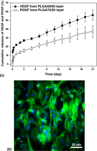
The regeneration of human tissues with complex anatomy such as gastrointestinal (GI) tract remains greatly challenging since it requires appropriate cell microenvironments with well-defined structural and biochemical cues. In this investigation, bilayer scaffolds consisting of different polymer nanofibers with orthogonal fiber orientations were prepared, in which vascular endothelial growth factor (VEGF) and platelet-derived growth factor (PDGF) were encapsulated separately. The bilayer scaffolds have similar architecture to the anatomy of the GI tract and can achieve dual releases of VEGF and PDGF in sequential and sustained manners, which hold promise as appropriate cell microenvironments for promoting the regeneration of the GI tract.

Type
Research Letters
Copyright
Copyright © The Author(s) 2019 

Access options

Get access to the full version of this content by using one of the access options below. (Log in options will check for institutional or personal access. Content may require purchase if you do not have access.)

References

1.Bitar, K.N. and Zakhem, E.: Tissue engineering and regenerative medicine as applied to the gastrointestinal tract. Curr. Opin. Biotechnol. 24, 909 (2013).10.1016/j.copbio.2013.03.021Google Scholar
2.Webber, M.J., Khan, O.F., Sydlik, S.A., Tang, B.C., and Langer, R.: A perspective on the clinical translation of scaffolds for tissue engineering. Ann. Biomed. Eng. 43, 641 (2015).Google Scholar
3.Yang, G., Li, X., He, Y., Ma, J., Ni, G., and Zhou, S.: From nano to micro to macro: electrospun hierarchically structured polymeric fibers for biomedical applications. Prog. Polym. Sci. 81, 80 (2018).Google Scholar
4.Zhao, Q., Wang, J., Cui, H., Chen, H., Wang, Y., and Du, X.: Programmed Shape-morphing Scaffolds Enabling Facile 3D Endothelialization. Adv. Funct. Mater. 28, 1801027 (2018).Google Scholar
5.Zhao, Q., Cui, H., Wang, J., Chen, H., Wang, Y., Zhang, L., Du, X., and Wang, M.: Regulation effects of biomimetic hybrid scaffolds on vascular endothelium remodeling. ACS Appl. Mater. Interfaces 10, 23583 (2018).Google Scholar
6.Tsao, C.J., Pandolfi, L., Wang, X., Minardi, S., Lupo, C., Evangelopoulos, M., Hendrickson, T., Shi, A., Storci, G., Taraballi, F., and Tasciotti, E.: Electrospun patch functionalized with nanoparticles allows for spatiotemporal release of VEGF and PDGF-BB promoting in vivo neovascularization. ACS Appl. Mater. Interfaces 10, 44344 (2018).Google Scholar
7.Han, F., Jia, X., Dai, D., Yang, X., Zhao, J., Zhao, Y., Fan, Y., and Yuan, X.: Performance of a multilayered small-diameter vascular scaffold dual-loaded with VEGF and PDGF. Biomaterials 34, 7302 (2013).Google Scholar
8.Zhao, Q., Lu, W.W., and Wang, M.: Modulating the release of vascular endothelial growth factor by negative-voltage emulsion electrospinning for improved vascular regeneration. Mater. Lett. 193, 1 (2017).Google Scholar
9.Ferrara, N., Gerber, H., and LeCouter, J.: The biology of VEGF and its receptors. Nat. Med. 9, 669 (2003).10.1038/nm0603-669Google Scholar
10.Wells, J.M. and Spence, J.R.: How to make an intestine. Development 141, 752 (2014).Google Scholar
11.Ye, H., Zhang, K., Kai, D., Li, Z., and Loh, X.J.: Polyester elastomers for soft tissue engineering. Chem. Soc. Rev. 47, 4545 (2018).10.1039/C8CS00161HGoogle Scholar
12.Szentivanyi, A., Chakradeo, T., Zernetsch, H., and Glasmacher, B.: Electrospun cellular microenvironments: understanding controlled release and scaffold structure. Adv. Drug Deliv. Rev. 63, 209 (2011).10.1016/j.addr.2010.12.002Google Scholar
13.Nair, L.S. and Laurencin, C.T.: Biodegradable polymers as biomaterials. Prog. Polym. Sci. 32, 762 (2007).Google Scholar
14.Zhou, Y., Zhao, Q., Tsai, N.L.Y., and Wang, M.: Bicomponent nanofibrous scaffolds with dual release of anticancer drugs and biomacromolecules. MRS Commun. 9, 413 (2019).Google Scholar
15.Freudenberg, U., Zieris, A., Chwalek, K., Tsurkan, M.V., Maitz, M.F., Atallah, P., Levental, K.R., Eming, S.A., and Werner, C.: Heparin desulfation modulates VEGF release and angiogenesis in diabetic wounds. J. Control. Release 220, 79 (2015).Google Scholar
16.Zhao, Q. and Wang, M.: Strategies to incorporate polyelectrolyte in emulsion electrospun nanofibrous tissue engineering scaffolds for modulating growth factor release from the scaffolds. Mater. Lett. 162, 48 (2016).10.1016/j.matlet.2015.09.120Google Scholar
17.Serra, L., Doménech, J., and Peppas, N.A.: Drug transport mechanisms and release kinetics from molecularly designed poly (acrylic acid-g-ethylene glycol) hydrogels. Biomaterials 27, 5440 (2006).Google Scholar
18.Kobayashi, M., Lei, N.Y., Wang, Q., Wu, B.M., and Dunn, J.C.Y.: Orthogonally oriented scaffolds with aligned fibers for engineering intestinal smooth muscle. Biomaterials 61, 75 (2015).Google Scholar
19.Zhang, H., Jia, X., Han, F., Zhao, J., Zhao, Y., Fan, Y., and Yuan, X.: Dual-delivery of VEGF and PDGF by double-layered electrospun membranes for blood vessel. Biomaterials 34, 2202 (2013).Google Scholar