

The spinal cord serves as a kind of information relay center, transmitting signals between the brain and the rest of the body—damage to this center can lead to various issues, including paralysis. Between 250,000 and 500,000 people suffer a spinal-cord injury each year, according to the World Health Organization. For decades, researchers have sought to find ways to restore communication between mind and body in people with damaged spinal cords, and they have made some significant advances. Brain-computer interfaces and other technologies have allowed some people to control machines (robotic arms) with their thoughts and others to regain some function to their paralyzed hands.
In a leap forward for the field, an international team of researchers has created a new technology that allowed a rhesus macaque with a paralyzed leg (due to a spinal lesion) regain the ability to walk within a week, without training. The new technology, called a “brain-spine interface” and described recently in Nature (doi:10.1038/nature20118), works by bypassing severed nerves in the spinal cord. That is, a computer decodes locomotion-related signals from the monkey’s brain into intentions and sends instructions to an implanted pulse generator (situated below the spinal-cord site of injury), which stimulates sensory neurons involved in controlling the flexion and extension of the limbs—all in real time.
“For a long time, we’ve been focused on the pharmaceutical world and have neglected the possibility of technological devices that may provide very good therapy,” says David A. Borton, a neuroengineering professor at Brown University and one of the study’s lead authors. “We’re moving into an age where technology is getting small enough and safe enough that we could use it in ways that we haven’t thought of before.”
While the new work certainly builds on prior research in the field, it is distinct in significant ways. All previous demonstrations of brain interfaces have focused on the upper limbs, specifically recording signals from the hand and arm areas of the motor cortex (the region of the brain that is responsible for voluntary movement). Additionally, other systems have not stimulated nerve cells that ultimately control muscle movement; they have focused instead on directly stimulating muscles.
Movement in the legs that enables walking is controlled by neurological activity in the leg area of the motor cortex. Signals from this area travel down the spinal cord to its lumbar (lower) region where they activate motor neurons that coordinate muscle movement. If the spinal cord is damaged or severed, however, those brain signals are unable to reach their destination in the lumbar spinal cord, resulting in leg paralysis. Though both motor cortex and spinal neurons may be functional, their inability to communicate prevents leg movement.
“It is often thought that if you have an injury at some place in the spinal cord, nothing below it is functional,” Borton says. “But a lot of the spinal-cord circuits are still quite functional, so what this study relied on was reactivating those remaining circuits.”
The brain-spine interface is made up of several components. The first is a standard 96-channel microelectrode array that is placed over the leg area of the motor cortex and detects the spiking activity of 100 neurons. This array is connected to a lightweight wireless neurosensor; developed by Borton and his team at Brown in 2014, the low-power, high-efficiency neurosensor is composed of a 100-channel transmitter and four-antenna receiver. The device records and beams the neural activity to a computer, which decodes the signals into intentions in real time. The computer wirelessly sends instructions to an electrical spinal stimulator (a pulse generator) that is implanted in the lumbar spine, just below the site of spinal injury. Rather than activate motor neurons directly, which would result in muscle activity that is not necessarily coordinated, the implant stimulates the sensory inputs into the spinal cord. These inputs are part of the reflex circuits that assist with balance and coordinated muscle movement, and are integrated naturally into the normal gait cycle—people do not have to actively think about walking most of the time because the process is mostly controlled by the spinal cord and reflex circuits.
Importantly, to determine which neural circuits along the spinal cord the implant needed to stimulate, Borton’s colleagues at the École Polytechnique Fédérale Lausanne in Switzerland, led by neuroscientist Grégoire Courtine, developed spatiotemporal maps of motoneuron activation in healthy macaques. By monitoring the neuronal activity of monkeys during locomotion, the researchers were able to identify extension and flexion hotspots in the lumbar spinal cord. The researchers designed their spinal implant to specifically target these spinal-cord hotspots. Microfabrication of the implant involved spin-coating a 40-μm-thick polyimide film onto a silicon substrate, and using UV lithography to shape the polyimide-based bottom layer. The researchers created a conductive seed pattern by depositing a 200-nm-thick gold layer using vacuum evaporation and lithography. They also electroplated an additional gold layer for the electrodes and leads. They then spin-coated a 20-μm-thick cover layer of photosensitive polyimide to the bottom of the implant, and used ultraviolet lithography to structure the top layer of the implant (while also creating an opening for the electrodes and contact pads). After releasing the implant from the silicon substrate, they mounted it to a printed circuit board and connected surgical leads. Finally, they joined the implant to a vertebral orthosis, which they fashioned through three-dimensional laser sintering of medical grade titanium. This polyimide spinal implant is connected to the pulse generator.
The researchers also developed a decoder that tied activity in the motor cortex to hotspot activation and locomotion. They calibrated the decoder with healthy macaques and found that the system was able to accurately predict motor cortex activity associated with the flexion and extension of leg muscles.
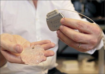
The brain-spine interface works by first decoding signals from the motor cortex into intentions and then using electrical pulses to target specific circuits of the spinal cord that ultimately drive locomotion. Shown here: a pulse generator and a silicon model of a primate’s brain with an attached microelectrode array. Credit: Alain Herzog, EPFL.
The researchers’ colleagues at the China Academy of Medical Sciences implanted the system in two macaques with spinal-cord lesions that disabled the use of their right hind legs. The first macaque, which had a less severe lesion, was able to walk again on a treadmill and normal ground—with nearly normal locomotor patterns—in just six days after using the brain-spine interface, so long as the device was turned on. The second macaque recovered the ability to walk after two weeks of using the technology.
Gaurav Sharma, a research scientist at the nonprofit research and development organization Battelle, thinks the work is “fantastic” and further shows how neural signals can be rerouted to help people with paralysis. “The study is exciting as it shows what is possible in the future and has important implications in the field of rehabilitation and neuroplasticity,” says Sharma, who recently helped develop neural bypass technology that allowed a man with quadriplegia regain hand function. “Translation of this work for human use, however, is still many years away and will not be as straightforward. Human locomotion is more complex [than macaque locomotion], as we not only need to support our weight while walking but also need to coordinate gait during locomotion.”
Borton agrees that much work is necessary before the brain-spine interface is ready for human testing and use, and stresses that its first application will likely be in the rehabilitation setting for people whose spinal cords are not completely severed. “It could be used to help those neurons remaining after the injury to strengthen their connection and regain some function,” he says.
