


Introduction
Tourmalines are complex borosilicates that have been studied extensively in terms of their crystal structure and crystal chemistry (e.g. Foit, Reference Foit1989; Hawthorne, Reference Hawthorne1996; Hawthorne and Henry, Reference Hawthorne and Henry1999; Ertl et al., Reference Ertl, Hughes, Pertlik, Foit, Wright, Brandstatter and Marler2002; Novák et al., Reference Novák, Povondra and Selway2004; Bosi and Lucchesi, Reference Bosi and Lucchesi2007; Bosi, Reference Bosi2013, Reference Bosi2018; Henry and Dutrow, Reference Henry and Dutrow2011; Cempírek et al., Reference Cempírek, Houzar, Novák, Groat, Selway and Šrein2013; Bačík and Fridrichová, Reference Bačík and Fridrichová2020). In accordance with Henry et al. (Reference Henry, Novák, Hawthorne, Ertl, Dutrow, Uher and Pezzotta2011), the general chemical formula of tourmaline is written as: XY3Z6T6O18(BO3)3V3W, where X = Na+, K+, Ca2+, □ (= vacancy); Y = Al3+, Fe3+, Cr3+, V3+, Mg2+, Fe2+, Mn2+, Li+; Z = Al3+, Fe3+, Cr3+, V3+, Mg2+, Fe2+; T = Si4+, Al3+, B3+; B = B3+; V = (OH)–, O2–; and W = (OH)–, F–, O2–. Note that the non-italicised letters X, Y, Z and B represent groups of cations hosted in the [9]X, [6]Y, [6]Z, [4]T and [3]B crystallographic sites (letters italicised). As for the letters V and W, they represent groups of anions accommodated at the [3]-coordinated O3 and O1 crystallographic sites, respectively. The dominance of specific ions at one or more sites of the structure gives rise to a range of distinct mineral species.
A formal description of the new tourmaline species princivalleite is presented here. The mineral is named after Francesco Princivalle (b. 1956), Professor of Mineralogy at the Department of Mathematics and Geosciences, University of Trieste, Italy, for his contributions to the understanding of the crystal chemistry and geothermometery of several group minerals such as spinels, olivines and pyroxenes. The new species and the name (symbol Pva) have been approved by International Mineralogical Association's Commission on New Minerals, Nomenclature and Classification (IMA2020-056, Bosi et al., Reference Bosi, Pezzotta, Skobgy, Altieri, Hålenius, Tempesta and Cempírek2020). Holotype material is deposited in the collections of the Natural History Museum of Milano, Italy, catalogue number M38850.
Occurrence
The holotype specimen was collected in 2003 by one of the authors (FP), along the cut of a small road at the eastern side of the Curiglia Village, Veddasca Valley, Luino area, Varese, Lombardy, Italy (46°03′30.74′′N, 8°48′24.47′′E, ~730 m above the sea level). The Veddasca Valley is characterised by rocks belonging to the ‘Serie del Laghi’, a structural unit which is part of the tectonic unit known as Massiccio del Laghi, comprising the central-western sector of the crystalline basement of the Southern Alps (Pezzotta and Pinarelli, Reference Pezzotta and Pinarelli1994). The ‘Serie dei Laghi’ consists of paragneiss (pelites, sandstones, greywackes and metatufites) and scattered orthogneissic bodies, affected by amphibolite-facies Hercynian metamorphism (Boriani et al., Reference Boriani, Giobbi Origoni and Borghi1990).
During a detailed field mapping performed by one of the authors in 1990–1991, a few narrow, not-metamorphic, veins of pegmatite were discovered nearby the Curiglia village in the Veddasca valley. These pegmatites, which have never been reported in literature, crosscut some heavily deformed metamorphic rocks (flaser gneiss) perpendicularly to their foliation, and probably originated by some minor partial melting that occurred during the uplift of the tectonic units, during the latest stages of the Hercynian metamorphic event. At the centre of one of these small veins (2 to 3 cm wide and a few metres long), vertically crosscutting the flaser gneiss at Curiglia, there are some quite common occurrences of dispersed grains and crystals up to 1 cm long, of azure princivalleite and oxy-schorl. The pegmatitic vein is composed of muscovite aggregates, with blades mostly oriented perpendicular to the walls, with quartz, albitic plagioclase and minor K-feldspar. In addition to tourmaline, other accessories are rare small pyrite crystals and violet glassy cordierite grains.
Appearance, physical and optical properties
The princivalleite crystals show subhedral habits, up to ~10 mm and are azure with a vitreous lustre (Fig. 1). It has a white streak and shows no fluorescence. It has a Mohs hardness of ~7 and is brittle with a conchoidal fracture. The calculated density, based on the empirical formula and unit-cell volume refined from single-crystal X-ray diffraction (XRD) data, is 3.168 g/cm3. In thin section, princivalleite is transparent; in transmitted light, pleochroism was not visible in the thin-section fragment investigated. Princivalleite is uniaxial (–) with refractive indices ω = 1.650(5) and ɛ = 1.635(5) measured by the immersion method using white light from a tungsten source. The mean index of refraction, density, and chemical composition led to an excellent compatibility index (1 – Kp/Kc = 0.024) (Mandarino, Reference Mandarino1981).
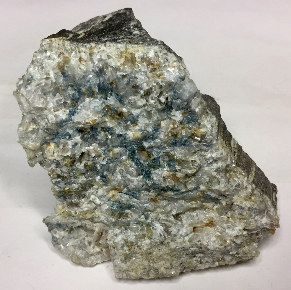
Fig. 1. Photo of the azure princivalleite in reflected light: crystalline aggregates in pegmatitic vein in gneiss. Field of view ca. 12 cm × 12 cm. Sample deposited in the collections of the Natural History Museum of Milano, Italy (photo by F. Pezzotta).
Experimental methods and results
General comment
The present crystal structure refinement (SREF), electron microprobe (EMP) and μ-laser induced breakdown spectroscopy (μ-LIBS) data were all obtained from the same crystal fragment. However, complementary experimental data were recorded from coexisting crystals. Small differences in composition occur between these princivalleite crystals (see text).
Microprobe analysis
Electron microprobe analysis was obtained using a wavelength dispersive spectrometer (WDS mode) with a Cameca SX50 instrument at the ‘Istituto di Geologia Ambientale e Geoingegneria (Rome, Italy), CNR’, operating at an accelerating potential of 15 kV, a sample current of 15 nA and 10 μm beam diameter. Minerals and synthetic compounds were used as standards: wollastonite (Si and Ca), magnetite (Fe), rutile (Ti), corundum (Al), vanadinite (V) fluorophlogopite (F), periclase (Mg), jadeite (Na), orthoclase (K), sphalerite (Zn), rhodonite (Mn) and metallic Cr and Cu. The PAP routine was applied (Pouchou and Pichoir, Reference Pouchou, Pichoir, Heinrich and Newbury1991). The composition (mean of 6 spot analyses) is given in Table 1. Titanium, V, Cr and K were below detection limits (<0.03 wt.%).
Table 1. Electron microprobe data (WDS mode) and atoms per formula unit (apfu) normalised to 31 anions for princivalleite.

a Calculated by stoichiometry, (Y+Z+T) = 15.00 apfu.
b Fe oxidation state determined by Mössbauer spectroscopy.
c Li Determined by μ-Laser Induced Breakdown Spectroscopy.
Micro-laser induced breakdown spectroscopy
Lithium analysis was performed using 110 mJ of energy per pulse by a double pulse Q-Switched (Nd-YAG, λ = 1064 nm) laser with a 1 μs delay between the two pulses. The small spot size (7–10 μm) was obtained using a petrographic optical microscope (objective lens = 10X, NA = 0.25 and WD = 14.75 mm). The μ-LIBS spectra were acquired using an AvaSpec Fiber Optic Spectrometer (390–900 nm with 0.3 nm resolution) with a delay of 2 μs after the second pulse and were integrated for 1 ms. Quantitative data were obtained by generating a linear regression using the main Li emission line intensity (670.706 nm corresponding to resonance transition 1s 2 2s > 1s 2 2p) particularly sensitive to Li amounts. The linear fit was made according to Bosi et al. (Reference Bosi, Celata, Skogby, Hålenius, Tempesta, Ciriotti, Bittarello and Marengo2021) and revealed amounts of Li2O = 0.12 wt.% (Table 1) in line with that estimated by the Pesquera et al. (Reference Pesquera, Gil-Crespo, Torres-Ruiz, Torres-Ruiz and Roda-Robles2016) approach (0.18 Li2O wt.%).
Mössbauer spectroscopy
To determine the Fe3+/ΣFe ratio of princivalleite, a crystal fragment was ground under acetone and analysed using 57Fe Mössbauer spectroscopy with a conventional spectrometer system equipped with a 10 mCi point source and operated in constant acceleration mode. Data were collected over 1024 channels and were folded and calibrated against the spectrum of an α-Fe foil. The spectrum (Fig. 2) was fitted using the software MossA (Prescher et al., Reference Prescher, McCammon and Dubrowinsky2012) with three absorption doublets consistent with Fe2+ (Table 2). No indications of absorption due to Fe3+ was observed.

Fig. 2. Mössbauer spectrum of princivalleite. Fitted absorption doublets assigned to Fe2+ are indicated in blue colour. Diamonds denote measured spectrum, and black curve represents summed fitted spectra.
Table 2. Mössbauer parameters for princivalleite obtained at room-temperature.

δ = centroid shift, ΔEQ = quadrupole splitting, FWHM = full width at half-maximum.
Single-crystal infrared spectroscopy
Polarised Fourier-transform infrared (FTIR) absorption spectra were measured on a 35 μm thick doubly polished single-crystal section oriented parallel to the c-axis. A Bruker Vertex spectrometer attached to a Hyperion 2000 microscope and equipped with a halogen lamp source, a CaF2 beamsplitter, a ZnSe wiregrid polariser and an InSb detector was used to collect spectra in the range 2000–13000 cm–1 at a resolution of 2 cm–1. Spectra recorded in polarised mode parallel to the crystallographic c-axis show a significant band at 3365 cm–1, a very intense band around 3500 cm–1, two weaker bands at 3632 and 3644 cm–1, and two very weak bands at 3662 and 3671 cm–1 (Fig. 3). As observed typically for tourmaline spectra in the (OH) range, the main band is off-scale for the E||c direction due to excessive absorption. Spectra obtained perpendicular to the c-axis show considerably weaker bands.

Fig. 3. Polarised FTIR spectra for princivalleite, vertically off-set for clarity. The main band is truncated at ~2 absorbance units in the E||c direction due to excessive absorption. Note comparatively low intensities of bands above 3600–3650 cm–1 corresponding to very small (OH) contents at W [≡ the O(1) site]. Sample thickness 35 μm.
Note that the band at 3365 cm–1 is consistent with the presence of minor Al along with Si in [4]-fold coordination (Nishio-Hamane et al., Reference Nishio-Hamane, Minakawa, Yamaura, Oyama, Ohnishi and Shimobayashi2014), whereas the comparatively weak bands above 3600–3650 cm–1, which is the region where bands due to (OH) at the W position (≡ O1 site) are expected (e.g. Gonzalez-Carreño et al., Reference Gonzales-Carreño, Fernández and Sanz1988; Bosi et al., Reference Bosi, Skogby, Lazor and Reznitskii2015b), indicate small amounts of W(OH). On the basis of previous investigations of Bosi et al. (Reference Bosi, Skogby, Agrosì and Scandale2012, Reference Bosi, Skogby and Balić-Žunić2016, Reference Bosi, Celata, Skogby, Hålenius, Tempesta, Ciriotti, Bittarello and Marengo2021) and Watenphul et al. (Reference Watenphul, Burgdorf, Schlüter, Horn, Malcherek and Mihailova2016), the main broad FTIR band at ~3500 cm–1 is probably caused by the occurrence of the atomic arrangements 3[Y(Mn2+,Al)ZAlZAl]–O3(OH)3, whereas the bands above 3600 cm–1 may be caused by the arrangements Y[(Li,Fe2+,Mn2+)(Mn2+,Al)Al]–O1(OH)–X(□), where □ = vacancy.
Optical absorption spectroscopy (OAS)
Polarised optical absorption spectra of princivalleite (Fig. 4) were acquired at room temperature on the same polished crystal that was used for the collection of infrared spectra. An AVASPEC-ULS2048X16 spectrometer, connected via a 400 μm UV fibre cable to a Zeiss Axiotron UV-microscope, was used. A 75 W Xenon arc lamp was used as the light source and Zeiss Ultrafluar 10× lenses served as objective and condenser. An UV-quality Glan-Thompson prism, with a working range from 40000 to 3704 cm–1 was used as a polariser.

Fig. 4. Polarised optical absorption spectra of princivalleite in the UV and visible region.
The recorded spectra show two broad absorption bands at 13500 and 8900 cm–1. The weak polarisation of these bands is explained by the absence of Fe3+ (e.g. Mattson and Rossman, Reference Mattson and Rossman1987) in the sample and consequently the bands mark pure d-d transitions in [6]-coordinated Fe2+. This assignment agrees with the Fe valency and site distribution observed from Mössbauer spectra of the sample. Additional sharp absorption bands observed in the E||c-spectrum in the range 6700–7000 cm–1 mark overtones of the fundamental (OH)-stretching modes. Weak and relatively sharp absorption bands at ~18000, ~22500, ~24000 and ~27500 cm–1 are related to spin-forbidden electronic transitions in [6]-coordinated Mn2+ (e.g. Hålenius et al., Reference Hålenius, Bosi and Skogby2007).
Single-crystal structure refinement
A representative azure crystal of princivalleite from Veddasca Valley was selected for X-ray diffraction measurements on a Bruker KAPPA APEX-II single-crystal diffractometer (Sapienza University of Rome, Earth Sciences Department), equipped with a CCD area detector (6.2 × 6.2 cm active detection area, 512 × 512 pixels) and a graphite-crystal monochromator, using MoKα radiation from a fine-focus sealed X-ray tube. The sample-to-detector distance was 4 cm. A total of 1621 exposures (step = 0.4°, time/step = 20 s) covering a full reciprocal sphere with a redundancy of ~12 was collected using ω and φ scan modes. Final unit-cell parameters were refined using the Bruker AXS SAINT program on reflections with I > 10 σ(I) in the range 5° < 2θ < 75°. The intensity data were processed and corrected for Lorentz, polarisation and background effects using the APEX2 software program of Bruker AXS. The data were corrected for absorption using a multi-scan method (SADABS, Bruker AXS). The absorption correction led to an improvement in R int. No violation of R3m symmetry was detected.
Structure refinement was done using the SHELXL-2013 program (Sheldrick, Reference Sheldrick2015). Starting coordinates were taken from Bosi et al. (Reference Bosi, Andreozzi, Agrosì and Scandale2015a). The variable parameters were: scale factor, extinction coefficient, atom coordinates, site-scattering values (for X, Y and Z sites) and atomic-displacement factors. The fully ionised-oxygen scattering factor and neutral-cation scattering factors were used. In detail, the X site was modelled using the Na scattering factor. The occupancy of the Y site was obtained considering the presence of Al versus Mn, and the Z site with Al versus Fe. The T, B and anion sites were modelled, respectively, with Si, B and O scattering factors and with a fixed occupancy of 1, because refinement with unconstrained occupancies showed no significant deviations from this value. The position of the H atom bonded to the oxygen at the O3 site in the structure was taken from the difference-Fourier map and incorporated into the refinement model; the O3–H3 bond length was restrained (by DFIX command) to be 0.97 Å with the isotropic displacement parameter constrained to be equal to 1.2 times that obtained for the O3 site. There were no correlations greater than 0.7 between the parameters at the end of the refinement. Table 3 lists crystal data, data-collection information, and refinement details; Table 4 gives the fractional atom coordinates, equivalent isotropic-displacement parameters and Table 5 shows selected bond lengths. The crystallographic information file has been deposited with the Principal Editor of Mineralogical Magazine and is available as Supplementary material (see below).
Table 3. Single-crystal X-ray diffraction data details for princivalleite.

Notes: R int = merging residual value; R 1 = discrepancy index, calculated from F-data; wR 2 = weighted discrepancy index, calculated from F 2-data; GooF = goodness of fit. Refined as an inversion twin.
Table 4. Fractional atom coordinates, isotropic (*) or equivalent-isotropic displacement parameters (in Å2) and site occupancy factors (s.o.f.) for princivalleite.

* Isotropic displacement parameters (U iso) for H(3) constrained to have a U iso 1.2 times the U eq value of the O(3) oxygen atom.
Table 5. Selected bond lengths (Å) for princivalleite.

Powder X-ray diffraction
A powder X-ray diffraction pattern for princivalleite was collected using a Panalytical X'pert powder diffractometer equipped with an X'celerator silicon-strip detector. The range 5–80° (2θ) was scanned with a step-size of 0.017° with the sample mounted on a background-free Si holder using sample spinning. The diffraction data (for CuKα = 1.54059 Å), corrected using Si as an internal standard, are listed in Table 6. The program UnitCell (Holland and Redfern, Reference Holland and Redfern1997) was used to refine unit-cell parameters in the trigonal system: a = 15.8851(3) Å, c = 7.1041(2) Å and V = 1522.46(5) Å3.
Table 6. Powder X-ray diffraction patterns for princivalleite.

Notes: only the reflections with I ≥ 5% are listed. The six strongest reflections are given in bold.
Determination of number of atoms per formula unit (apfu)
In agreement with the structure-refinement results, the boron content was assumed to be stoichiometric (B3+ = 3.00 apfu). Both the site-scattering results and the bond lengths of B and T are consistent with the B site being fully occupied by boron and no amount of B3+ at the T site (e.g. Bosi and Lucchesi, Reference Bosi and Lucchesi2007). The iron oxidation state was determined by Mössbauer spectroscopy. In accordance with these results, together with results from optical absorption spectroscopy and Fe and Mn redox potential arguments, all Mn was considered as Mn2+. Lithium was determined by μ-LIBS. The (OH) content and the formula were then calculated by charge balance with the assumption (T + Y + Z) = 15 apfu and 31 anions. The excellent agreement between the number of electrons per formula unit (epfu) derived from EMP data and SREF (223.2 and 223.0 epfu, respectively) supports the stoichiometric assumptions.
Site populations
The princivalleite site populations at the X, B, T, O3 (≡ V) and O1 (≡ W) sites follow the standard site preference suggested for tourmaline (e.g. Henry et al., Reference Henry, Novák, Hawthorne, Ertl, Dutrow, Uher and Pezzotta2011) and are coherent with the information from FTIR absorption spectra (Fig. 3). In particular, the presence of 0.40 Al apfu at the T site is consistent with observed <T–O> = 1.624 Å, which is larger than the expected value for <TSi–O> = 1.619(1) Å (Bosi and Lucchesi, Reference Bosi and Lucchesi2007). The site populations at the octahedrally coordinated Y and Z sites were optimised according to the procedure of Bosi et al. (Reference Bosi, Reznitskii, Hålenius and Skogby2017), and by fixing the minor elements Zn and Li at the Y site.
The resulting empirical crystal-chemical formula is:
X(Na0.54Ca0.11□0.35)Σ1.00Y(Al1.82Mn2+0.84Fe2+0.19Zn0.07Li0.08)Σ3.00Z(Al5.85Fe2+0.13Mg0.02)Σ6.00 [T(Si5.60Al0.40)Σ6.00O18] (BO3)3O(3)[(OH)2.71O0.29]Σ3.00 O(1)[O0.66F0.22(OH)0.12]Σ1.00
A comparison between the values of refined site-scatterings and those calculated from this site population is reported in Table 7. The agreement between the refined and calculated values is very good, and validates the distribution of cations over the X, Y, Z and T sites in the empirical structural formula of princivalleite. This site population is also supported by the comparison of weighted bond valence sums and mean formal charge calculated from the empirical structural formula (Table 8).
Table 7. Refined site-scattering values and optimised site-populations for princivalleite.

Table 8. Weighted bond valences (valence units) for princivalleite.

Note: Weighted bond valence according to Bosi (Reference Bosi2014). Bond valence parameters from Brown and Altermatt (Reference Brown and Altermatt1985).
a Mean Formal Charge (or weighted atomic valence) from the empirical crystal-chemical formula.
For classification purposes, the empirical crystal-chemical formula was recast in its ordered form following Henry et al. (Reference Henry, Novák, Hawthorne, Ertl, Dutrow, Uher and Pezzotta2011):
X(Na0.54Ca0.11□0.35)Σ1.00Y(Al1.67Mn2+0.84Fe2+0.32Zn0.07Mg0.02Li0.08)Σ3.00ZAl6T[(Si5.60Al0.40)Σ6.00O18] (BO3)3 V[(OH)2.71O0.29]Σ3.00W[O0.66F0.22(OH)0.12]Σ1.00.
End-member formula and relation to other species
The composition of the present sample is consistent with an oxy-tourmaline belonging to the alkali group (Henry et al., Reference Henry, Novák, Hawthorne, Ertl, Dutrow, Uher and Pezzotta2011): it is Na-dominant at the X position of the general formula of tourmaline, oxy-dominant at W with O2– > (F + OH) and Al3+ dominant at Z.
With regard to the Y position, the formula electroneutrality requires that the total charge at Y is +7 in the end-member formula: Na(Y 3)Σ7+Al6(Si6O18)(BO3)3(OH)3O. In accord with the dominant-valency rule and the valency-imposed double site-occupancy (Bosi et al., Reference Bosi, Biagioni and Oberti2019a, Reference Bosi, Hatert, Hålenius, Pasero, Miyawaki and Mills2019b), the possible charge and atomic arrangements compatible with the Y-site population in the ordered formula are:
Y(2+23+)Σ7+ → Y(2+23+)0.625 → (2+1.250Al3+0.625) = 1.875 apfu (limited by 2+ contents)
Y(3+21+)Σ7+ → Y(3+21+)0.080 → (Al3+0.160Li+0.080) = 0.240 apfu (limited by Li contents)
As a result, and in accordance with the dominant-valency rule, the proportion of the arrangement (2+1.250Al3+0.625) is greater than the proportion of (Al3+0.160Li+0.080). In accordance with the dominant-constituent rule, Mn2+ prevails among the 2+-cations (0.84 Mn apfu > 0.32 Fe > 0.07 Zn > 0.02 Mg). Thus, the atomic arrangement (Mn2+2Al3+) is the dominant one: (Mn2+2Al3+)0.42 = 1.26 apfu. The end-member composition may hence be represented as Na(Mn2Al)Al6(Si6O18)(BO3)3(OH)3O. As no tourmalines are currently approved with this composition, it can be identified as a new species.
Princivalleite is related to oxy-schorl and darrellhenryite by the substitutions Mn2+ ↔ Fe2+ and 2Mn2+ ↔ Al3+ + Li+. The properties of these three tourmalines are compared in Table 9, whereas the position of the princivalleite holotype sample in the ternary diagram for the (Fe2+2Al)–(Mn2+2Al)–(Al2Li) subsystem is displayed in Fig. 5. This figure also shows the chemical variability of princivalleite to oxy-schorl and darrellhenryite by the occurrence of two additional samples from the same batch of tourmalines from the Veddasca rock sample (Fig. 1) and samples from Uvildy, Chelyabinsk region, Russia, and Pikárec, Czech Republic (Cempírek et al., Reference Cempírek, Groat, Scott, Novák and Škoda2015; Bosi et al., Reference Bosi, Pezzotta, Altieri, Andreozzi, Ballirano, Tempesta, Cempírek, Škoda, Filip, Čopjaková, Novák, Kampf, Scribner, Groat and Evans2022). The chemical composition of these four samples is reported in Table 10. Moreover, the chemical composition of the yellow Mn-tourmaline identified as tsilaisite by Nuber and Schmetzer (Reference Nuber and Schmetzer1984) is WO-dominant; thus, it corresponds to princivalleite (Fig. 5). It is most likely that the locality of this yellow tourmaline should be the Canary mining area in the Lundazi District of eastern Zambia (for details, see Laurs et al., Reference Laurs, Simmons, Rossman, Fritz, Koivula, Anckar and Falster2007), although it is important to point out that other tourmalines from this locality are actually Mn-rich elbaite or fluor-elbaite samples (Laurs et al., Reference Laurs, Simmons, Rossman, Fritz, Koivula, Anckar and Falster2007; Simmons et al., Reference Simmons, Falster and Laurs2011).

Fig. 5. Plot of princivalleite compositions on the (Fe2+2Al)–(Mn2+2Al)–(Al2Li) diagram. Black circles represent the coexisting samples from same batch of tourmalines from the Veddasca rock sample (Italy); black triangle and black diamond represent princivalleite samples from Uvildy (Russia) and Pikárec (Czech Republic), respectively; black star is the yellow Mn-tourmaline from Zambia (Nuber and Schmetzer, Reference Nuber and Schmetzer1984) identified as princivalleite in this study.
Table 9. Comparative data for princivalleite, oxy-schorl and darrellhenryite.

a Na(Mn2Al)Al6(Si6O18)(BO3)3(OH)3O
b Na(Fe2Al)Al6(Si6O18)(BO3)3(OH)3O
c Na(LiAl2)Al6(Si6O18)(BO3)3(OH)3O
Table 10. Chemical composition of princivalleite (Prn) and oxy-schorl (Osch) from: Veddasca Valley, Varese, Lombardy, Italy; Uvildy, Chelyabinsk region, Russia; and Pikárec, Czech Republic.

Atoms per formula unit (apfu) normalised to 31 anions. Standard deviations for oxides and F are in brackets.
a Calculated by stoichiometry (see text). bDetermined by μ-LIBS. cDetermined by LA-ICP-MS.
‘–’ = below detection limits
Petrogenesis of princivalleite
Formation of Mn-dominant tourmalines (e.g. tsilaisite, fluor-tsilaisite and celleriite; Bosi et al., Reference Bosi, Skogby, Agrosì and Scandale2012, Reference Bosi, Andreozzi, Agrosì and Scandale2015a, Reference Bosi, Pezzotta, Altieri, Andreozzi, Ballirano, Tempesta, Cempírek, Škoda, Filip, Čopjaková, Novák, Kampf, Scribner, Groat and Evans2022) requires specific geochemical conditions that are rare in nature: ideally, high activity of Al and Mn combined with low activity of Fe, Li and Mg. For example, Simmons et al. (Reference Simmons, Falster and Laurs2011) suggested that the original pegmatite-forming melt (preferably a B-rich peraluminous melt) of tsilaisitic and Mn-rich elbaitc tourmalines must be relatively low in Fe and enriched in Mn and B, moreover, during the early stages of crystallisation Fe must be removed, but abundant B and Mn must still be available when tourmaline crystallises.
The system, at the stage of growth of princivalleite, must be also depleted in F; this condition is not commonly achieved as Mn-enrichment in pegmatites is typically followed by an increase of F content in the system and in the Mn-rich tourmalines formed (e.g. Selway et al., Reference Selway and Novák1999; Dixon et al., Reference Dixon, Cempírek and Groat2014). Manganese enrichment in late-stage pocket tourmaline is a characteristic feature of elbaite-subtype pegmatites (e.g., Novák and Povondra, Reference Novák and Povondra1995; Novotný and Cempírek, Reference Novotný and Cempírek2021; Bosi et al., Reference Bosi, Pezzotta, Altieri, Andreozzi, Ballirano, Tempesta, Cempírek, Škoda, Filip, Čopjaková, Novák, Kampf, Scribner, Groat and Evans2022) that, compared to lepidolite-subtype (Selway et al., Reference Selway and Novák1999) or transitional pegmatites (Dixon et al., Reference Dixon, Cempírek and Groat2014; Roda-Robles et al., Reference Roda-Robles, Simmons, Pesquera, Gil-Crespo, Nizamoff and Torres-Ruiz2015), contain lower amounts of F. The latter typically remains below 0.5 apfu in tourmaline until the hydrothermal-metasomatic stage of pegmatite crystallisation, which is characterised by fluor-elbaite to fluor-liddicoatite compositions (e.g. Novotný and Cempírek, Reference Novotný and Cempírek2021; Zahradníček, Reference Zahradníček2012; Flégr, Reference Flégr2016). This is the case of the princivalleite occurrences in the Uvildy and Pikárec pegmatites (Table 10), whereas F in princivalleite from Veddasca valley could have been limited by the abundant crystallisation of muscovite in the pegmatitic vein.
Princivalleite from Veddasca Valley is Li-poor and relatively Fe2+-rich, with Fe2+ contents that are sometimes higher than those of Mn2+, leading to the oxy-schorl compositions. Despite princivalleite being an oxy-species, the occurrence of Mn and Fe in oxidation state +2 indicates that its formation is not constrained by oxidising conditions. Princivalleite originated in a B-rich and peraluminous anatectic pegmatitic melt formed in situ, poor in Fe and characterised by reducing conditions, as evidenced by the occurrence of rare pyrite and only Fe2+ in tourmaline. Such reducing conditions are related to a low fugacity of oxygen in the late-stage metamorphic fluids derived by the flaser gneiss, which promoted the formation of very low levels of melting. The formation of this type of vein could also be compatible with the crystallisation of batches of ‘silicate-rich fluids’, as in the model proposed by Thomas and Davidson (Reference Thomas and Davidson2012). The Mn-enrichment in tourmaline was allowed by the lack of formation other minerals competing for this element such as garnet.
The Mn-enrichment is unusual for anatectic pegmatites (cf. Cempírek and Novák Reference Cempírek and Novák2006, Cempírek et al. Reference Cempírek, Novák, Ertl, Hughes, Rossman and Dyar2006); we therefore assume that micas (especially biotite) in the protolith metapelite were enriched in Mn, possibly due to admixture of volcanosedimentary component; this might also be indicated by relatively elevated ZnO contents in princivalleite (Tables 1 and 10). Another explanation might be an unexposed magmatic source of melt as in the case of (apparently anatectic) kyanite-bearing Li-rich pegmatites at Virorco, Argentina (Galliski et al. Reference Galliski, Marquez-Zavalia, Lira, Cempírek and Škoda2012).
Acknowledgements
Chemical analyses were done with the kind assistance of M. Serracino to whom the authors express their gratitude. F.B. acknowledges funding by Sapienza University of Rome (Prog. Università 2020) and by the Italian Ministry of Education (MIUR)–PRIN 2020, ref. 2020WYL4NY. J.C. acknowledges funding from project GAČR 19-05198S. Comments by the Structural Editor (P. Leverett) and an anonymous reviewer are very appreciated.
Supplementary material
To view supplementary material for this article, please visit https://doi.org/10.1180/mgm.2022.3


 Open access