


Specific EM Stain for Amyloid In Situ (Not Isolated Fibrils)
Microscopy Listserver
Does anyone know of a specific EM stain for amyloid in situ (not isolated fibrils)? Lee Cohen-Gould lcgould@med.cornell.edu
An osmium/potassium ferricyanide mix in buffer helps to better visualize the amyloid and make it easier to measure. Michael Pidgeon mikepidg@gmail.com
In the TEM, appearance and measuring of the fibrils has usually been enough. For a light microscopy sample of about 0.35 to 0.5 μm thick sections we had some success etching with KOH in methanol and running an adapted PAS protocol. Times, temperatures and concentrations had to be played with, but I have done it more than once in the past. I recommend running several slides, as some variance in outcome occurred. Lou Miller lamiller@illinois.edu
I agree that you see them in TEM, and maybe you can localize at the light microscopy level first with this method: https://journals.sagepub.com/doi/pdf/10.1177/14.10.725 Nan Skogaker nan.t.skogaker@ntnu.no
Uranyl Acetate Alternatives
3D EM Listserver
Dear all, It has become difficult to possess uranyl acetate salts. Hence, we would like to have feedback concerning alternatives for staining sections and negative staining. Along this line, we would appreciate your experience concerning gadolinium acetate tetrahydrate as a stain. Other suggestions are welcome! Best, Denis Chretien denis.chretien@univ-rennes1.fr
I've tried ammonium molybdate and methylamine tungstate with acceptable results on different membrane proteins. Obviously, the contrast is reduced compared to uranyl acetate, but management of the waste materials is greatly simplified. Zuben Brown zb2218@cumc.columbia.edu
I have used sodium silicotungstate on a variety of samples. Also, phosphotungstic acid is a classic. Like Zuben, I have used ammonium molybdate and methylamine tungstate. Both need to be made fresh. They can't sit on the shelf for a long time like uranyl acetate can. None have as much contrast as uranyl acetate except for the sodium silicotungstate, which can be outstanding. But it can cause some complexes to fall apart. Sharon G. Wolf sharon.wolf@weizmann.ac.il
I use NanoVan or NanoW. NanoVan is great for negative staining if phosphates omit the use of uranyl stains. But it is a vanadium stain and therefore much weaker than uranyl salts. I keep it in the fridge for years. Dietmar Riedel driedel@gwdg.de
For section staining, I have used 2% aqueous samarium acetate (prepared essentially the same as 2% UA). Samarium acetate waste (at our institution) is just added to some absorbent pellets and then put through our in-house incinerator. I use 1% Samarium acetate in en bloc staining of TEM samples before embedding in resin. It seems to work just fine there too. However, the stain is somewhat weak for sections and if I need something a bit stronger, I do use 2% UA. For en bloc staining, it seems to be about equal to UA. I have never tried the SA for negative staining. Cindi L. Schwartz cindi.schwartz@nih.gov
There are some examples and images in: https://doi.org/10.3791/57199 with a range of lanthanide based stains and UA. Might be worth a quick look. Neil Ranson n.a.ranson@leeds.ac.uk
After 35 years of negative staining experience, my go-to stain is sodium silicotungstate. It provides good contrast with a fine grain and therefore reveals details not obvious with uranyl salts. It is great for really small molecules and has none of the problems of radioactivity for handling and disposal. I use it with all the conventional buffers including PBS, but it's of no use when detergent is present. UK suppliers sell the acid form (not sure about anywhere else), and to achieve the best effects the solution must be neutral. I prefer to prepare a batch of the salt from the acid solution and precipitate it out to keep the solid for later use. It keeps for years that way. I then prepare a 1% solution of the salt form, which also keeps for many months. If anyone would like the recipe for the conversion to the salt form, I'd be happy to supply it. Lesley Calder lesley.calder@crick.ac.uk
If the problem is acquiring uranyl acetate solutions but not its other salts, then uranyl formate would be our suggestion. It has finer granule than the acetate salt, but is stable for only a couple of days. Alpay Burak Seven alburse@gmail.com
Temperature Probe for Live Imaging of Mouse Embryos
Confocal Listserver
I have a user who is imaging mouse embryos in small droplets on our spinning disk confocal. After months of troubleshooting, we still see the embryos dying, or simply never dividing. We've ruled out various factors such as laser intensity, dyes, etc., and are left with temperature as a potential factor. We have a live-cell chamber that regulates and monitors CO2 and the temperature of the lid, base, and objective heating ring, but we don't have any concrete way of monitoring the actual temperature of the sample. We would like to look into acquiring a temperature probe small enough to fit into the chamber and make direct contact with a drop of media to run some tests and to also monitor the actual temperature during the experiment. Do any of you have a good suggestion or recommendation based on something that has worked well for you? Alternatively, I would welcome any tidbit of wisdom about cells failing to divide in the live imaging of mouse embryos should one feel inclined to offer some advice. Thank you. Mathew Duguay mathew.duguay@ladydavis.ca
Perhaps a VAHEAT system would be helpful. It controls and monitors the temperature of a sample on the slide and in the field of view. https://www.boselec.com/product-category/microscope-temperature-control-vaheat/. Rick Mannello rmannello@boselec.com
Years ago (15?) I received some small round pieces of plastic at a conference (I believe) which would change color at certain temperatures, like black is too cold, green is 37° and red is too hot. Maybe 5 mm in diameter, thin like a piece of paper and probably can be cut down to any desired size. Maybe something like that would work for you? Unfortunately, I do not recall the producer or distributor. In my mind it is connected to a Bioptechs chamber, but I am not sure if it was provided by them or if I just got it around the same time. You said you ruled out imaging as such, so I assume you kept unstained embryos under the microscope without imaging (just checking with brightfield with the 10x), and they are still not dividing? Steffen Dietzel lists@sdietzel.de
The temperature dots come from Bioptechs. If you drop me an email, I can possibly get you some (being their distributor). Mika Ruonala mika@icit.bio
For a slightly different application we have used a thermal imaging camera. You can buy these separately, but perhaps the most economical solution is to buy/use a CAT S62 mobile phone that has an integrated thermal camera. Gabor Csucs gabor.csucs@scopem.ethz.ch
The CAT mobile phones are a good possibility; they're pretty obsolete now in terms of their operating system and specs, but they still have a mostly unique FLIR camera in them for the higher-end models. I've seen them for sale with various discounts as vendors try to unload them. It would at least let you see if there is a strong temperature gradient across your sample. It is possible you have a local hot/cold spot that is disrupting the environment. Craig Brideau craig.brideau@gmail.com
We have imaged 2-cell to 16-cell mouse embryos in DIC and fluorescence modes. The key for us was to use an overlay of mineral oil on the droplet of medium to prevent evaporation. The mineral oil needs to be equilibrated with the gas mixture before use. We used 5% CO2, 5% O2, a balance of N2. The O2 is critical, as gas flow without O2 will remove O2 from the embryo medium. Imaging dishes were set up the previous evening in a gassed incubator. Temperature was maintained across the entire microscope with a chamber. Your temperature setup sounds sufficient. Jim Denegre mdijmd@gmail.com
James´ reply sounds reasonable that it's not temperature after all that's killing the embryos. If you want to check it, use the image itself to measure the temperature. I was approached many years ago by a user who wanted to measure the temperature in a device too small for any sensor, and it worked nicely with fluorescence lifetime imaging (FLIM). It is easy to calibrate the fluorescence lifetime versus temperature in a large device, then you can use it as a pixel-precise temperature sensor. If I remember correctly, they tried several standard dyes, I think FITC worked well, but they ended up with another one which worked even better. If you don't have access to a FLIM microscope, you might find a ratiometric dye that does the same job. You can always test it in a droplet without embryo, to avoid any other effects like calcium or pH. Martin Spitaler spitaler@biochem.mpg.de
I have used the IT-18 probe for making such measurements: https://www.wpiinc.com/blog/post/choosing-a-temperature-probe. Pair it with a Type T thermocouple thermometer having sufficient range, resolution, and accuracy, for example, from amazon.com or coleparmer.com. Jeff Reese jeff.reece@nih.gov
Maybe something as simple as this will solve your problem: https://uk.rs-online.com/web/p/multimeters/1231938. If you need something more sophisticated, have a look at this: www.oscilas.com. You can attach a LoRaWan sensor and look at temperatures in a mobile phone. Nuno Moreno moreno@igc.gulbenkian.pt
It might not be the immediate solution you're looking for, but I'd like to provide our good experience with an open-source solution originally developed for monitoring energy production equipment. It works well for temperature monitoring. It's cheap, and as it now implements MQTT, in principle, any kind of IoT device can be attached, like, for example, a tiny thermocouple: https://openenergymonitor.org/. Julio Nateos-Langerak julio.mateos-langerak@igh.cnrs.fr
Beside the temperature, I would also suspect CO2 and/or O2 concentrations as possible culprits. In a previous lab, someone was struggling with her cells not dividing until we realized that there was a small crack in the tubing that sent the CO2 enriched air to the imaging chamber. Check all lines and, if possible, monitor CO2 in the chamber (and not just in the regulator). Someone else mentioned O2. Embryonic tissue needs lots of oxygen, and the embryos might be starved for it. Elke Kuester-Schoeck elke.kuster@gmail.com
Thank you for all the ideas and suggestions. I never knew things like temperature dots existed, nor would I have ever thought to try a multimeter or a thermal camera on a cell phone. Some of the probes mentioned seem to fit the bill quite well. “You said you ruled out imaging as such, so I assume you kept unstained embryos under the microscope without imaging (just checking with brightfield with the 10x), and they are still not dividing?” To answer your question, yes, exactly. The user also managed to generate some mouse embryonic fibroblasts and imaged them, and they divided successfully, though after 24 hours of imaging they weren't looking too good anymore. So, I'm thinking that a factor like temperature may be too harsh for the embryos but moderately tolerated by the fibroblasts. Thanks again. Mathew Duguay mathew.duguay@ladydavis.ca
Fluorescent Dyes for Long-Term Cell Culture
Confocal Listserver
We would like to run an experiment in which 3 different cell populations are each labeled with one fluorescent dye and then cultured together for 2-3 weeks prior to fixation. These are primary cells that cannot easily be transfected. The dyes need to have the following properties: Ability to label live cells without short- and long-term toxicity; maintain a reasonable level of fluorescence over several weeks; do not leak from one cell to the next; and can be fixed. Cytostain from ABCam claims up to 9 cell generations without losing fluorescence and no transfer between cells so we will test these dyes. Does anyone know of other alternatives that we could test? Thanks. Sylvie Le Guyader sylvie.le.guyader@ki.se
There are some dyes that come to mind, but a 2 to 3 week duration is very long. The first are the CellTracker dyes that diffuse through the cell membranes. Some require cleaving by esterases to become activated (but some don't need cleavage) and they then bind to intracellular proteins via thiol binding. Because they bind the proteins, they are retained long-term and are fixable. Carboxyfluorescein diacetate succinimidyl ester (CFDA-SE) also works this way (but binds to amine groups instead of thiols). We typically say that this is good for about a week of tracking, but really it depends upon the proliferation rate, since with each proliferation the daughter cells will have approximately half the intensity of the mother cell (in fact, CFDA-SE is used for this mechanism in flow cytometry to determine number of proliferations). Eventually the intensity will be too low to detect over background. Cytotoxicity typically isn't a problem with these dyes.
Another option is the Qtracker cell labeling reagents. These utilize quantum dot (Qdot) nanoparticles, which are hugely brighter than organic dyes. They work via endocytosis and then are sequestered in endosomes, giving a punctate label to the cells. They have been shown to be non-toxic at the recommended concentration and fixable. And, because they are so bright, they are detectable in cells for a much longer period. They are a bit more picky with mounting media, though, and it is best to use the lowest available excitation wavelength (because their excitation curve is not a bell curve, but rather exponentially higher absorbance lower excitation), so usually UV or 405 nm is used. There is a record of Qdots being used for imaging even a month after labeling. I do NOT recommend lipophilic cyanine dyes, like DiI C18, for this. They are used for tracking, but they usually last only 3 to 5 days since they just “float” in the lipid bilayer. Jason A. Kilgore jason.kilgore@thermofisher.com
Freeze Substitution Temperature Fluctuation
Microscopy Listserver
I am currently performing a freeze-substitution protocol on biological tissues using the Leica AFS2, and at the time of this posting my samples are 40 hours into an 80-hour-long -90oC step. I refilled the LN2 to last over the weekend, and during the filling step the chamber temperature reached -136oC before returning back to -90oC. The entire duration of the fluctuation was 15 minutes. Given that the liquid acetone at -90oC had replaced the vitrified water in the cells over the course of those 40 hours, the cells would have had frozen nitrogen for 15 minutes in the cells. My question is, are the samples now ruined as a result of the nitrogen solidification? My understanding is that water is the only non-metal that will increase in volume when frozen, so what should I expect from the frozen nitrogen with respect to changing cellular dimensions and morphology? Sam Livingston sam.livingston@botany.ubc.ca
I was a bit confused by you saying the nitrogen would have frozen (as its freezing point is -210oC); but I guess you are worried about the acetone having solidified in the tissue (freezing point -95oC). Firstly, the temperature sensor might be somewhere that responds more quickly to temperature changes than where the tissue is, so maybe the tissue didn't actually experience as much of a drop as you recorded (depends on how much acetone and other material that lies between the nitrogen and tissue). Were you able to see if any acetone in the chamber froze (it looks white when frozen)? It is not only the ‘expansion’ aspect of ice that causes damage, but the formation of crystals that puncture structures; hence the need for rapid freezing to keep crystals small. I would assume that slowly formed acetone crystals would be similarly damaging. Ben Micklem ben.micklem@bndu.ox.ac.uk
As Ben Micklem stated in his post, recorded temperature is from the temperature sensor. Whether this is the real temperature inside the sample is another story and matter of a number of variables. The sensor - this is for sure - is NOT inside the sample. The sample itself is the ultimate sensor of what happened during the procedure which you had programmed. The computer inside the AFS2 (including its sensors) does not have direct contact with the sample, hence does not have any knowledge of what happens to the samples. With regard to ruined samples, it is mainly the anomaly of water molecules which, at and below the temperature of melting, form crystals in which the water molecules need more space than in the liquid form of water. This expansion occurs upon freezing and ice crystal formation. The second effect, which is the bad one, is that during freezing of an aqueous suspension in LN2, or upon any slow freezing, the water molecules form crystals SLOWLY, and the solutes (= ions) are expelled from the aqueous suspension forming phases with high solute concentrations. Eutecticum. THIS is VERY bad. If the sample melts due to increased temperature, locally high salt/solute concentrations will be present and this will ultimately result in damaged cells and organelles. This does not happen in acetone, to the best of my knowledge. I bet that at the end of the day you will have fairly nice samples. Proceed as usual and write your protocol carefully. Reinhard Rachel reinhard.rachel@biologie.uni-regensburg.de
I once had the acetone FS cocktail freeze as I had overfilled the Dewar and the overflowing LN2 cooled the cryochamber drastically for several minutes, very similar to what happened to you. I was worried at the time but continued to process the samples. In the end, they looked very nice and showed no ultrastructural damage. As Reinhard suggested, make a note in your protocol but the samples should be fine. Ulla Neumann neumann@mpipz.mpg.de
Thank you very much for your advice and insight on my post. As you've all correctly pointed out, I did intend to say the acetone may have frozen inside the cells, not the nitrogen. I'm much more assured of the eventual product based on your information and look forward to sharing the results. Sam Livingston sam.livingston@botany.ubc.ca
Liquid Nitrogen Supply Lines
3D EM Listserver
Does anyone have a good reason for not using copper pipes to supply liquid nitrogen to cryo-microscopes? We are looking to move our liquid nitrogen tank further away from our Arctica. We want to run an insulated line from the new location to where the tank stood and then use a flexible hose to connect from there to the instrument itself. We can make this happen almost immediately if we use copper, but it will take months and months if we ask for stainless steel. Thanks in advance for any advice. David Gene Morgan dagmorga@indiana.edu
Not a comment on copper lines per se, but one thing to consider is the length and volume of the new setup from the scope and how this will impact filling times. We had a longer than spec hose ship with our Glacios and were not filling the Dewars by 20% in the specified time, causing it to time out and not fill at all. We had to replace the hose with a shorter one to avoid this issue. Probably worth checking with your microscope engineer to make sure this doesn't happen to you. Claire Atkinson claire.elizabeth.atkinson@gmail.com
Claire, Thanks. That is something I had not thought about. David Gene Morgan dagmorga@indiana.edu
I'm not an expert at cryo engineering so take this with a grain of salt, but I looked into this a while back for our facility and can share what I remember (we still haven't implemented anything, so I can't say from experience). From information I found online, it seems that copper piping is OK for liquid nitrogen and has actually been used that way for a long time. There are a few special points, for example, the joints need to be silver brazed, not soldered, to deal with temperature contraction. Traditionally, closed-cell urethane foam with a PVC outer jacket (as a vapor shield) was used to insulate cryogenic copper pipes. However, supposedly the repeated chill cycles cause this insulation to break down and lose performance over a few years. Whether this is a huge problem for you probably depends on how long the pipes are and how expensive the nitrogen supply is. A more modern and lower-maintenance (but more expensive) insulation method is to use vacuum-insulated lines, but I think this goes back to the stainless steel you were trying to avoid. Thomas Cleveland thomas.cleveland@gmail.com
Background Reconstruction
Confocal Listserver
Does anyone know how to subtract an uneven background from an image with large objects and a small background area? Fully automated methods (rolling ball) do not work on such images. Ideally, I would like to manually pick a number of points that I think represent the background in various parts of the image, then run a 2D interpolation, and then subtract the interpolated image from the original image. I think this problem has been resolved in Photoshop, but I don't know if it works on images greater than 8 bits. Thank you! Mike Model mmodel@kent.edu
I think that Fit_Polynomial (https://imagejdocu.tudor.lu/plugin/filter/fit_polynomial/start) in ImageJ might work. There is also a package called Gwyddion (http://gwyddion.net/) that has a rich collection of leveling methods, including the ability to create a polynomial background interpolated from the masked areas only. It is geared towards scanning probe microscopy, but it may work. Aryeh Weiss aryeh@cc.huji.ac.il
I still employ Fovea Pro Photoshop add-ons from Reindeer Graphics/John Russ (rebranded as “Quantitative Image Analysis”?) to do polynomial-fit background subtraction and it works fantastically. Sounds like the ImageJ plugin should do the same but throwing this out there as back-up. I also love Fovea Pro Fourier-based pattern matching. I use it to find rare/duplicate events in large images. I haven't come across anything else that performs similarly. No commercial interest. Esteban Fernandez g.esteban.fernandez@gmail.com
Nonuniform -Background Removal (https://forum.image.sc/t/nonuniform-background-removal/26626/4) should do exactly what you want. Find the link to Nonuniform_Background_Removal.jar in the discussion thread. It will use the regions that you define and can fit to a plane or cubic surface. Aryeh Weiss aryeh@cc.huji.ac.il
In my opinion and experience the most correct method is hardware-based, also known as control image subtraction/normalization. Purely software-based methods always make some assumptions that may be quite approximate. Of course, if no control image is available (very typical when working with images collected by others) there remains no other choice. For correct background removal, one first needs to identify its causes and their contribution. These typically are hardware and sample imperfections. The first may be removed by subtracting/normalizing a control/calibration image, and the latter requires a control sample (unstained/unlabeled). In practice, background removal/correction may depend on the imaging application, for example, typically non-uniform variation of staining/labeling, or in the case of environment-sensitive indicators, the baseline level may be considered background. In summary, an imaging experiment needs to be well-designed from the very beginning, otherwise post-acquisition correction possibilities are limited. Arvydas Matiukas matiukaa@upstate.edu
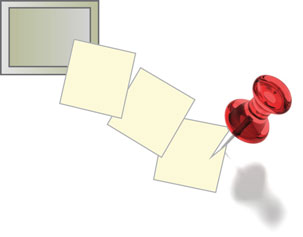
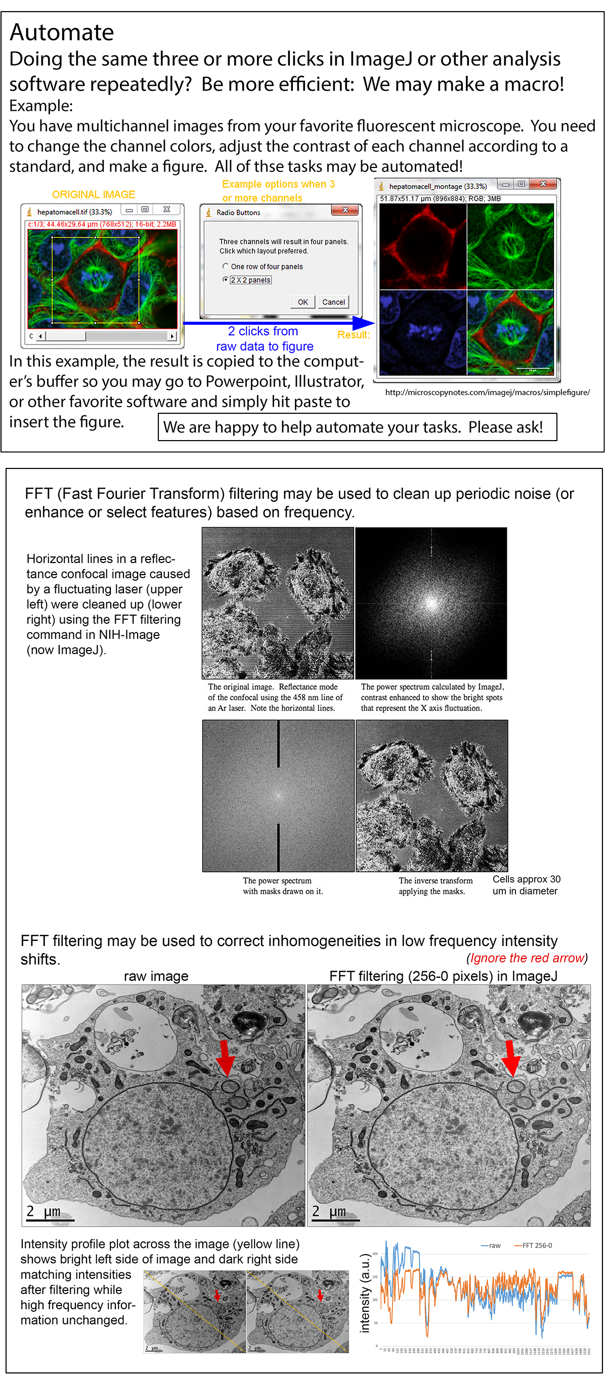
If the change in background is much lower frequency than the objects of interest, perhaps you could separate it out using FFT filtering. This depends on how quantitative you need the intensities to be in the resulting image. Examples can be found at http://microscopynotes.com/imagej/fft/index.html and http://microscopynotes.com/users/retreat2019/05-06.png. Michael Cammer michael.cammer@med.nyu.edu
There are some great software packages that can perform such tasks with ease including ImagePro 10 from Media Cybernetics and MetaMorph from Molecular Devices. I am sure others will identify open-source solutions, but these other merit as well. Mika Ruonala mika@icit.bio
I feel compelled (sorry) to remind you that all operations that mess with the original data (each pixel being a data point) should follow the MSA policy on the ethics of digital imaging. Basically, you can adjust the levels and contrast, but you must report anything else you do to alter the image in the Materials and Methods section of a paper. And, yeah, there are some great programs out there. Just remember to report it. Tina Carvalho tinacarv@hawaii.edu
Data Server
Confocal Listserver
We would like to build a data server of about 10 TB to store and distribute the imaging data of a central facility. What would be the best option for data storage, internal 10TB space or Google Drive? Does Google Drive compress the data while uploading and downloading? Regards. Manish Kumar sndrpmanish@gmail.com
This is an interesting topic. IMO the best solution would be a NAS storage (for example, a Synology NAS). This way you can have the data on a physical hard drive and can easily extend it (more storage, RAID system for Backup and/or faster speed, etc.). Cloud solutions are also a great tool. In my experience Google Drive compresses upon downloading to minimize traffic, but data are otherwise uncompressed. I would personally opt for a NAS storage which you manage (storage size, backup, access external/internal, etc.). Nino Karpf nino.karpf@gmail.com
The Microscopy Today, July 2021, NetNotes has a section on NAS and other solutions for data storage and access. This is an open access issue: https://www.cambridge.org/core/services/aop-cambridge-core/content/view/1B268DB93611602FE4F7B75D035F914B/S1551929521000821a.pdf/netnotes.pdf. There is also an article in the July 2020 issue of Microscopy Today on this topic: Practical Guide to Storage of Large Amounts of Microscopy Data: https://www.cambridge.org/core/services/aop-cambridge-core/content/view/C8D9065CC792B564FC3F0B9F8BBA42AD/S1551929520001029a.pdf/netnotes.pdf. I also recommend every issue's Dear Abbe and Stephen Carmichael columns. If you have the ‘need for speed’ on your microscope acquisition computers, and maybe the central file server, you may find of use my suggestions at http://confocal.jhu.edu/mctips/pc_tips_2021. George McNamara geomcnamara@earthlink.net
As others have suggested, a RAID-striped NAS array (we use https://www.synology.com/en-ca/products/DS2419+II) is a good way to go. You can hook it up to the network and access from anywhere, and as it uses HDDs it's reasonably cheap to expand the storage as needed. This will also be at least a bit faster than Google drive for accessing your data, though not as fast as the setup George describes. The Synology NAS I mentioned also has extensive management tools so you can actually have a bit more control of the server (setting user groups, access to individual folders, etc.) quite quickly - more so than on Google drive. I have no affiliation with Synology; I just like it! Chris Law centre-for-microscopy@concordia.ca
I've worked with a few different NAS options, but went with UnRAID for my latest build. UnRAID is a compact operating system that allows hanging any drives off a suitable computer to easily create network shares. It uses a parity disk method rather than traditional RAID striping, so it is a bit more robust. I've seen too many striped RAIDs completely fail, and when the RAID fails there is no easy way to retrieve data from the remaining disks. The parity disk method, on the other hand, assigns a single disk in the group to store parity data, from which any missing drives can be reconstructed. There are also options to have two parity disks for the paranoid. If the RAID fails, or the host computer fails, you can still read the individual disks (plug them into another machine, etc.) and recover data as it is not striped. If the parity drive(s) fail, they can be replaced and the UnRAID system will construct a new parity drive. If another disk fails, it can be replaced and the UnRAID system will reconstruct the drive with the parity information. UnRAID is mainly designed for cheaper platter drives, so is best suited for large amounts of moderate-speed storage. It can be accelerated by adding one or two SSDs to the stack and designating them as “cache” in the UnRAID system. A “mover” process can be tweaked to copy from the “cache” drives to the “storage” drives when convenient, although the default settings work well enough for my purposes. For complete protection, two SSDs can be assigned as redundant cache, so if a cache drive fails during a cache process it will not result in data loss. No commercial interest in UnRAID; I just observed two “real” RAID systems completely fail so wished to try something different. Craig Brideau craig.brideau@gmail.com
I use an 8 TB Synology server that is, importantly, off-site in another building so if I blow up the facility the data are safe. Tina Carvalho tinacarv@hawaii.edu
In order to work with many collaborators, I recently purchased a Synology NAS DS1520+ and would like to share my experience with you. This NAS has 14TB of storage and 1GB of SSD cache. The array is formatted with the Btrfs file system using the Synology Hybrid RAID5 technology. For software it has Synology Drive, which is similar to Google Drive. Public and NAS users can easily share files. When data are accidentally deleted or attacked by ransomware, snapshot replication is ideal. I also appreciate Synology's ability to index files. It allows me to find files much faster. I hope this helps. Wulin Teo wulin.teo@gmail.com
I strongly suggest you check with your IT section as to what they recommend. We have implemented a variety of solutions based on suggestions from our IT team. Of course, this depends strongly on what support you have and what level of financial/technical resources you have access to. We found Synology is a bit limiting in flexibility and throughput, so are using a server for the facility. We have an internal data server for transferring acquired data for internal users. This is not accessible outside the campus and saves us the trouble of confidentiality issues which arise out of using third party solutions. Our IT team takes care of the backup and archival issues. We used to use a Synology system for backing up data acquired by external users. For cases where the user agrees, we transfer data through Google drive or One drive. In cases where the user wants absolute confidentiality, we transfer the files through hard media like thumbdrives. Feroz Mustafa ferozm@ccamp.res.in
Benchtop Scanning Electron Microscopy on a Ship
Microscopy Listserver
Does anyone have practical experience with using a benchtop SEM onboard a research ship? Or any SEM on a boat for that matter? I can think of a dozen or more challenges that would need to be overcome but it only takes one to “sink” the operation. Thanks. Scott Whittaker whittaks@si.edu
There is a benchtop SEM installed on the International Ocean Discovery Program (IODP) drill ship *Resolution*. It works well in calm waters. Stefanie Brachfeld brachfelds@mail.montclair.edu
I don't have practical experience, but one of our customers had a benchtop SEM installed on their ship. They were happy, I suppose, because they never complained of any major issues. Rohan Prakash rohan.prakash14@gmail.com
You might consider the Mochii™ SEM that was described at the Microscopy & Microanalysis meetings, I think in 2020 and 2019. It is on the International Space Station. You can find information at NASA.gov. The company is Voxa based in Seattle. Roseann Csencsits csencsits100@gmail.com
I know of one organization in Germany operating a “normal” SEM in a truck for environmental control (in case of emergency situations). It seems to be quite stable, but they run the SEM only when the truck is stopped, so a slightly different story regarding vibrations/oscillations found on a ship. I remember some “SEM-on-ship” projects I heard of in the past. If I remember right, it was called “Jetscan” and they used ZEISS EVOs on aircraft carriers to investigate jet engine debris (https://www.selectscience.net/products/zeiss-jetscan/?prodID=171859#tab-2). As it is military-related, it may be difficult to get detailed Information, but it seemed to work. Ferenc (no details on contact information)
The system described in your links looks like a model designed to compete with one developed at US Steel many years ago. That research section was spun off as the RJLee group and continued with automated particle analysis. They developed and marketed the Personal-SEM. The company name changed to Aspex and was bought by FEI. They had what they called their Explorer system, which was crated up and shipped to Air Force bases around the world. I'm inclined to believe such a robust system would work aboard ship. I would pay attention to the vibration isolation. If there is not enough built-in, you will have to provide your own. Warren Straszheim wesaia@iastate.edu
I think the biggest possible pitfall is damage to a turbo pump. Make sure the SEM manufacturer verifies that the turbo can run on a rocking boat. It might require some sort of isolation table to limit possible damage. Neal Magdefrau neal@emitllc.com
LaB6 Question
3D EM Listserver
Does anyone worry about keeping a spare LaB6 filament for several years before using it? David Gene Morgan dagmorga@indiana.edu
I keep ours in a normal cabinet (so no desiccator) for several years without any trouble. Wim Hagen hagen@embl.de
I store ours for years in a desiccator. But I agree with Wim, the cathode is sealed by vacuum and therefore they should be protected. Dietmar Riedel driedel@gwdg.de
Our engineers strongly advise against it. They prefer that we order the filament just before installing it. Yaroslav Tsybovsky yaroslav.tsybovsky@nih.gov
I do not have a problem with my LaB6 filaments that I buy up to one year in advance. That way I am always sure to have one in stock in case of an unexpected failure of the current filament. I leave it in the vacuum seal, however. Marcus Fislage marcus.fislage@vub.be
