


Microscopy Facility Map
The Royal Microscopy Society has created a map of core microscopy facilities for the UK and beyond. The data include resources and skills within several imaging facilities around the globe. To list a facility or to submit updates go to https://www.rms.org.uk/network-collaborate/facilities-database.html.
Royal Microscopy Society

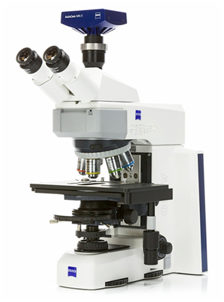
Smart Microscopes for Biomedical Use
Acquisition of fluorescence images has never been easier. ZEISS has combined the Axioscope 5 with the high-performance Colibri 3 LED light source and the sensitive, stand-alone microscope Axiocam 202 mono camera to create the perfect setup for easy multichannel fluorescence documentation. By selecting the relevant channels and pressing Snap, the system automatically adjusts the exposure time, acquires the image, switches channels, and starts again.
ZEISS
3D-Micromac AG Appoints Hartmut Schubert as Chief Technology Officer
The Management Board of 3D-Micromac AG has appointed Hartmut Schubert to the position of Chief Technology Officer. In this role, he is responsible for the strategic management of the company's technology division. “My career stages have always been characterized by strong company growth through technical innovation, digitalization, business development, and change management. I look forward to contributing these experiences to the continued successful development at 3D-Micromac,” said Schubert.
3D-Micromac

New Nikon Center of Excellence
It is a great pleasure to announce the opening of the Nikon Center of Excellence (NCofE) for Neuro-NanoImaging at the Aix-Marseille University Institute of NeuroPhysiopathology. Located in the Institute imaging facility, the NCofE for Neuro-NanoImaging focuses on how the latest super-resolution microscopy techniques can help in understanding brain cells and their dysfunctions.
Institute of NeuroPhysiopathology (INP)
Release of Bio-Formats 6.8.0
The OME Team of the University of Dundee has released Bio-Formats 6.8.0, which includes the following improvements: 1) updated ordering of reader.txt, 2) an updated bfconvert tool for multiple tile size granularity when writing, and 3) a new non-sequential option to enable writing in non-sequential order.
The OME Team (University of Dundee)
https://docs.openmicroscopy.org/bio-formats/6.8.0/about/whats-new.html

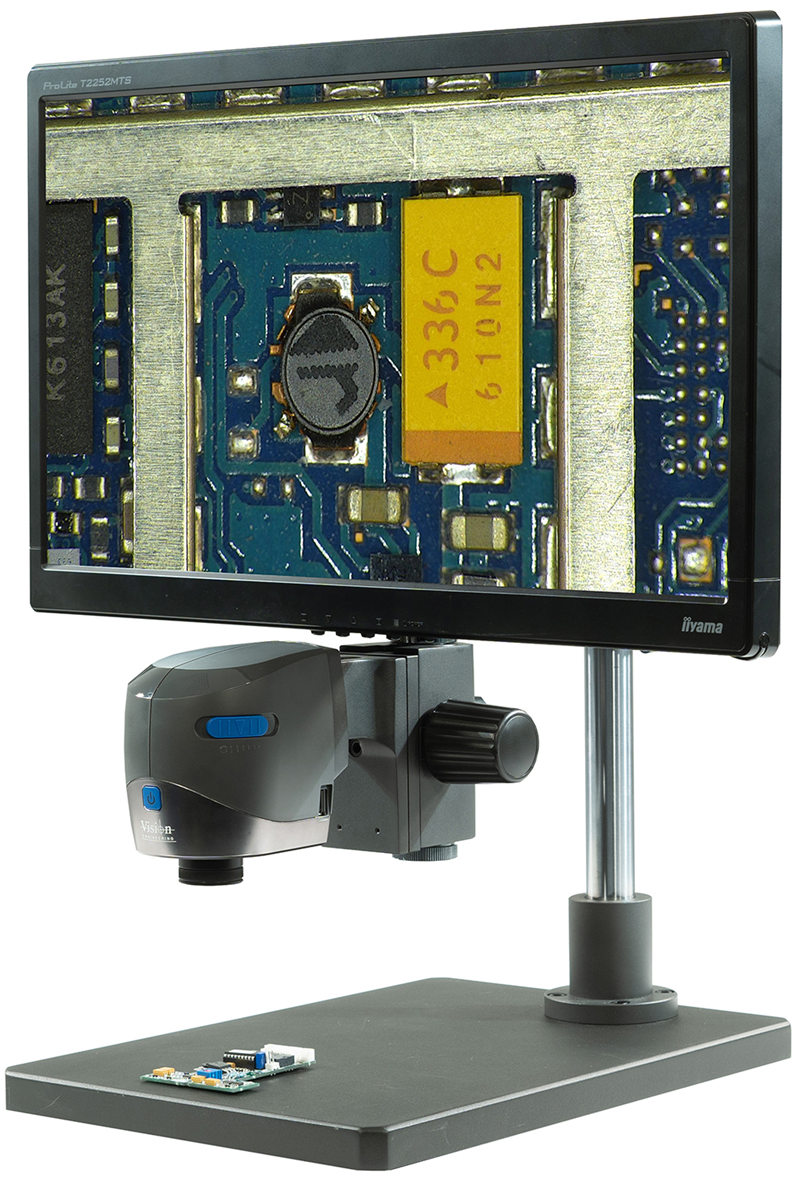
A Compact Digital Microscope
Vision Engineering has introduced the VE Cam, a compact digital microscope for a wide range of applications, including circuit board examination and tracing of problems in a wide range of industrial applications. The VE Cam 50 has a 50 mm field-of-view, and the VE CAM 80 has an 80 mm field-of-view. Both offer power, speed, and efficiency in a compact digital imaging package.
Vision Engineering
CAMECA Announces Collaboration with University of Sydney
CAMECA, a world-leading supplier of microanalytical and metrology instrumentation, is partnering with the University of Sydney on acquisition of the Invizo 6000 atom probe tomography (APT) instrument. The Invizo 6000 introduces breakthrough technologies including a patented electrostatic design that enables simultaneous increased field-of-view and enhanced mass resolving power, a deep UV laser to promote enhanced ion emission, advanced dual-beam beam delivery optics to improve specimen symmetry, and a new extraction electrode design.
CAMECA

Pfeiffer Vacuum Opens New Silicon Valley Innovation Center
Pfeiffer Vacuum, a leading provider of high-tech vacuum solutions for the semiconductor, analytical, industrial, and research and development markets, has opened a new 10,000-square-foot Silicon Valley Innovation Center (SVIC) in San Jose, CA. The facility will create several new high-tech jobs.
Pfeiffer Vacuum

Harness the Power of Nanoscopy with Single-Molecule Localization Microscopy (SMLM)
Olympus has partnered with Abbelight to deliver multimodal imaging solutions that expand research possibilities with PALM, STORM, and single-particle tracking. Abbelight SAFe 360 features include epi, HiLo, and TIRF illumination modes; ultimate 3D localization precision; spectral demixing for multicolor imaging; and live 3D reconstruction.
Olympus and Abbelight

ibidi Awarded Export Prize Bavaria
For its excellent crisis strategy during the pandemic, ibidi GmbH has been awarded the Export Prize Bavaria: Special Edition 2021, in the category “Successful Order Processing in Corona Times.” The award has been presented since 2007 to small- and medium-sized companies that work with foreign markets. The 2021 award recognizes efforts to solve the unique travel and supply chain challenges caused by the coronavirus in 2020.
ibidi

Covalent Metrology and JEOL Announce Partnership
Covalent Metrology, a leading North American provider of analytical services, announces its partnership with JEOL, a global leader in the development of cutting-edge scientific instruments used in microscopy, analytical chemistry, and materials characterization. JEOL's state-of-the-art metrology instruments will be located within Covalent's Silicon Valley lab.
JEOL and Covalent Metrology

Metrohm Companies Unite in New Northeast Hub
Metrohm is excited to announce the opening of a new office in New Jersey. The new facility will house development, production, and manufacturing space for Metrohm Group companies Innovative Photonic Solutions and B&W Tek. It is in the heart of the U.S. pharmaceutical market and will also serve as a field office for Metrohm USA.
Metrohm

New EBSD.com Website Keeps Pace with Technological Advances
Since 2003, Oxford Instruments has used the website www.ebsd.com to inform and educate researchers about the electron backscatter diffraction (EBSD) technique. Following significant updates in both 2011 and 2016, Oxford Instruments is delighted to announce that the ebsd.com website has now been completely rewritten and redesigned, providing readers with an up-to-date overview of the EBSD technique.
Oxford Instruments

Using Optical Spectroscopy to Study Microscopic Biological Samples
Microspectrometers are used in biology to study the spectral response of microscopic structures. For studies ranging from understanding vision to pharmaceutical development to quantifying protein crystals, CRAIC Technologies provides UV-visible-NIR, fluorescence, photoluminescence, and Raman microspectroscopy instruments. CRAIC Technologies systems provide spectral ranges from the deep UV to the NIR for both imaging and spectroscopy.
CRAIC

Bruker Introduces Fast BioAFM
Bruker has released the JPK NanoWizard® V BioAFM, which offers a high level of automation and ease of use for life science atomic force microscopy research. NanoWizard V is a fast, automated BioAFM that enables rapid, quantitative mechanical measurements and analysis of dynamics on samples ranging in size from sub-molecular to cells and tissues. The automated setup, alignment, and re-adjustment of system parameters opens new possibilities for long-term, self-regulating experiments on mechanobiological dynamics.
Bruker

DigitalMicrograph 3.5 is Now Available
Gatan's version 3.5 of DigitalMicrograph is now available for download. This latest software can help push the boundaries of what can be done with electron microscopy, with state-of-the-art tools to enable advanced imaging applications and streamlined routine workflows.
Gatan

Ultrafast Lasers
The short pulse durations and high peak powers of ultrafast lasers make them ideal for a wide range of applications, including materials processing, medical lasers, nonlinear imaging, and microscopy. Ultrafast lasers are sensitive to chromatic dispersion, increasing pulse durations and reducing ultrafast system performance. Learn more about ultrafast dispersion and how to combat it at www.edmundoptics.com.
Edmund Optics

DiATOME® ultra-Diamond Knives at Ted Pella, Inc.
Ted Pella, Inc. now provides DiATOME ultra-Diamond Knives featuring the highest-quality diamonds and optimal crystal orientation for guaranteed perfect ultrathin sections and a durable cutting edge. ultra-Diamond knives provide a horizontal boat, allowing water to completely fill the boat to facilitate section collection, and a hydrophilic surface making it easier to wet the cutting edge, even with a low water level.
Ted Pella, Inc.

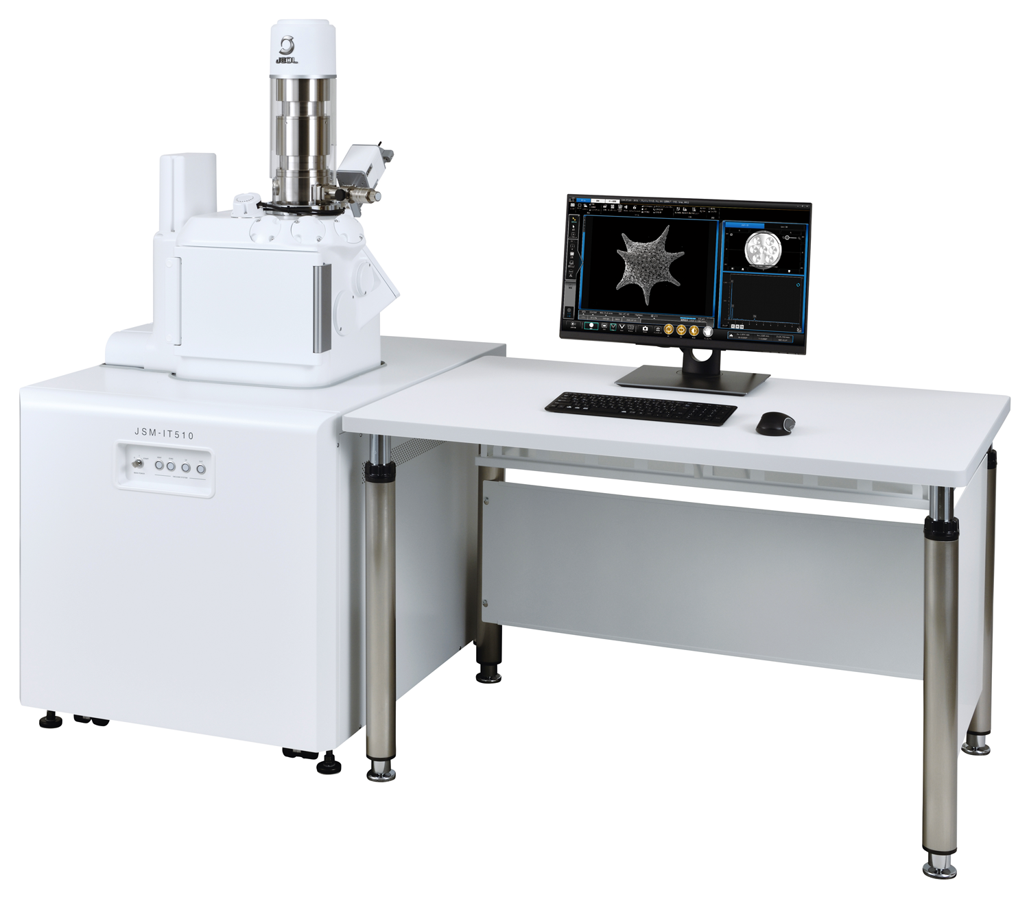
New JEOL SEM Enhances Productivity
A new scanning electron microscope from JEOL provides faster and easier acquisition of both SEM images and EDS data analysis. The new large-chamber JSM-IT510 (Tungsten or LaB6) SEM features advanced automation that enhances productivity, including: “Simple SEM” automated image collection, Live 3D, Live EDS, automated X-ray generation depth calculation, auto functions from alignment to focus, and ZeroMag seamless navigation from optical to SEM imaging.
JEOL
JAI Cameras Now Supported by Eye Vision
Eye Vision Technology, GmbH has integrated JAI cameras into the Eye Vision image processing software. Now, cameras from over 70 manufacturers are available for direct use with the Eye Vision 4 software. Simply select any camera and take pictures with it. A complete list of the integrated, and thus supported, cameras can be selected after the hardware installation in the hardware configurator or requested directly from EVT.
EVT Eye Vision

CosMx Spatial Molecular Imager (SMI)
The CosMx Spatial Molecular Imager (SMI) takes the next leap in spatial biology by offering high-plex in situ analysis with true single-cell to subcellular resolution in intact formalin-fixed paraffin-embedded (FFPE) and fresh frozen tissue samples. From the leader in spatial biology, the CosMx SMI sets the bar in data quality and key performance metrics.
nanoString

HEMCO Vented Tabletop Workstation Hood Model 24800
Typical uses of the HEMCO 24800 include histology, venting for hot plates, stable environment for microscopes and student workstations, sample weighing stations, and handling of pharmaceuticals. Constructed of chemically resistant, lightweight, advanced composites, it can be moved easily as procedures or workflow change. The work surface is recessed to contain spillage, and a three-inch diameter outlet collar is provided for duct connection.
HEMCO
www.hemcocorp.com/ductless.html

Park Systems Introduces a New Class of Atomic Force Microscopes
Effortlessly get the sharpest, clearest, highest-resolution images and measurements, one sample after another, on various applications. Boost progress and scientific discoveries through unprecedented speed and accuracy as the Park FX40 AFM autonomously images and acquires data powered by its artificial intelligence, robotics, and machine learning capability while cameras automatically align the laser beams and photodetectors. These innovations allow investigators to focus on the research and not the tool.
Park Systems

Olympus Self-Learning Microscopy Delivers Fast, Efficient Image Analysis
The scanR imaging system provides fully automated image acquisition and data analysis. New software enhances the system's deep-learning technology with instance segmentation, the ability to detect and delineate distinct objects of interest, such as cells and nuclei, in an image. The result is faster, more efficient image analysis. This system offers reliable image segmentation, easier calibration, collaboration, and pretrained models.
Olympus

Spicer Consulting SC28 Monitoring System
The SC28 monitoring system allows labs to detect magnetic fields and other disturbances that can negatively affect image quality of electron-beam instruments. Ideal for use with electron microscopes, electron-beam lithography tools, and CD-SEM, it provides continuous measurements of magnetic fields and vibrations, as well as acoustics, temperature, and humidity. The SC28 can even be configured to send alerts when environmental readings are outside of user-determined operating conditions.
Spicer Consulting

ZEISS LSM Plus and ZEISS Airyscan Joint Deconvolution Introduced
ZEISS has introduced ZEISS LSM Plus and ZEISS Airyscan Joint Deconvolution for confocal microscopy. ZEISS laser scanning microscopes (LSM) provide a wide range of detection modes with high sensitivity and spectral flexibility for multi-fluorescent experiments. ZEISS LSM Plus and ZEISS Airyscan Joint Deconvolution significantly improve data quality in confocal imaging, independent of the chosen detector, imaging mode, and emission range.
ZEISS

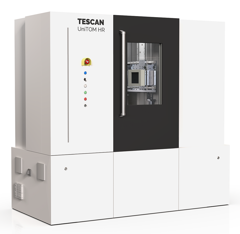
Tescan UniTOM HR Micro CT
Spatial resolution meets temporal resolution. As pioneers in dynamic micro-CT, TESCAN has been pushing the limits of temporal resolution for lab-based micro-CT down to seconds, enabling true dynamic imaging experiments to provide unique research insight. Building on that expertise, the Tescan UniTOM HR is the first dynamic micro-CT system to offer sub-micron spatial resolution without compromising temporal resolution and in situ capabilities.
Tescan
Nanosoft Introduces New Cryo Tools
Nanosoft has introduced new tools and services to enhance cryo-research capabilities, including a foam vitrification dewar that is compatible with the Vitrobot (ThermoFisher), the Quick Lid that protects frozen grids in their grid boxes during vitrification sessions, a cryo-tweezer/Vitrobot tweezer repair service, and a clipping tool repair service.
Nanosoft

HORIBA Microscopy Spectroscopy System (SMS)
New Standard Microscope Spectroscopy (SMS) systems enable standard microscopes to be fitted with a spectrometer and a detector to add spectroscopy capabilities. The SMS platform brings flexibility and modularity to performing spectroscopy on standard microscope systems, without compromising the microscope’s imaging functionality. The SMS systems can combine spectroscopies on one platform, enabling techniques like Raman, steady state and time-resolved photoluminescence, reflectance/transmittance, electroluminescence, photocurrent, and darkfield scattering.
HORIBA Scientific

Interherence's Stage-Top Incubator
The VAHEAT stage-top incubator offers new possibilities for microscopy of temperature-sensitive processes, which were not possible to image before. This is the first stage-top incubator on the market that controls the temperature directly in the field-of-view. VAHEAT is extremely fast, precise, very easy to use, and compatible with many microscopy techniques including super-resolution and microfluidics applications.
Interherence

Large SEM Sample Handling
RAVE Scientific Universal vise-type sample holders are quick, easy to use, and cleaner than using conductive adhesives or glue. The vise holds the sample between moving jaws, enabling quick sample loading and providing the rigidity needed for high-resolution imaging. They are especially useful when similar samples or a series of the same sample need to be examined.
Rave Scientific

New Large-Area Hybrid Pixel Detector for TEM
The new ASI CheeTah M3 Mega detector provides high-resolution imaging of large protein samples. Its innovative features include noiseless acquisition, single-electron sensitivity, and high dynamic range enabling acquisition of the cleanest data possible. The large 1024 × 1024 pixel area and increased readout speed makes imaging ultrafast and painless.
Amsterdam Scientific Instruments

