


The fourth Microscopy Today Micrograph Awards competition was successful once again. The premise of these competitions is that scientific micrographs can be interesting in their own right as images with visual impact. This year submissions came from 20 countries and 18 US states. Evaluation by the judges was accomplished without knowing the names or affiliations of the submitters. Of the 25 finalist micrographs (shown on the July cover and in the Microscopy Today micrograph gallery at https://www.microscopy.org/awards/micrograph_gallery_2022/), eight were from the US and 17 were from other countries.
In this article we show the prize winners in each category: Published category, for micrographs published in the previous year; Open category, for unpublished still micrographs; and the Video category, for clips of movies taken through a microscope or of animations of reconstructed images. The images in this article are the first, second, and third prize winners in each category, as well as the winner of the People's Choice Award and an interesting image comparison at the end of this article.
Finalists and prize winners were selected by a panel of judges led by Robert Simmons. The People's Choice Award was selected via public voting at the micrograph awards gallery on the MSA website. The judging panel for the 2022 competition was comprised of five judges, all of whom bring their own special expertise. This year Robert Simmons (Chief Judge), Charles Lyman (Senior Editor), and Bob Price (Editor-in-Chief) were joined by Beth Richardson and Donovan Leonard. Beth Richardson is an award-winning microscopist and long-time member of MSA. She ran the EM lab for the Department of Plant Pathology at the University of Georgia for 19 years, specializing in high-pressure freezing of biological samples. After laboratory consolidation at UGA, she acted as EM Lab Coordinator for Georgia Electron Microscopy, one of UGA's core facilities, until her retirement in 2019. During her career she won awards in several micrograph competitions. She continues to enjoy photography, and her work has been displayed in several Georgia galleries. She is known for her ability to generate excellent technical images with an appreciation for the beauty often found in scientific specimens. Donovan Leonard is a Senior Technical Staff Member in the Manufacturing Sciences Division at Oak Ridge National Laboratory and is currently serving as a Director for MSA. Leonard comments that “viewing materials surfaces or diffraction patterns continually reminds me to appreciate the inherent beauty in our natural world. The feelings of wonder, bewilderment, and excitement felt acquiring micrographs also remind me of what I feel when experiencing a sunrise/sunset at the beach or soaking in a Caravaggio at a museum.” This group of scientists/artists provides the broad knowledge base needed to address the scientific, technical, and esthetic aspects of our competition.
The original idea for the Microscopy Today Micrograph Awards competition was proposed by Robert and Camille Simmons. In 2017 they suggested that Microscopy Today sponsor a micrograph contest emphasizing both the scientific and artistic merit of micrographs. During 2018 a set of specifications for micrograph submission software was developed, and Nestor Zaluzec produced the required software capable of dealing with hundreds of submitted images.
The judging process is in two steps: after the scientific relevance of an image has been established, judges evaluate its visual impact by asking the following: Can the micrograph stand on its own as a captivating image without requiring knowledge of the subject or the type of microscopy employed? In other words, would the image look good on a living room wall or in a museum?
Another goal of our competition is to honor images that may not be eligible or competitive in other micrograph contests. All types of micrographs are welcome in this competition, whether they were acquired with a light microscope, electron microscope, X-ray microscope, scanning probe microscope, or some other microanalytical tool. Also, many worthy micrographs are published in journals or magazines without a thought of entering them in a competition. By honoring published images in a separate category we hope to encourage microscopists to think about image composition and visual impact early in experiment planning and during image acquisition.
Understanding mechanisms and processes often requires dynamic imaging acquired by in situ microscopy of all types. Thus, we have established a separate category for video micrographs. This category also includes digital animations of reconstructed three-dimensional datasets, for example, from cryo-electron microscopy, which is providing new insights into cellular and molecular structures.
Our micrograph contest is also driven by image quality from a technical standpoint. Sharpness of image details is important. Imaging of three-dimensional objects with a large depth-of-field was once the exclusive domain of the scanning electron microscope. But with focus-stacking software, light micrographs now can be in sharp focus over a considerable depth-of-field. Our judges evaluate submitted micrographs on large high-resolution monitors that can reveal lack of sharpness, as well as other image problems. We request that submitted images have both inherent sharpness and sufficient pixel density to be presented in an 11″ x 14″ format suitable for hanging in an exhibition. Often image sharpness can be maintained by acquiring the micrograph at a lower magnification than might be required for research purposes. Acquisition at high pixel density is now available to most microscopists, since the cost of suitable cameras has decreased dramatically over the last decade. An excellent micrograph with only a modest pixel density is not necessarily eliminated from the competition, but justification may be needed for the image to be competitive.
So, what makes a winning micrograph? Microscopes reveal interesting features and patterns in objects that are not visible to the naked eye. Some microscopists encounter these images accidentally during the pursuit of other goals, and other microscopists actively seek specific subjects, environmental conditions, or specimen preparation methods that make acquisition of a great image more likely. Whereas the composition of the image is always important, there are other considerations. Micrographs initially acquired by electron microscopy or scanning probe microscopy are monochrome, which does not prevent an image from winning. However, it is now common to “improve” images with post-acquisition processing to add color to features or to change the background color behind the subject. While such manipulation is acceptable, to be successful the microscopist must make good decisions along the way. Choosing to use artificial color in an image is an artistic choice which should be made carefully. While color may enhance a good image, it will not make up for lack of quality in the original. Color choices are important, and discordant colors can reduce the appeal of an image as easily as a lack of sharpness. Post-processing should be undertaken carefully with an eye to improving the appeal or clarity of an image. Of course, it is important to read and carefully follow the instructions for submission. A complete entry is much easier for the judges to evaluate.
The editors and judges of Microscopy Today thank all entrants to this year's competition and welcome their submissions to the next contest. The submission site will re-open on or about October 1, 2022, and will close on March 10, 2023.
Published Category
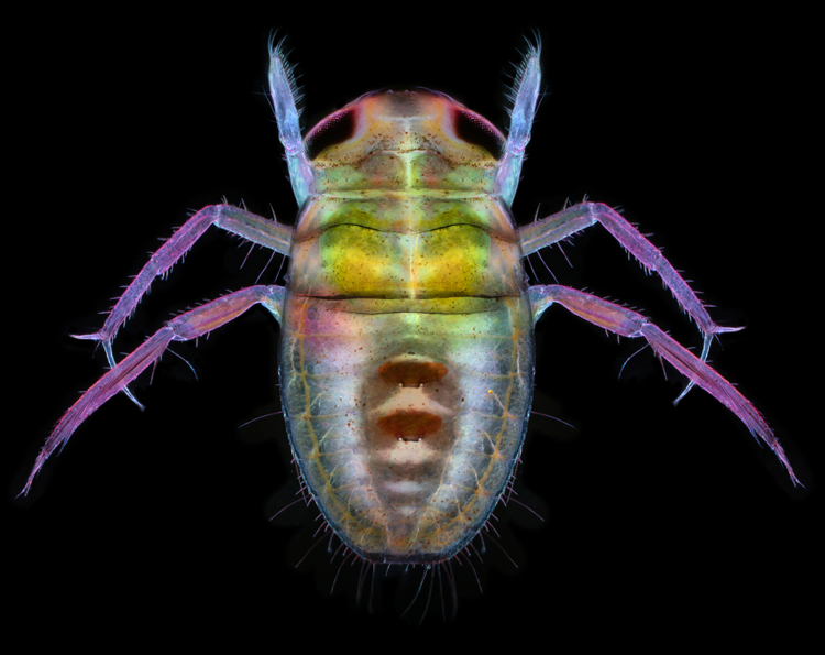
Published 1st Prize. Water boatman. After collection from surface water, the anesthetized insect was imaged in a focus stack. Polarized dark-field microscopy. Published in the 2022 Olympus Life Science Calendar. Image by Karl Gaff, Karl Gaff Microscopy, Dublin, Ireland.
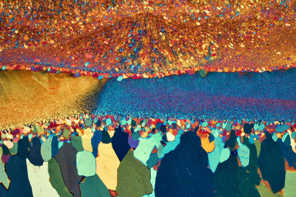
Published 2nd Prize. Agate from Brazil. This thin section of agate exhibits both fine grains (top) and coarse grains (bottom). Polarized light microscopy. Published in the 2022 ZEISS Microscopy Calendar. Image by Bernardo Cesare, University of Padova, Padova, Italy.

Published 3rd Prize. Cyanobacteria colony. Gloeotricha sp. collected from Lake Mälaren, Stockholm, Sweden. This species is often found in large summer blooms, causing the water to become toxic. Fluorescence microscopy with acridine orange stain. Published in Microscopy & Analysis (Front Cover), January/February 2022, and in the 2021 Olympus Life Science Calendar. Image by Håkan Kvarnström, Bromma, Sweden.
Open Category

Open 1st Prize. Sphagnum moss with desmid. Commonly known as peat moss, this lake sample shows a desmid (Micrasterias fimbriata) in the center. Dark-field polarized light microscopy. Image by Marek Mis, Marek Mis Photography, Suwalki, Poland.

Open 2nd Prize. Radiolarian. Spherical mineral skeleton of a radiolarian, a unicellular protist. When radiolaria die, their shells sink to the ocean floor and contribute to ocean sediment. Scanning electron microscopy. Multiple SEM backscatter images were artificially colored and combined. Image by Elizabeth King, NUANCE Center, Northwestern University, Evanston, IL.

Open 3rd Prize. Magnet microstructure. Solidification microstructure at a laser melt-pool in a sintered CoSm permanent magnet. Grains of varying orientation revealed by electron backscatter diffraction (EBSD). Scanning electron microscopy. Image by Felix Trauter, Aalen University, Aalen, Germany.
Video Category

Video 1st Prize. Intestinal immune cells. Cells from optically cleared small intestinal villi: lymphocytes (red), epithelial cells (green), leucocytes (cyan), and alpha-SMA (yellow). Fluorescence microscopy. Image by Frederic Fercoq, CRUK Beatson Institute, Glasgow, Scotland, UK.

Video 2nd Prize. Domain contrast. Demagnetization and magnetization of an electrical iron sheet as the material goes through a hysteresis loop. The magnetooptical Kerr Effect is used to show changes in the domain structure. Light microscopy. Image by Dominic Hohs, Aalen University, Aalen, Germany.
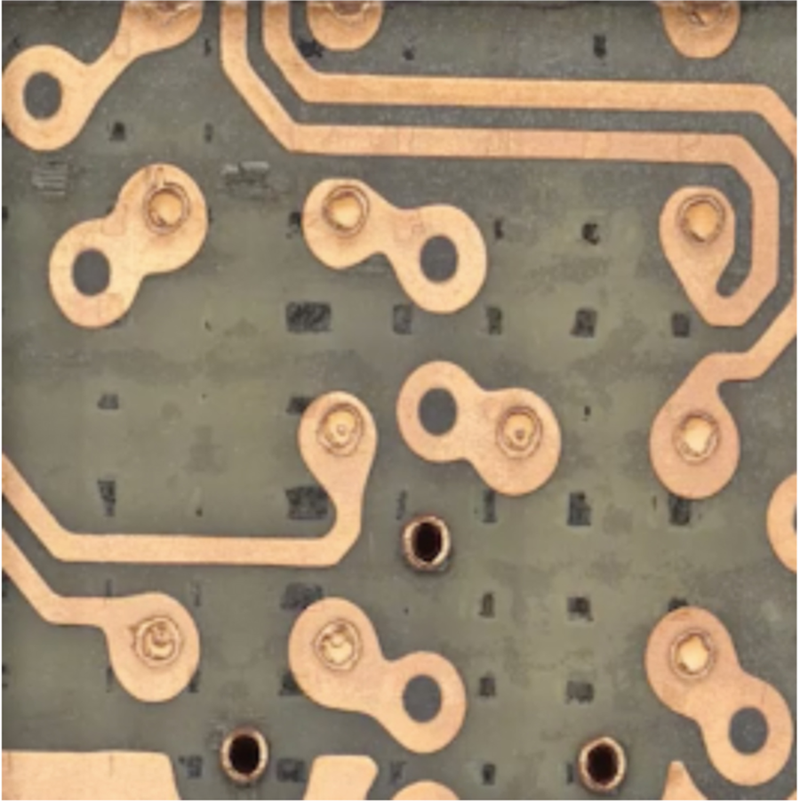
Video 3rd Prize. Printed circuit board. Pulsed laser ablation was used for serial sectioning in failure analysis and reverse engineering. Light microscopy image juxtaposed with X-ray computed tomography of corresponding sections. X-ray computed tomography. Image by Pouya Tavousi, University of Connecticut, Storrs, CT.
People's Choice Award

Agate from Brazil. This thin section of agate exhibits both fine grains (top) and coarse grains (bottom). Polarized light microscopy. Published in the 2022 ZEISS Microscopy Calendar. Image by Bernardo Cesare, University of Padova, Padova, Italy.
Special Honors

Large and small microscopes. (left) Cave bear enamel. Diverse apatite nanocrystal orientations (different colors) within enamel rods from a cave bear (Ursus spelaeus). X-ray photoemission electron microscopy at the Advanced Light Source Synchrotron, arguably one of the largest and most expensive microscopes in the world. Image submitted by Cayla Stifler, University of Wisconsin, Madison, WI. (right) Crystallization. Formation of potassium permanganate crystals. Movie acquired with a Foldscope, a paper microscope personally owned by over 1.5 million adults and children—arguably the smallest, and certainly the least expensive, microscope in the world. Light microscopy. Image by Sameer Shaikh, Azam Colony, Parbhani, India.
