Hostname: page-component-76fb5796d-5g6vh Total loading time: 0 Render date: 2024-04-26T04:30:15.274Z Has data issue: false hasContentIssue false

Visualization of Bacterial Colonization and Cellular Layers in a Gut-on-a-Chip System Using Optical Coherence Tomography

Published online by Cambridge University Press:  27 October 2020

Lu Yuan
Affiliation:
Department of Biomedical Engineering, University of Groningen, University Medical Center Groningen, 9713 AV Groningen, The Netherlands
Pim de Haan
Affiliation:
University of Groningen, Groningen Research Institute of Pharmacy, Pharmaceutical Analysis, 9713 AV Groningen, The Netherlands TI-COAST, 1098 XH Amsterdam, The Netherlands
Brandon W. Peterson
Affiliation:
Department of Biomedical Engineering, University of Groningen, University Medical Center Groningen, 9713 AV Groningen, The Netherlands
Ed D. de Jong
Affiliation:
Department of Biomedical Engineering, University of Groningen, University Medical Center Groningen, 9713 AV Groningen, The Netherlands
Elisabeth Verpoorte
Affiliation:
University of Groningen, Groningen Research Institute of Pharmacy, Pharmaceutical Analysis, 9713 AV Groningen, The Netherlands
Henny C. van der Mei*
Affiliation:
Department of Biomedical Engineering, University of Groningen, University Medical Center Groningen, 9713 AV Groningen, The Netherlands
Henk J. Busscher
Affiliation:
Department of Biomedical Engineering, University of Groningen, University Medical Center Groningen, 9713 AV Groningen, The Netherlands
*
*Author for correspondence: Henny C. van der Mei, E-mail: h.c.van.der.mei@umcg.nl
Get access

Abstract

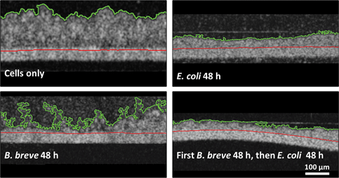
Imaging of cellular layers in a gut-on-a-chip system has been confined to two-dimensional (2D)-imaging through conventional light microscopy and confocal laser scanning microscopy (CLSM) yielding three-dimensional- and 2D-cross-sectional reconstructions. However, CLSM requires staining and is unsuitable for longitudinal visualization. Here, we compare merits of optical coherence tomography (OCT) with those of CLSM and light microscopy for visualization of intestinal epithelial layers during protection by a probiotic Bifidobacterium breve strain and a simultaneous pathogen challenge by an Escherichia coli strain. OCT cross-sectional images yielded film thicknesses that coincided with end-point thicknesses derived from cross-sectional CLSM images. Light microscopy on histological sections of epithelial layers at the end-point yielded smaller layer thicknesses than OCT and CLSM. Protective effects of B. breve adhering to an epithelial layer against an E. coli challenge included the preservation of layer thickness and membrane surface coverage by epithelial cells. OCT does not require staining or sectioning, making OCT suitable for longitudinal visualization of biological films, but as a drawback, OCT does not allow an epithelial layer to be distinguished from bacterial biofilms adhering to it. Thus, OCT is ideal to longitudinally evaluate epithelial layers under probiotic protection and pathogen challenges, but proper image interpretation requires the application of a second method at the end-point to distinguish bacterial and epithelial films.

Type
Biological Applications
Copyright
Copyright © The Author(s), 2020. Published by Cambridge University Press

Access options

Get access to the full version of this content by using one of the access options below. (Log in options will check for institutional or personal access. Content may require purchase if you do not have access.)

References

Borthakur, A, Gill, RK, Tyagi, S, Koutsouris, A, Alrefai, WA, Hecht, GA, Ramaswamy, K & Dudeja, PK (2008). The probiotic Lactobacillus acidophilus stimulates chloride/hydroxyl exchange activity in human intestinal epithelial cells. J Nutr 138, 13551359.CrossRefGoogle ScholarPubMed
Bousi, E, Charalambous, I & Pitris, C (2010). Optical coherence tomography axial resolution improvement by step-frequency encoding. Opt Express 18, 1187711890.CrossRefGoogle ScholarPubMed
Candela, M, Perna, F, Carnevali, P, Vitali, B, Ciati, R, Gionchetti, P, Rizzello, F, Campieri, M & Brigidi, P (2008). Interaction of probiotic Lactobacillus and Bifidobacterium strains with human intestinal epithelial cells: Adhesion properties, competition against enteropathogens and modulation of IL-8 production. Int J Food Microbiol 125, 286292.CrossRefGoogle ScholarPubMed
De Haan, P, Ianovska, MA, Mathwig, K, Van Lieshout, GAA, Triantis, V, Bouwmeester, H & Verpoorte, E (2019). Digestion-on-a-chip: A continuous-flow modular microsystem recreating enzymatic digestion in the gastrointestinal tract. Lab Chip 19, 15991609.CrossRefGoogle ScholarPubMed
Dreszer, C, Wexler, AD, Drusová, S, Overdijk, T, Zwijnenburg, A, Flemming, HC, Kruithof, JC & Vrouwenvelder, JS (2014). In-situ biofilm characterization in membrane systems using optical coherence tomography: Formation, structure, detachment and impact of flux change. Water Res 67, 243254.CrossRefGoogle ScholarPubMed
Duffy, DC, McDonald, JC, Schueller, OJA & Whitesides, GM (1998). Rapid prototyping of microfluidic systems in poly(dimethylsiloxane). Anal Chem 70, 49744984.CrossRefGoogle ScholarPubMed
Fortunato, L & Leiknes, T (2017). In-situ biofouling assessment in spacer filled channels using optical coherence tomography (OCT): 3D biofilm thickness mapping. Bioresour Technol 229, 231235.CrossRefGoogle ScholarPubMed
Fortunato, L, Qamar, A, Wang, Y, Jeong, S & Leiknes, T (2017). In-situ assessment of biofilm formation in submerged membrane system using optical coherence tomography and computational fluid dynamics. J Membr Sci 521, 8494.CrossRefGoogle Scholar
Fukuda, S, Toh, H, Hase, K, Oshima, K, Nakanishi, Y, Yoshimura, K, Tobe, T, Clarke, JM, Topping, DL, Suzuki, T, Taylor, TD, Itoh, K, Kikuchi, J, Morita, H, Hattori, M & Ohno, H (2011). Bifidobacteria can protect from enteropathogenic infection through production of acetate. Nature 469, 543549.CrossRefGoogle ScholarPubMed
Gao, K, Wang, C, Liu, L, Dou, X, Liu, J, Yuan, L, Zhang, W & Wang, H (2017). Immunomodulation and signaling mechanism of Lactobacillus rhamnosus GG and its components on porcine intestinal epithelial cells stimulated by lipopolysaccharide. J Microbiol Immunol Infect 50, 700713.CrossRefGoogle ScholarPubMed
Graf, BW & Boppart, SA (2010). Imaging and analysis of three-dimensional cell culture models. Methods Mol Biol 591, 211227.CrossRefGoogle ScholarPubMed
Guarner, F & Malagelada, J-R (2003). Gut flora in health and disease. Lancet 361, 512519.CrossRefGoogle ScholarPubMed
Hou, J, Wang, C, Rozenbaum, RT, Gusnaniar, N, De Jong, ED, Woudstra, W, Geertsema-Doornbusch, GI, Atema-Smit, J, Sjollema, J, Ren, Y, Busscher, HJ & Van der Mei, HC (2019). Bacterial density and biofilm structure determined by optical coherence tomography. Sci Rep 9, 9794.CrossRefGoogle ScholarPubMed
Huh, D, Kim, HJ, Fraser, JP, Shea, DE, Khan, M, Bahinski, A, Hamilton, GA & Ingber, DE (2013). Microfabrication of human organs-on-chips. Nat Protoc 8, 21352157.CrossRefGoogle ScholarPubMed
Huh, D, Matthews, BD, Mammoto, A, Montoya-Zavala, M, Hsin, HY & Ingber, DE (2010). Reconstituting organ-level lung functions on a chip. Science 328, 16621668.CrossRefGoogle ScholarPubMed
Jalili-Firoozinezhad, S, Prantil-Baun, R, Jiang, A, Potla, R, Mammoto, T, Weaver, JC, Ferrante, TC, Kim, HJ, Cabral, JMS, Levy, O & Ingber, DE (2018). Modeling radiation injury-induced cell death and countermeasure drug responses in a human gut-on-a-chip article. Cell Death Dis 9, 223.CrossRefGoogle Scholar
Janjaroen, D, Ling, F, Monroy, G, Derlon, N, Mogenroth, E, Boppart, SA, Liu, WT & Nguyen, TH (2013). Roles of ionic strength and biofilm roughness on adhesion kinetics of Escherichia coli onto groundwater biofilm grown on PVC surfaces. Water Res 47, 25312542.CrossRefGoogle ScholarPubMed
Kim, HJ, Huh, D, Hamilton, G & Ingber, DE (2012). Human gut-on-a-chip inhabited by microbial flora that experiences intestinal peristalsis-like motions and flow. Lab Chip 12, 21652174.CrossRefGoogle ScholarPubMed
Kim, HJ & Ingber, DE (2013). Gut-on-a-chip microenvironment induces human intestinal cells to undergo villus differentiation. Integr Biol 5, 11301140.CrossRefGoogle ScholarPubMed
Kim, HJ, Li, H, Collins, JJ & Ingber, DE (2016). Contributions of microbiome and mechanical deformation to intestinal bacterial overgrowth and inflammation in a human gut-on-a-chip. Proc Natl Acad Sci 113, E7E15.Google Scholar
Kleerebezem, M & Vaughan, EE (2009). Probiotic and gut lactobacilli and bifidobacteria: Molecular approaches to study diversity and activity. Annu Rev Microbiol 63, 269290.CrossRefGoogle ScholarPubMed
Miskinyte, M, Sousa, A, Ramiro, RS, de Sousa, JAM, Kotlinowski, J, Caramalho, I, Magalhães, S, Soares, MP & Gordo, I (2013). The genetic basis of Escherichia coli pathoadaptation to macrophages. PLoS Pathog 9, e1003802.CrossRefGoogle ScholarPubMed
Mitsuoka, T (1990). Bifidobacteria and their role in human health. J Ind Microbiol 6, 263267.CrossRefGoogle Scholar
Miyoshi, Y, Okada, S, Uchimura, T & Satoh, E (2006). A mucus adhesion promoting protein, MapA, mediates the adhesion of Lactobacillus reuteri to caco-2 human intestinal epithelial cells. Biosci Biotechnol Biochem 70, 16221628.CrossRefGoogle ScholarPubMed
O'Callaghan, A & Van Sinderen, D (2016). Bifidobacteria and their role as members of the human gut microbiota. Front Microbiol 7, 925.CrossRefGoogle ScholarPubMed
Ohland, CL & MacNaughton, WK (2010). Probiotic bacteria and intestinal epithelial barrier function. Am J Physiol Gastrointest Liver Physiol 298, G807G819.CrossRefGoogle ScholarPubMed
Otsu, N (1979). Threshold selection method from gray-level histograms. IEEE Trans Syst Man Cybern 9, 6266.CrossRefGoogle Scholar
Polat, A, Hassan, S, Yildirim, I, Oliver, LE, Mostafaei, M, Kumar, S, Maharjan, S, Bourguet, L, Cao, X, Ying, G, Hesar, EM & Zhang, YS (2019). A miniaturized optical tomography platform for volumetric imaging of engineered living systems. Lab Chip 19, 550561.CrossRefGoogle ScholarPubMed
Proença, JT, Barral, DC & Gordo, I (2017). Commensal-to-pathogen transition: One-single transposon insertion results in two pathoadaptive traits in Escherichia coli-macrophage interaction. Sci Rep 7, 4504.CrossRefGoogle ScholarPubMed
Rao, SSC, Rehman, A, Yu, S & de Andino, NM (2018). Brain fogginess, gas and bloating: A link between SIBO, probiotics and metabolic acidosis article. Clin Transl Gastroenterol 9, 162.CrossRefGoogle Scholar
Reid, G, Younes, JA, Van der Mei, HC, Gloor, GB, Knight, R & Busscher, HJ (2011). Microbiota restoration: Natural and supplemented recovery of human microbial communities. Nat Rev Microbiol 9, 2738.CrossRefGoogle ScholarPubMed
Sattler, ECE, Poloczek, K, Raphaela, K & Welzel, J (2013). Confocal laser scanning microscopy and optical coherence tomography for the evaluation of the kinetics and quantification of wound healing after fractional laser therapy. J Am Acad Dermatol 69, 165173.CrossRefGoogle ScholarPubMed
Sridharan, G & Shankar, A (2012). Toluidine blue: A review of its chemistry and clinical utility. J Oral Maxillofac Pathol 16, 251255.CrossRefGoogle ScholarPubMed
Tang, S, Sun, C-H, Krasieva, TB, Chen, Z & Tromberg, BJ (2007). Imaging subcellular scattering contrast by using combined optical coherence and multiphoton microscopy. Opt Lett 32, 503505.CrossRefGoogle ScholarPubMed
Tropini, C, Earle, KA, Huang, KC & Sonnenburg, JL (2017). The gut microbiome: Connecting spatial organization to function. Cell Host Microbe 21, 433442.CrossRefGoogle ScholarPubMed
Tuo, Y, Yu, H, Ai, L, Wu, Z, Guo, B & Chen, W (2013). Aggregation and adhesion properties of 22 Lactobacillus strains. J Dairy Sci 96, 42524257.CrossRefGoogle ScholarPubMed
Xi, C, Marks, DL, Schlachter, S, Luo, W & Boppart, SA (2006). High-resolution three-dimensional imaging of biofilm development using optical coherence tomography. J Biomed Opt 11, 034001.CrossRefGoogle ScholarPubMed
Supplementary material: File

Yuan et al. supplementary material

Figure S1

Download Yuan et al. supplementary material(File)
File 2 MB