Hostname: page-component-7c8c6479df-27gpq Total loading time: 0 Render date: 2024-03-28T15:23:43.545Z Has data issue: false hasContentIssue false

The Prospect of Spatially Accurate Reconstructed Atom Probe Data Using Experimental Emitter Shapes

Published online by Cambridge University Press:  05 November 2021

Jonathan Op de Beeck*
Affiliation:
Imec, Kapeldreef 75, 3001 Leuven, Belgium Quantum Solid-State Physics Group, KU Leuven, Celestijnenlaan 200D, 3001 Leuven, Belgium
Jeroen E. Scheerder
Affiliation:
Imec, Kapeldreef 75, 3001 Leuven, Belgium
Brian P. Geiser
Affiliation:
CAMECA Instruments Inc., 5470 Nobel Drive, Madison, WI 53711, USA
Joseph H. Bunton
Affiliation:
CAMECA Instruments Inc., 5470 Nobel Drive, Madison, WI 53711, USA
Robert M. Ulfig
Affiliation:
CAMECA Instruments Inc., 5470 Nobel Drive, Madison, WI 53711, USA
David J. Larson
Affiliation:
CAMECA Instruments Inc., 5470 Nobel Drive, Madison, WI 53711, USA
Paul van der Heide
Affiliation:
Imec, Kapeldreef 75, 3001 Leuven, Belgium
Wilfried Vandervorst
Affiliation:
Imec, Kapeldreef 75, 3001 Leuven, Belgium Quantum Solid-State Physics Group, KU Leuven, Celestijnenlaan 200D, 3001 Leuven, Belgium
Claudia Fleischmann
Affiliation:
Imec, Kapeldreef 75, 3001 Leuven, Belgium Quantum Solid-State Physics Group, KU Leuven, Celestijnenlaan 200D, 3001 Leuven, Belgium
*
*Corresponding author: Jonathan Op de Beeck, E-mail: jonathan.opdebeeck@imec.be
Get access

Abstract

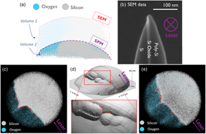
Reliable spatially resolved compositional analysis through atom probe tomography requires an accurate placement of the detected ions within the three-dimensional reconstruction. Unfortunately, for heterogeneous systems, traditional reconstruction protocols are prone to position some ions incorrectly. This stems from the use of simplified projection laws which treat the emitter apex as a spherical cap, although the actual shape may be far more complex. For instance, sampled materials with compositional heterogeneities are known to develop local variations in curvature across the emitter due to their material phase specific evaporation fields. This work provides three pivotal precursors to improve the spatial accuracy of the reconstructed volume in such cases. First, we show scanning probe microscopy enables the determination of the local curvature of heterogeneous emitters, thus providing the essential information for a more advanced reconstruction considering the actual shape. Second, we demonstrate the cyclability between scanning probe characterization and atom probe analysis. This is a key ingredient of more advanced reconstruction protocols whereby the characterization of the emitter topography is executed at multiple stages of the atom probe analysis. Third, we show advances in the development of an electrostatically driven reconstruction protocol which are expected to enable reconstruction based on experimental tip shapes.

Type
Development and Computation
Copyright
Copyright © The Author(s), 2021. Published by Cambridge University Press on behalf of the Microscopy Society of America

Access options

Get access to the full version of this content by using one of the access options below. (Log in options will check for institutional or personal access. Content may require purchase if you do not have access.)

References

Adachi, T, Tomita, M, Kuroda, T & Nakamura, S (1986). AP-FIM study of Si oxide and Si-Si oxide interface. J Phys Colloques 47, C7-315–C7-319.CrossRefGoogle Scholar
Ashton, M, Mishra, A, Neugebauer, J & Freysoldt, C (2020). Ab initio description of bond breaking in large electric fields. Phys Rev Lett 124, 176801.CrossRefGoogle ScholarPubMed
Barnes, JP, Grenier, A, Mouton, I, Barraud, S, Audoit, G, Bogdanowicz, J, Fleischmann, C, Melkonyan, D, Vandervorst, W, Duguay, S, Rolland, N, Vurpillot, F & Blavette, D (2018). Atom probe tomography for advanced nanoelectronic devices: Current status and perspectives. Scr Mater 148, 9197.CrossRefGoogle Scholar
Bas, P, Bostel, A, Deconihout, B & Blavette, D (1995). A general protocol for the reconstruction of 3D atom probe data. Appl Surf Sci 87–88, 298304.CrossRefGoogle Scholar
Beinke, D, Oberdorfer, C & Schmitz, G (2016). Towards an accurate volume reconstruction in atom probe tomography. Ultramicroscopy 165, 3441.CrossRefGoogle ScholarPubMed
Beinke, D & Schmitz, G (2019). Atom probe reconstruction with a locally varying emitter shape. Microsc Microanal 25, 280287.CrossRefGoogle ScholarPubMed
Bogdanowicz, J, Gilbert, M, Innocenti, N, Koelling, S, Vanderheyden, B & Vandervorst, W (2013). Light absorption in conical silicon particles. Optics Express 21, 38913896.CrossRefGoogle ScholarPubMed
Bogdanowicz, J, Kumar, A, Fleischmann, C, Gilbert, M, Houard, J, Vella, A & Vandervorst, W (2018). Laser-assisted atom probe tomography of semiconductors: The impact of the focused-ion beam specimen preparation. Ultramicroscopy 188, 1923.CrossRefGoogle ScholarPubMed
Bogdanowicz, J & Vandervorst, W (2014). On the understanding of local optical resonance in elongated dielectric particles. J Quant Spectrosc Radiat Transf 146, 175180.CrossRefGoogle Scholar
Cairney, JM, McCarroll, I, Chen, Y-S, Eder, K, Sato, T, Liu, Z, Rosenthal, A & Wepf, R (2019). Correlative UHV-cryo transfer suite: Connecting atom probe, SEM-FIB, transmission electron microscopy via an environmentally-controlled glovebox. Microsc Microanal 25, 24942495.CrossRefGoogle Scholar
Canet-Ferrer, J, Coronado, E, Forment-Aliaga, A & Pinilla-Cienfuegos, E (2014). Correction of the tip convolution effects in the imaging of nanostructures studied through scanning force microscopy. Nanotechnology 25, 395703.CrossRefGoogle ScholarPubMed
CDI (n.d.). CNT probe (accessed March 27, 2021).Google Scholar
Cuduvally, R, Morris, RJH, Ferrari, P, Bogdanowicz, J, Fleischmann, C, Melkonyan, D & Vandervorst, W (2020). Potential sources of compositional inaccuracy in the atom probe tomography of InxGa1-xAs. Ultramicroscopy 210, 112918.CrossRefGoogle Scholar
De Geuser, F & Gault, B (2017). Reflections on the projection of ions in atom probe tomography. Microsc Microanal 23, 238246.CrossRefGoogle ScholarPubMed
Di Russo, E, Houard, J, Langolff, V, Moldovan, S, Rigutti, L, Deconihout, B, Blavette, D, Bogdanowicz, J & Vella, A (2018). Optical shaping of a nano-scale tip by femtosecond laser assisted field evaporation. Appl Phys Lett 112, 143103.CrossRefGoogle Scholar
Fleischmann, C, Paredis, K, Melkonyan, D & Vandervorst, W (2018). Revealing the 3-dimensional shape of atom probe tips by atomic force microscopy. Ultramicroscopy 194, 221226.CrossRefGoogle ScholarPubMed
Fletcher, C, Moody, MP & Haley, D (2019). Fast modelling of field evaporation in atom probe tomography using level set methods. J Phys D: Appl Phys 52, 435305.CrossRefGoogle Scholar
Fletcher, C, Moody, MP & Haley, D (2020). Towards model-driven reconstruction in atom probe tomography. J Phys D: Appl Phys 53, 475303.CrossRefGoogle Scholar
Gault, B, Saxey, DW, Ashton, MW, Sinnott, SB, Chiaramonti, AN, Moody, MP & Schreiber, DK (2016). Behavior of molecules and molecular ions near a field emitter. New J Phys 18, 33031.CrossRefGoogle Scholar
Ge, X, Chen, N, Zhang, W & Zhu, F (1999). Selective field evaporation in field-ion microscopy for ordered alloys. J Appl Phys 85, 34883493.CrossRefGoogle Scholar
Geiser, BP, Larson, DJ, Gerstl, S, Reinhard, D, Kelly, TF, Prosa, TJ & Olson, D (2009 a). A system for simulation of tip evolution under field evaporation. Microsc Microanal 15, 302303.CrossRefGoogle Scholar
Geiser, BP, Larson, DJ, Oltman, E, Gerstl, S, Reinhard, D, Kelly, TF & Prosa, TJ (2009 b). Wide-field-of-view atom probe reconstruction. Microsc Microanal 15, 292293.CrossRefGoogle Scholar
Gerstl, SSA & Wepf, R (2015). Methods in creating, transferring, & measuring cryogenic samples for APT. Microsc Microanal 21, 517518.CrossRefGoogle Scholar
Grenier, A, Duguay, S, Barnes, JP, Serra, R, Haberfehlner, G, Cooper, D, Bertin, F, Barraud, S, Audoit, G, Arnoldi, L, Cadel, E, Chabli, A & Vurpillot, F (2014). 3D analysis of advanced nano-devices using electron and atom probe tomography. Ultramicroscopy 136, 185192.CrossRefGoogle ScholarPubMed
Haley, D, Bagot, PAJ & Moody, MP (2018). Extending continuum models for atom probe simulation. Mater Charact 146, 299306.CrossRefGoogle Scholar
Haley, D, Petersen, T, Ringer, SP & Smith, GDW (2011). Atom probe trajectory mapping using experimental tip shape measurements. J Microsc 244, 170180.CrossRefGoogle ScholarPubMed
Hatzoglou, C & Vurpillot, F (2019). A mesoscopic field evaporation model. Microsc Microanal 25, 286287.CrossRefGoogle Scholar
Karahka, M & Kreuzer, HJ (2013). Field evaporation of oxides: A theoretical study. Ultramicroscopy 132, 5459.CrossRefGoogle ScholarPubMed
Kelly, TF, Miller, MK, Rajan, K & Ringer, SP (2013). Atomic-scale tomography: A 2020 vision. Microsc Microanal 19, 652664.CrossRefGoogle ScholarPubMed
Koelling, S, Innocenti, N, Schulze, A, Gilbert, M, Kambham, AK & Vandervorst, W (2011). In-situ observation of non-hemispherical tip shape formation during laser-assisted atom probe tomography. J Appl Phys 109, 104909.CrossRefGoogle Scholar
Kuchibhatla, SVNT, Shutthanandan, V, Prosa, TJ, Adusumilli, P, Arey, B, Buxbaum, A, Wang, YC, Tessner, T, Ulfig, R, Wang, CM & Thevuthasan, S (2012). Three-dimensional chemical imaging of embedded nanoparticles using atom probe tomography. Nanotechnology 23, 215704.CrossRefGoogle ScholarPubMed
Larson, DJ, Gault, B, Geiser, BP, De Geuser, F & Vurpillot, F (2013). Atom probe tomography spatial reconstruction: Status and directions. Curr Opin Solid State Mater Sci 17, 236247.CrossRefGoogle Scholar
Larson, DJ, Geiser, BP, Prosa, TJ, Gerstl, SSA, Reinhard, DA & Kelly, TF (2011). Improvements in planar feature reconstructions in atom probe tomography. J Microsc 243, 1530.CrossRefGoogle ScholarPubMed
Larson, DJ, Geiser, BP, Prosa, TJ & Kelly, TF (2012). On the use of simulated field-evaporated specimen apex shapes in atom probe tomography data reconstruction. Microsc Microanal 18, 953963.CrossRefGoogle ScholarPubMed
Lee, JH, Lee, BH, Kim, YT, Kim, JJ, Lee, SY, Lee, KP & Park, CG (2014). Study of vertical Si/SiO2 interface using laser-assisted atom probe tomography and transmission electron microscopy. Micron 58, 3237.CrossRefGoogle ScholarPubMed
Ling, YT, Bogdanowicz, J, Fleischmann, C & Vandervorst, W (2018). A layer-by-layer reconstruction method including field of view effects, missing atoms and laser effects (Presented at APT&M).Google Scholar
Macauley, C, Heller, M, Rausch, A, Kümmel, F & Felfer, P (2021). A versatile cryo-transfer system, connecting cryogenic focused ion beam sample preparation to atom probe microscopy. PLoS One 16, e0245555.CrossRefGoogle ScholarPubMed
Marquis, EA, Geiser, BP, Prosa, TJ & Larson, DJ (2010). Evolution of tip shape during field evaporation of complex multilayer structures. J Microsc 241, 225233.CrossRefGoogle Scholar
Melkonyan, D, Fleischmann, C, Arnoldi, L, Demeulemeester, J, Kumar, A, Bogdanowicz, J, Vurpillot, F & Vandervorst, W (2017). Atom probe tomography analysis of SiGe fins embedded in SiO2: Facts and artefacts. Ultramicroscopy 179, 100107.CrossRefGoogle ScholarPubMed
Miller, MK & Forbes, RG (2014). Atom-Probe Tomography: The Local Electrode Atom Probe. Boston, US: Springer.CrossRefGoogle Scholar
Miller, MK & Hetherington, MG (1991). Local magnification effects in the atom probe. Surf Sci 246, 442449.CrossRefGoogle Scholar
Müller, M, Saxey, DW, Smith, GDW & Gault, B (2011). Some aspects of the field evaporation behaviour of GaSb. Ultramicroscopy 111, 487492.CrossRefGoogle ScholarPubMed
Nanosensors (n.d.). SSS-FMR (accessed January 25, 2020).Google Scholar
Oberdorfer, C, Eich, SM & Schmitz, G (2013). A full-scale simulation approach for atom probe tomography. Ultramicroscopy 128, 5567.CrossRefGoogle ScholarPubMed
Oberdorfer, C, Stender, P, Reinke, C & Schmitz, G (2007). Laser-assisted atom probe tomography of oxide materials. Microsc Microanal 13, 342346.CrossRefGoogle ScholarPubMed
Op de Beeck, J, Fleischmann, C, Vandervorst, W & Paredis, K (2020). Nanoscale localization of an atom probe tip through electric field mapping. J Phys Chem C 124, 63716378.CrossRefGoogle Scholar
Op de Beeck, J, Labyedh, N, Sepúlveda, A, Spampinato, V, Franquet, A, Conard, T, Vereecken, PM & Celano, U (2018). Direct imaging and manipulation of ionic diffusion in mixed electronic–ionic conductors. Nanoscale 10, 1256412572.CrossRefGoogle ScholarPubMed
Perea, DE, Gerstl, SSA, Chin, J, Hirschi, B & Evans, JE (2017). An environmental transfer hub for multimodal atom probe tomography. Adv Struct Chem Imaging 3, 12.CrossRefGoogle ScholarPubMed
Placko, D & Kundu, T (2007). Basic Theory of Distributed Point Source Method (DPSM) and Its Application to Some Simple Problems. Hoboken, US: John Wiley & Sons.CrossRefGoogle Scholar
Prosa, TJ, Strennen, S, Olson, D, Lawrence, D & Larson, DJ (2019). A study of parameters affecting atom probe tomography specimen survivability. Microsc Microanal 25, 425437.CrossRefGoogle ScholarPubMed
Rice, KP, Ulfig, RM, Maier, U & Passey, RG (2019). Cryogenic UHV specimen preparation for APT: A transfer solution. Microsc Microanal 25, 528529.CrossRefGoogle Scholar
Rolland, N, Larson, DJ, Geiser, BP, Duguay, S, Vurpillot, F & Blavette, D (2015 a). An analytical model accounting for tip shape evolution during atom probe analysis of heterogeneous materials. Ultramicroscopy 159, 195201.CrossRefGoogle ScholarPubMed
Rolland, N, Vurpillot, F, Duguay, S & Blavette, D (2015 b). A meshless algorithm to model field evaporation in atom probe tomography. Microsc Microanal 21, 16491656.CrossRefGoogle ScholarPubMed
Sha, G, Cerezo, A & Smith, GDW (2008). Field evaporation behavior during irradiation with picosecond laser pulses. Appl Phys Lett 92, 43503.CrossRefGoogle Scholar
Shariq, A, Mutas, S, Wedderhoff, K, Klein, C, Hortenbach, H, Teichert, S, Kücher, P & Gerstl, SSA (2009). Investigations of field-evaporated end forms in voltage- and laser-pulsed atom probe tomography. Ultramicroscopy 109, 472479.CrossRefGoogle ScholarPubMed
Shinde, D, Arnoldi, L, Devaraj, A & Vella, A (2016). Laser-material interaction during atom probe tomography of oxides with embedded metal nanoparticles. J Appl Phys 120, 164308.CrossRefGoogle Scholar
Spampinato, V, Dialameh, M, Franquet, A, Fleischmann, C, Conard, T, van der Heide, P & Vandervorst, W (2020). A correlative ToF-SIMS/SPM methodology for probing 3D devices. Anal Chem 92, 1141311419.CrossRefGoogle ScholarPubMed
Stephenson, LT, Moody, MP, Liddicoat, PV & Ringer, SP (2007). New techniques for the analysis of fine-scaled clustering phenomena within atom probe tomography (APT) data. Microsc Microanal 13, 448463.CrossRefGoogle ScholarPubMed
Stephenson, LT, Szczepaniak, A, Mouton, I, Rusitzka, KAK, Breen, AJ, Tezins, U, Sturm, A, Vogel, D, Chang, Y, Kontis, P, Rosenthal, A, Shepard, JD, Maier, U, Kelly, TF, Raabe, D & Gault, B (2018). The Laplace project: An integrated suite for preparing and transferring atom probe samples under cryogenic and UHV conditions. PLoS One 13, e0209211.CrossRefGoogle ScholarPubMed
Suchorski, Y, Ernst, N, Schmidt, WA, Medvedev, VK, Kreuzer, HJ & Wang, RLC (1996). Field desorption and field evaporation of metals: In memoriam professor J.H. Block. Prog Surf Sci 53, 135153.CrossRefGoogle Scholar
Thompson, K, Larson, DJ & Ulfig, RM (2005). Pre-sharpened and flat-top microtip coupons: A quantitative comparison for atom-probe analysis studies. Microsc Microanal 11, 882883.CrossRefGoogle Scholar
van der Heide, P, Mathotkin, I, Vandervorst, W & Fleischmann, C (2019). APT tip shape modifications during analysis, its implications, and the potential to measure tip shapes in real time via soft-X-ray ptychography. Microsc Microanal 25, 25042505.CrossRefGoogle Scholar
Vella, A (2013). On the interaction of an ultra-fast laser with a nanometric tip by laser assisted atom probe tomography: A review. Ultramicroscopy 132, 518.CrossRefGoogle ScholarPubMed
Vella, A, Silaeva, EP, Houard, J, Itina, TE & Deconihout, B (2013). Probing the thermal response of a silicon field emitter by ultra-fast laser assisted atom probe tomography. Ann Phys 525, L1L5.CrossRefGoogle Scholar
Vurpillot, F, Bostel, A & Blavette, D (1999). The shape of field emitters and the ion trajectories in three-dimensional atom probes. J Microsc 196, 332336.CrossRefGoogle ScholarPubMed
Vurpillot, F, Bostel, A & Blavette, D (2000). Trajectory overlaps and local magnification in three-dimensional atom probe. Appl Phys Lett 76, 31273129.CrossRefGoogle Scholar
Vurpillot, F, Gaillard, A, Da Costa, G & Deconihout, B (2013). A model to predict image formation in atom probe tomography. Ultramicroscopy 132, 152157.CrossRefGoogle Scholar
Vurpillot, F, Gruber, M, Da Costa, G, Martin, I, Renaud, L & Bostel, A (2011). Pragmatic reconstruction methods in atom probe tomography. Ultramicroscopy 111, 12861294.CrossRefGoogle ScholarPubMed
Vurpillot, F, Houard, J, Vella, A & Deconihout, B (2009). Thermal response of a field emitter subjected to ultra-fast laser illumination. J Phys D: Appl Phys 42, 125502.CrossRefGoogle Scholar
Vurpillot, F, Larson, D & Cerezo, A (2004). Improvement of multilayer analyses with a three-dimensional atom probe. Surf Interface Anal 36, 552558.CrossRefGoogle Scholar
Vurpillot, F & Oberdorfer, C (2015). Modeling atom probe tomography: A review. Ultramicroscopy 159, 202216.CrossRefGoogle ScholarPubMed
Wang, H, Houard, J, Arnoldi, L, Hideur, A, Silaeva, EP, Deconihout, B & Vella, A (2016). Effect of the laser pulse width on the field evaporation behavior of metals and oxides. Ultramicroscopy 160, 1822.CrossRefGoogle ScholarPubMed
Wilkes, TJ, Smith, GDW & Smith, DA (1974). On the quantitative analysis of field-ion micrographs. Metallography 7, 403430.CrossRefGoogle Scholar
Zanuttini, D, Blum, I, Rigutti, L, Vurpillot, F, Douady, J, Jacquet, E, Anglade, P-M & Gervais, B (2017). Simulation of field-induced molecular dissociation in atom-probe tomography: Identification of a neutral emission channel. Phys Rev A 95, 61401.CrossRefGoogle Scholar
Supplementary material: File

Op de Beeck et al. supplementary material

Op de Beeck et al. supplementary material

Download Op de Beeck et al. supplementary material(File)
File 2.7 MB