Hostname: page-component-7479d7b7d-8zxtt Total loading time: 0 Render date: 2024-07-12T03:08:38.242Z Has data issue: false hasContentIssue false

Mapping the Distribution of Special Structured Pulmonary Vasculatures in the Dromedary Camel (Camelus dromedarius): Histological and Histochemical Study

Published online by Cambridge University Press:  23 March 2022

Dalia Mohamedien*
Affiliation:
Department of Histology, Faculty of Veterinary Medicine, South Valley University, Qena 83523, Egypt
Salma A. Mohamed
Affiliation:
Department of Anatomy and Embryology, Faculty of Veterinary Medicine, South Valley University, Qena 83523, Egypt
Ahmed O. Salem
Affiliation:
Department of Anatomy and Histology, Faculty of Veterinary Medicine, Assuit University, Assuit 71526, Egypt
Mohamed R. Fath El-Bab
Affiliation:
Department of Anatomy and Histology, Faculty of Veterinary Medicine, Assuit University, Assuit 71526, Egypt
*
*Corresponding author: Dalia Mohamedien, E-mail: daliamahmoud.histology@gmail.com
Get access

Abstract

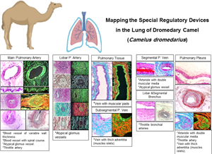
Dromedary camel (Camelus dromedarius) is adapted to survive the harsh environments. It has some key adaptation peculiarities in various organs. In this study, we aimed to map the distribution pattern of unique regulatory devices along the course of the pulmonary vessels using histological and histochemical analyses. Arteries with variable wall thickness and spirally oriented course were recorded within the adventitia of the main pulmonary artery. Throttle arteries and glomus bolsters were found within the wall of the lobar pulmonary artery. The bronchial artery was located within the wall of all bronchi reaching the subsegmental branches and it had elastic longitudinal muscular intima bolsters. Arteries with double muscular media were demonstrated in the pulmonary pleura. These bolsters are suggested to play a complicated role that allows for hemodynamic, humeral, and thermoregulatory activities. The lumen of some subsegmental pulmonary veins revealed occasional constrictions arising from the corresponding muscular pad-like protrusions of the tunica media. These veins may possess occlusive or constrictive mechanisms and their obstruction induces engorgement of the associated capillary bed in addition to restricting venous outflow. Collectively, these data strongly recommend a crucial role for the special regulatory devices in preserving the camel pulmonary function in the harsh desert environment.

Type
Micrographia
Copyright
Copyright © The Author(s), 2022. Published by Cambridge University Press on behalf of the Microscopy Society of America

Access options

Get access to the full version of this content by using one of the access options below. (Log in options will check for institutional or personal access. Content may require purchase if you do not have access.)

Footnotes

Current address: Department of Developmental Biology, Graduate School of Integrated Science for Life, Hiroshima University, Higashi-Hiroshima 739-8526, Japan.

References

Al-Hamad, WH & Khamas, WA (2002). Anatomical and histological features of the pulmonary tissue of the one-humped camel (Camelus dromedaries). J Camel Pract Res 9, 3136.Google Scholar
Dahme, E (1957). €Uber die Beurteilung der Angiopathien bei chronischsklerosierenden Nierenerkrankungen des Hundes. Eine vergleichend morphologische Studie zum Problem der spontanen und experimentellen Arteriosklerose in der Tierheilkunde. Arch Exp Vet Med 11(611–636 u), 752812.Google Scholar
Fath-Elbab, MR & Abou-Elhamd, AS (2016). Special cutaneous vascular elements in one-humped camel (Camelus dromedarius). J Adv Vet Anim Res 3, 106111.10.5455/javar.2016.c137CrossRefGoogle Scholar
Fath El-Bab, MR (1970). A histological study on the respiratory system of camel. PhD Thesis. Egypt: Faculty of Veterinary Medicine, Assiut University.Google Scholar
Gross, CA, Reddy, CK & Dazzo, FB (2010). CMEIAS color segmentation: An improved computing technology to process color images for quantitative microbial ecology studies at single-cell resolution. Microb Ecol 59, 400414.10.1007/s00248-009-9616-7CrossRefGoogle ScholarPubMed
Hussein, MM (2020). Structural and functional characteristics of the special regulatory devices in the peripheral pulmonary circulation in rabbits. Protoplasma 257, 755766.10.1007/s00709-019-01459-yCrossRefGoogle ScholarPubMed
Knudsen, L & Ochs, M (2018). The micromechanics of lung alveoli: Structure and function of surfactant and tissue components. Histochem Cell Biol 150, 661676. doi:10.1007/s00418-018-1747-9.CrossRefGoogle ScholarPubMed
Marini, TJ, He, K, Hobbs, SK & Kaproth-Joslin, K (2018). Pictorial review of the pulmonary vasculature: From arteries to veins. Insights Imaging 9, 971987. doi:10.1007/s13244-018-0659-5CrossRefGoogle ScholarPubMed
Mohamedien, D (2009). Angioarchitecture of the lungs of the camel (Camelus dromedarius). M.V.Sc. Egypt: South Valley University.Google Scholar
Mokhtar, DM & Abd-Elhafez, EA (2015). Morphological studies on the peripheral circulation of the ovary in one-humped camel (Camelus dromedarius).Anat Histol Embryol 45(4), 319328.10.1111/ahe.12204CrossRefGoogle Scholar
Suvarna, KS, Layton, C & Bancroft, JD (2013). Bancroft's Theory and Practice of Histological Techniques, 7th ed. Oxford: Churchill Livingstone.Google Scholar
Tibary, A & El Allali, K (2020). Dromedary camel: A model of heat resistant livestock animal. Theriogenology 154, 203211. doi:10.1016/j.theriogenology.2020.05.046CrossRefGoogle Scholar
Townsley, MI (2013). Structure and composition of pulmonary arteries, capillaries and veins. Compr Physiol 2, 675709.Google Scholar